Published by the University of Cape Town (UCT) under the non-exclusive license granted to UCT by the author. A manual tracing protocol was specifically developed to calculate the volume of the most prominent subcortical gray matter structures implicated in SAD by previous functional research. Cortical thickness analyzes showed significant cortical thinning in the left isthmus of the cingulate gyrus, the left temporal pole, and the left superior temporal gyrus.
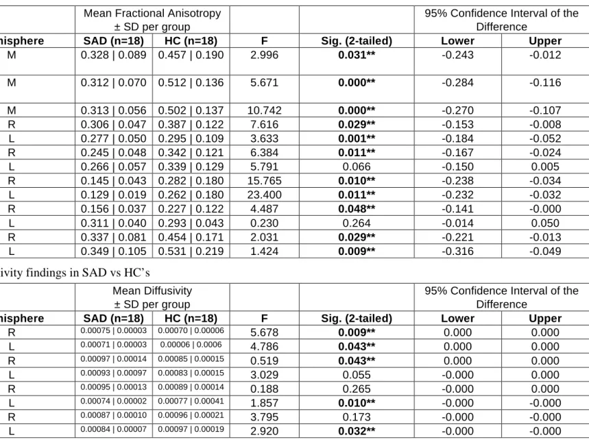
Analyzes of white matter tractography data demonstrated reduced FA in the genu, splenium, and tapetum of the corpus callosum.
C HAPTER I
Fear circuitry
Within the fear/anxiety circuit, amygdala activation is particularly associated with arousal and negative valence (Sergerie et al., 2008), and previous work on fear circuitry has noted a correlation between amygdala volume and craving (Baur et al., 2012). Hyperactivity of the fusiform gyrus (FFG), which has been implicated in an early SAD meta-analysis by Etkin and Wager (2007), may play a role in SAD. In addition to its contribution to face processing (Weiner and Grill-Spector, 2012), the FFG has also been consistently found to be hyperactive in response to emotional scene stimuli in SAD (Sabatinelli et al., 2011).
Therefore, hyperactivation of the FFG in patients with SAD may reflect both an increased sensitivity to facial stimuli and an increased reactivity of the emotional system as a whole in SAD.
Functional Anatomy of Regions of Interest i. Caudate Nucleus
The activity of the caudate nucleus in goal-directed behavior can be observed in actions that take place in a social context (see Montague et al., 2006, for a review). Monchi et al., (2006) did this in a functional imaging study with a similar card sorting task to the WCST. Such input interaction may be the basis of ensemble information processing (Buzsaki et al., 2004) in the nucleus accumbens.
The activation of the hippocampus involves a trisynaptic unidirectional circuit originating from the entorhinal cortex (Donkelaar et al., 2011).
Cortical Thickness
The medial temporal lobe memory system is necessary to link together distributed storage sites in the neocortex that represent an entire memory. The role of this system is only temporary: over time after learning, the memory stored in the neocortex gradually becomes independent of the structures of the medial temporal lobe (Squire 1987; Squire et al., 1991; Squire et al., 2007) . Hyperactivation of the limbic circuit including the hippocampus may mediate SAD symptoms (Hattingh et al., 2013) by influencing behavioral inhibition, anxious temperament and dispositional anxiety (Fox et al., 2014) which in turn may play a role in avoidance behavior seen in SAD.
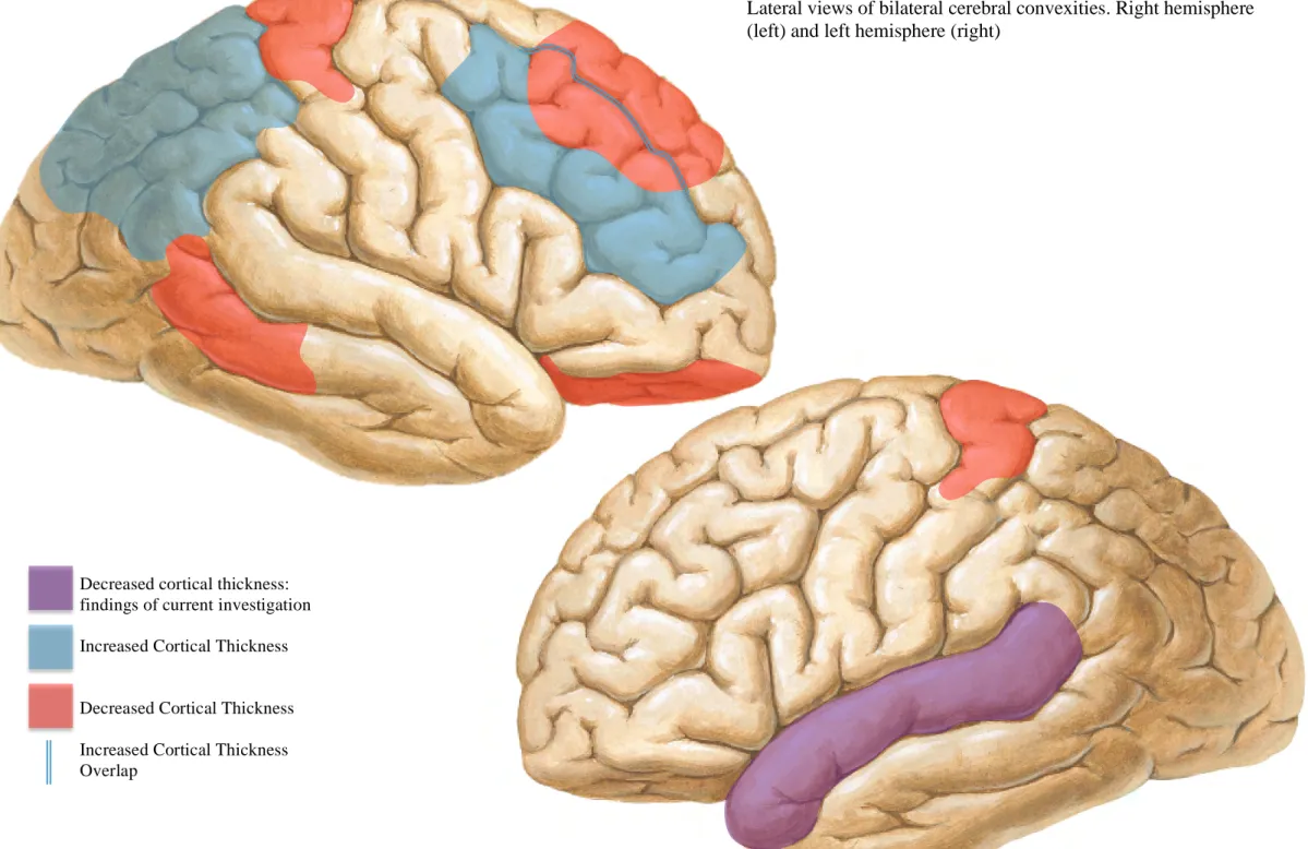
Increased thickness in the DLPFC and superior parietal cortex in SAD is consistent with models of disrupted, overactive, and dysregulated attentional networks in anxiety disorders (Sylvester et al., 2012).
White Matter
Manual Tracing
However, a potential limitation across all these studies is the quality of the clinical sample. In search of the trauma memory: a meta-analysis of functional neuroimaging studies of symptom elicitation in posttraumatic stress disorder (PTSD). The role of the amygdala in emotional processing: a quantitative meta-analysis of functional neuroimaging studies.
The extent and burden of mental and other brain disorders in Europe 2010.
C HAPTER II
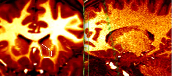
The anterior and posterior dimensions of the nucleus accumbens can best be appreciated in the sagittal plane. The anterior and posterior extent of the nucleus accumbens is best seen in the sagittal plane (Figure 9). The green guidelines intersect the most anterior side of the hippocampus in the axial plane.
C HAPTER III
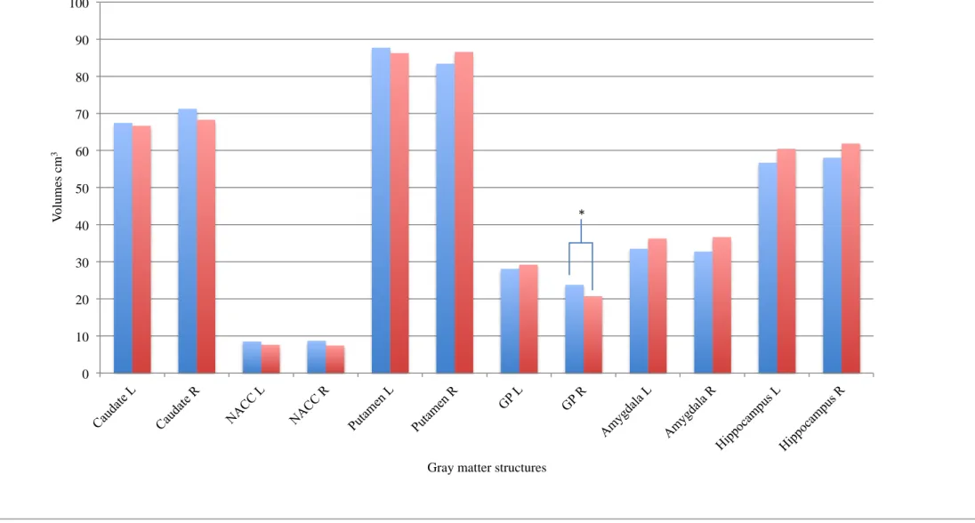
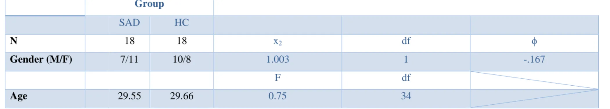
It has previously been noted that the nucleus accumbens itself shows increased functional connectivity in SAD (Brühl et al. 2014). The severity of social anxiety symptoms was assessed using the SPIN (Connor et al., 2000) and the Liebowitz Social Anxiety Scale (LSAS) (Liebowitz, 1987). Regarding the volumetric analysis, there were specific differences in the right globus pallidus.
The volume of the right globus pallidus was significantly increased in the SAD group (p = 0.022) compared to healthy controls. These data do not support the findings reported by Machado-de-Sousa et al. (2014) who found increased volume in the hippocampus and amygdala in SAD patients compared to controls. At the molecular level, dendritic architecture is mediated by brain-derived neurotrophic factor (BDNF) (Lakshminarasimhan et al., 2012).
BDNF expression in the globus pallidus has been previously reported (Kawamoto et al., 1999) under conditions of neurometabolic stress. A closer analysis of the samples enrolled here and in the study by Machado-de-Sousa et al. 2014) may help reduce the discrepancy between findings and shed light on limbic plasticity processes underlying social anxiety. We found structural differences in the right globus pallidus in individuals with SAD compared to controls.
Volumetric differences in the subcortical volumes of gray matter in participants with social anxiety disorder (SAD), healthy controls (HC). -Ferrari MC, Hallak JE, Trzesniak C, Filho AS, Machado-de-Sousa JP, et al. 2010) Neuroimaging in social anxiety disorder: a systematic review of the literature.
C HAPTER IV
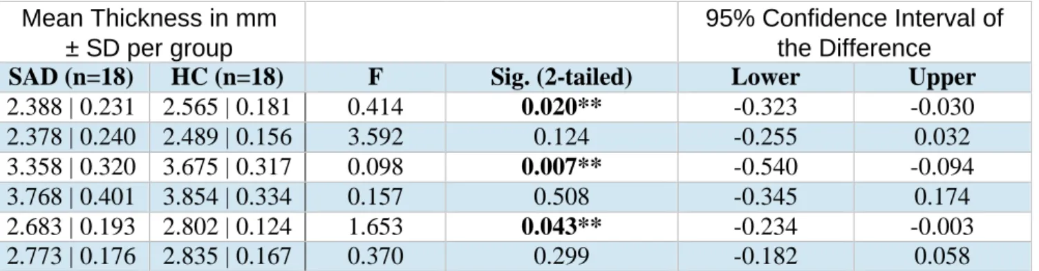
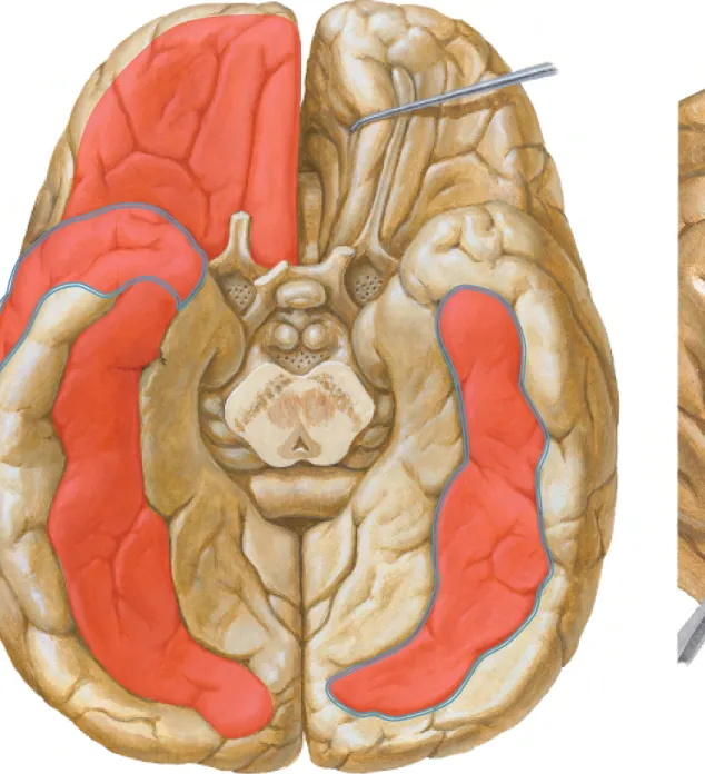
In the SAD group, a negative association was found between social anxiety symptom severity and thickness of the right rostral anterior cingulate cortex. Regarding the cortical thickness analysis, there were specific differences in the left isthmus of the cingulate gyrus (p = 0.020), the left superior temporal gyrus (p = 0.043) and the left temporal pole (p = 0.007). Specifically, socially anxious participants had significant decreases in left-sided cortical thickness in the isthmus of the cingulate gyrus, the superior temporal gyrus, and the temporal pole.
The findings of the current analyzes are in part consistent with what has been previously reported on cortical thickness in SAD. No other cortical thickness study has found involvement of the isthmus of the cingulate gurus or the superior temporal gyrus. In addition, cortical thinning of the superior temporal cortex has also been observed in individuals with other social information processing disorders, such as autism spectrum disorders (ASD) (Wallace et al. 2010).
Functional neuroimaging studies of SAD have shown hyperactivity in various regions of the cingulate cortex (Freitas-Ferrari et al. 2010), and cortical thickness differences may provide a link to the neuroplasticity and excitotoxic effects associated with this disorder. This study found reduced cortical thickness in the left isthmus of the cingulate gyrus, the left temporal pole, and the left superior temporal gyrus in individuals with social anxiety disorder compared to healthy controls. Midline sagittal sections demonstrating the medial aspects of the left (left) and right (right) hemispheres, respectively.
Katche C et al: Functional integrity of the retrosplenial cortex is essential for the consolidation and rapid recall of fear memory. Liao W, Qiu C, Gentili C, Walter M, Pan Z, Ding J, Zhang W et al (2010) Affective connectivity network of the amygdala in social anxiety disorder: a resting-state FMRI study.
C HAPTER V
Analyzes were corrected for multiple comparisons using threshold-free cluster enhancement (TFCE) (Smith et al., 2009). White matter regions were identified using the International Consortium of Brain Mapping (ICBM) DTI-81 white matter atlas ( Mori et al., 2008 ). Anatomical regions surrounding the white matter tracts were identified using the Harvard cortical and subcortical structural atlas (Makris et al., 2006).
Study Key findings and structures implicated Phan et al., 2009 D FA: R uncinate fasciculus near OFC. Liao et al., 2011 D FA: R arcuate fasciculus; C FA: L fronto-occipital fasciculus; C FA: Gen by CC Bauer et al., 2013 D Vol: L uncinate fasciculus; D Average fiber length: L uncinate fasciculus. The output from the hippocampus starts as the alveus, a thin layer of white matter overlying the hippocampus (Lovblad et al., 2014).
From here the loop is completed via projections back to the cingulum or cingulate cortex (Papez et al., 1937). The Papez circuit is mainly involved in the cortical control of emotions (Bear et al., 2006). An increased activity of the cingulate cortex in individuals with SAD compared to HC has previously been reported in SAD (Freitas-Ferrari et al., 2010).
Elevated FA in the fronto-occipital fasciculus has been previously reported by Liao et al., (2011). Decreased FA in the fronto-occipital fasciculus has been previously reported by Bauer et al., (2011).
C HAPTER VI
- Summary of the methodological approach
- Limitations
- Relating current findings to previous research
- Interpretation of current findings
- Future directions
- Concluding remarks
The discussion is organized as follows: 1) summary of the methodological approach used in the current study; This hypothesis was supported by the findings of reduced cortical thickness in the left isthmus of the cingulate gyrus, the left superior temporal gyrus and the left temporal pole in individuals with SAD compared to HCs. This hypothesis was supported by the findings of reduced FA in several elements of the Papez circuit - the cingulum and the fornix.
There is no evidence for increased FA in white matter in most of the existing studies (Phan et al., 2009; In this section, (a) I present a summary of the findings provided by the different techniques used in the current study.Using hand tracing to segment and calculate the volumes of the macroscopically visible subcortical gray matter structures in SAD, I found that the right globus pallidus was significantly enlarged in individuals with SAD compared to healthy controls.
In cortical thickness analyses, we found significant thinning in the left isthmus of the cingulate gyrus, the left superior temporal gyrus, and the left temporal pole. The DTI analyzes in the current study showed reduced structural integrity in several elements of Papez's circuitry – the cingulum and the fornix. Hyperactivity of the cortical components on the Papez circuit, the cingulate cortex, and the parahippocampal/entorhinal and hippocampal complex has been previously reported in SAD (Freitas-Ferrari et al., 2010).
Hyperactive cortical areas project through the limbic loop from the basal ganglia through the globus pallidus. Because the globus pallidus is the primary output nucleus (Standring et al., 2008) of the basal ganglia circuits, this increased work results in dendritic hypertrophy in the globus pallidi.