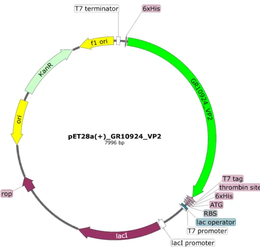
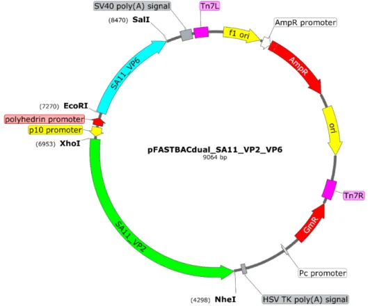
Die ORF's vir VP2 en VP6 is in die pFB-dubbelskenkerplasmied gekloneer vir dubbellaagdeeltjie (DLP) produksie. Die OLRe van VP2 en VP6 is in die pFB-dubbelskenkerplasmied gekloneer vir die produksie van dubbellaagdeeltjies (DLP).
An overview of rotavirus
Recombinantly expressed rotavirus VLPs can elicit humoral and cellular immune responses while being non-infectious in animal models (Fernandez et al., 1998). Chimeric RV-VLPs displaying VP7 and VP4 from human strains can be assembled onto a universal bilayer particle (DLP) backbone, providing an ideal platform for regionally or seasonally specific vaccine candidates (Jere et al., 2014).
Rotavirus classification
Serologic and genetic classification of rotavirus
In 2008, a genome-wide classification system based on the nucleotide sequence and appropriate sequence cutoffs was developed (Mattijnssens et al., 2008). The letter of the genotype name used in the abbreviation is derived from the key feature of the encoded protein (Matthijnssens et al., 2011), as shown in Table 2.
Rotavirus particles, genome structure, and proteins
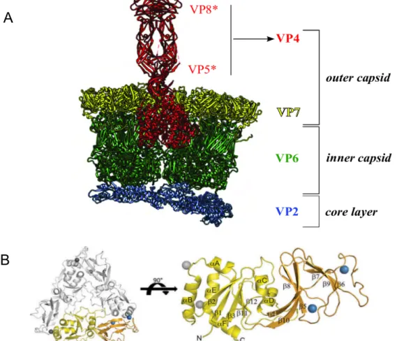
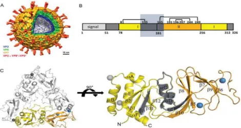
This three-layer particle shows the binding and interaction of structural proteins with each other. Removal of these Ca2+ ions causes destabilization of the VP7 outer layer (Aoki et al., 2009) (Figure 4 B).
Replication cycle of rotavirus
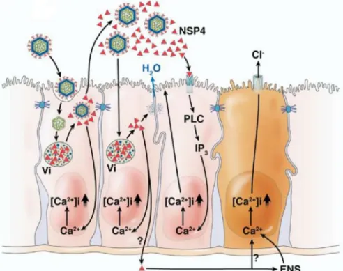
Rotavirus replication occurs in enterocytes of the small intestine, which are rich in proteases (Jayaram et al., 2004). Studies have shown that host cell lysis occurs in non-polarized cells, resulting in cell death (Altenburg et al., 1980).
Pathogenesis and immune response to rotavirus infection
Pathogenesis of rotavirus infection
Because VP7 forms calcium-dependent trimers, calcium is required for VP7 assembly at the DLP when VP7 trimers attach to VP6 trimers (Figure 4) (Chen et al., 2009). IRF3 is required to mediate interferon responses to rotavirus infections (Sen et al., 2011).
Immune response to rotavirus infection
Vaccine related research and development
SA11_VP2_BacO_Reference GGATCTGTA CGACGATGAC SA11_VP2_BacO_sequenced CTAGCWTGAC TGGYGGACAG CAAATGGGTC GGATCTGTA CGACGATGAC. SA11_VP4_BacO_Reference ATG GCCAGCCTGA TTTATCGTCA SA11__VP4_BacO_sequenced GATGACGATA AGGATCATCC CTTCACCATG GCCAGCCTGA TTTATCGTCA.
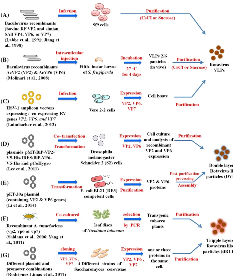
Rotavirus virus-like particles (VLPs) and expression systems
Expression in plant cells
The expressed VP2 and VP6 produced in the tomato fruit have molecular weights similar to those found in native rotaviruses (Figure 8). Similar results were obtained from a study with VP2/VP6 and VP2/VP6/VP7 VLPs produced in tobacco plants (Yang et al., 2011).
Expression in yeast cells
Expression in bacteria
This vector contains a trigger factor, a prokaryotic ribosome-associated chaperone, which increases protein folding during cold shock. The use of cold shock (or heat shock) proteins provides a unique expression system that could aid in the expression of challenging proteins, as illustrated by Naude's work.
Expression in insect cells
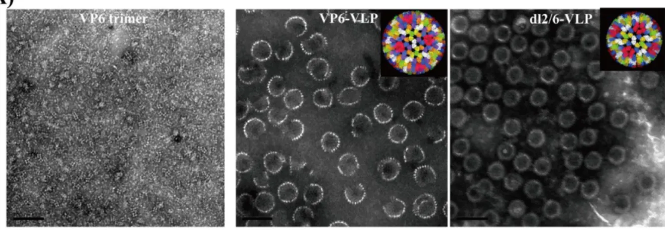
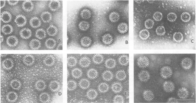
(C) Rotavirus-like particle consisting of VP2/VP6/VP4; (D) Rotavirus-like particle consisting of VP2/VP6/VP7; (E) VP2/VP6/VP4/VP7 TLP particle expressed in insect cells; (F) naturally occurring TLPs of SA11 (Crawford et al., 1994). Chimeric particles were produced using different African strains VP4/VP7 on a DS1-like backbone ( Jere et al., 2014 ).
Motivation and aims of the project
Main objectives of the project
To optimize the expression of single, bilayer, and trilayer VLP assembly by various means such as supplementing the medium with Ca2+, SF9 insect codon optimization, and varying the multiplicity of infection (MOI).
Introduction
This hypothesis was further supported by the fact that the peptide is not required for VLP formation and is excised from VP7 before assembly (during natural infection of eukaryotic cells) (Stirzaker et al., 1990). In 2017, a breakthrough was made in producing whole viruses using a plasmid-only reverse genetics system (Kanai et al., 2017).
Materials and Methods
- Design of plasmids for expression of rotavirus proteins in different
- Preparation and transformation of chemical competent Escherichia coli
- DNA transformation into commercial competent cells
- In-Fusion® HD cloning
- Plasmid isolation
- Spectrophotometric evaluation of nucleic acids
- Agarose gel electrophoresis
- Glycerol stocks for long term storage of bacterial samples
- DNA sequence determination
- Expression of rotavirus proteins using a pET expression system
- Cell lysis using BugBuster protein extraction reagent
- Sodium dodecyl sulphate polyacrylamide gel electrophoresis (SDS
A single colony from the overnight culture was used to inoculate 5 ml of LB and grown overnight with shaking at 225 rpm at 37°C. Samples were electrophoresed at 130 V until the bromophenol blue migrated to the bottom of the resolving gel.
Results and Discussion
Expression of rotavirus VP7-ER in different bacterial cell lines
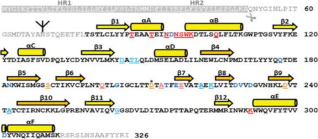
This protein truncation disrupted two disulfide bonds in the tertiary structure of the protein (Aoki et al., 2009) (Figure 15 B). Although the protein produced from this experiment was incomplete, its immunogenic epitopes were still intact and could be used in future work to establish an immunogenic response specific to RV_GR10924 VP7. Both domains are in the same colors as in A; Pattern of intrasubunit disulfide bonds with numbers corresponding to the positions of cysteine residues.
Ribbon diagram of the VP7 trimer (left), with one subunit and one Ca2+ ion pair in color. A single subunit, internal view, is shown on the right, with secondary structure elements labeled (Aoki et al., 2009).
Rotavirus VP2 and VP6 expression
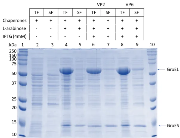
Although the protein's amino acid sequence contains information needed to adjust the protein's tertiary structure, some proteins misfold due to environmental stress and the lack of eukaryotic co-factors. GroEL molecular weight in its monomeric form is 60 kDa and is visible in lanes 4, 6 and 8. GroES molecular weight is 10 kDa but can be seen here at about 14 kDa in both the TF and SF.
No expression for VP2 at 102 kDa could be observed, although overexpression for chaperones could be seen. The addition of potassium phosphates to the media reduced cell death by preventing a decrease in the pH of the media. Since we made these cell lines competent using our one-step methods, a mutation could have occurred, which resulted in these two cell lines not expressing properly.
The highlighted region in lanes 2 through 5 indicates bands that cannot be observed in the uninduced lanes 8 and 9. With the supersaturated protein concentration in each lane, no definite expression can be observed in the sample lanes, nor in the positive control lanes.
Summary
48 Expression of GR10924 VP2/6 and SA11 VP2/6 could not be confirmed by SDS-PAGE analysis, despite the variety of expression conditions used, such as expression time, expression in bacteria co-expressing chaperones (Figure 16), and different media. in the experimental procedures. Due to further time constraints, bacterial expression was halted and thus VP4 expression was not attempted. Since our laboratory previously produced soluble VP6 fused to a trigger factor, the possibility of expressing VP2 and VP6 using this method needs to be re-examined.
Recent studies in our lab focused on expressing glycine-N-acyltransferase, GLYAT used the small ubiquitin-modifying protein (SUMO) tag to express GLYAT in bacteria. Due to the hydrophobic inner core and hydrophilic surface, proteins quickly fold correctly (Butt et al., 2005). The use of the SUMO tag for the expression of rotavirus structural protein should also be explored.
Introduction
Materials and Methods
- The principle of the Bac-to-Bac ® expression system
- Codon optimisation and construction of donor plasmids
- Transformation, purification, and validation of donor plasmids
- Transposition of pFBd expression cassettes into bacmid DNA
- Purification of recombinant bacmid DNA
- PCR analysis of recombinant DNA
- Sanger sequencing of recombinant bacmids
- Propagation of SF9 insect cells
- Transfecting insect cells
- Amplifying recombinant baculoviruses and expression of recombinant
- Viral plaque assay
- Plaque purification
- Viral titre and multiplicity of infection (MOI) determination
- Immunofluorescent monolayer assay (IFMA)
- Western blot analysis
- Isolation of rotavirus virus-like particles for transmission electron
The N-terminal region of the lacZα contains the attachment site for the mini-Tn7 element (mini-attTn7). However, if the transposition of the expression cassette has occurred, the lacZα peptide reading frame will be disrupted. A serial dilution of the transformation mixture was plated overnight at 37°C on LB agar plates containing 100 µg/ml carbenicillin.
This supernatant was centrifuged again to ensure that no white precipitate was present in the final reactions. Cells were detached from the bottom of the well and the supernatant was collected from each well and centrifuged at 500 x g for 5 min to pellet the cells and debris. Cells were detached from the bottom of the well and the supernatant was collected from each well and centrifuged at 500 x g for 5 min to pellet the cells and large debris.
After 1 hour of incubation, the medium containing the virus was removed and replaced with 2 ml of plating medium [50%]. Proteins in the blocking buffer bind to all remaining surfaces of the plate.
Results and Discussion
- Generating three SA11 recombinant bacmids
- Generation of recombinant baculoviruses expressing SA11 VP2/VP6
- Baculovirus protein expression of SA11 VP2/VP6
- Verification of assembly of SA11 VP2/VP6 DLPs
- Baculovirus expression of SA11 VP4 and SA11 VP7
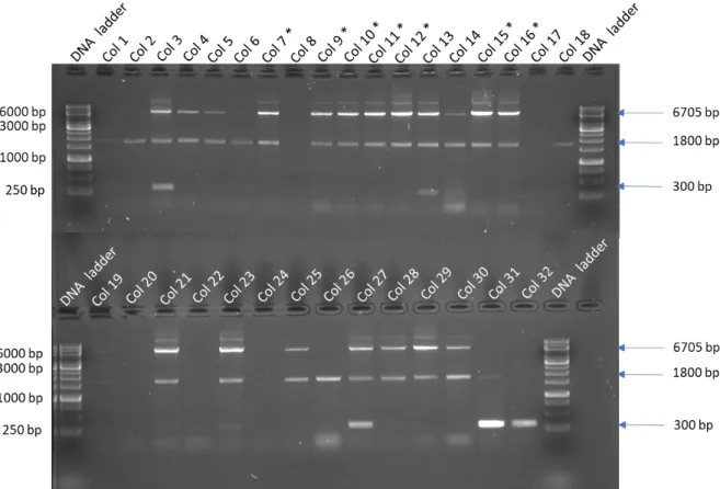
PCR analysis was performed on 30 colonies containing pFBd_SA11_VP2/VP6 to verify that recombinant bacmids were obtained and to identify colonies with a mixture of recombinant and wild-type bacmids (bull's eyes). Expected PCR amplicon size of recombinant SA11 VP2/VP6 bacmid is 6,705 bp and negative control is 300 bp. No blue colony containing wild-type bacmid was used as the VP2/VP6 PCR results already contained wild-type bacmid used in all the experiments.
However, the positive response to recombinant SA11 VP2/VP6 clearly indicates that the baculovirus expresses rotavirus SA11 VP2/VP6. After plates were selected, cells from 6-well serial dilution plates were used for IFMA to determine if it was possible to see if the selected plate contained recombinant VP2/VP6 (Figure 30). Soluble fraction; Lane 1: Protein molecular weight marker; Lane 2: Colony 11 plate 1 total fraction; Lane 3: Colony 11 plate 1 soluble fraction; Lane 4: Colony 11 plate 2 total fraction; Lane 5: Colony 11 plate 2 soluble fraction; Lane 6: Colony 15 plate 1 total fraction; Lane 7: Colony 15 plate 1 soluble fraction; Lane 8: Total fraction of wild-type baculovirus; Lane 9: Soluble fraction of wild-type baculovirus; Lane 10: Protein molecular weight marker.
Large-scale production of rotavirus VP2/VP6 was performed by making a 50 ml suspension culture of SF9 cells infected at a cell density of 1 x 10⁶ cells/ml with baculovirus expressing rotavirus SA11 VP2/VP6. The primary antibody used in this experiment was anti-SA11_RV DLP VP2/VP6 rabbit whole serum.
Summary
SA11_VP2_BacO_reference GATAAGGATC ATCCCTTCAC CATGGCCTAT CGTAAACGTG GTGCACGTCG SA11_VP2_BacO_sequenced GATAAGGATC ATCCCTTCAC CATGGCCTAT CGTAAACGTG GTGCACGTCG. SA11_VP2_BacO_reference TGAAACCAAT CTGAAACAGG ATGAACGTAT GCAAGAAAAA GAGGACAGCA SA11_VP2_BacO_sekventeret TGAAACCAAT CTGAAACAGG ATGAACGTAT GCAAGAAAAA GAGGACAGCA. SA11_VP2_BacO_reference GAATGTGAAC TTCCACAGCA ACTATAACGA ACGCATTAAT GATGCCGTTG SA11_VP2_BacO_sekventeret GAATGTGAAC TTCCACAGCA ACTATWACGA ACGCATTAAT GATGCCGTTG.
SA11_VP2_BacO_reference CCGGTGGAAA TTCGTCGTCT GGATATCTTT AATCTGATCG CAATGAACAT SA11_VP2_BacO_sequenced CCGGTGGAAA TTCGTCGTCT GGATATCTTT AATCTGATCG CAATGAACAT. SA11_VP2_BacO_reference GGAACAAATT GAACGTGCGA GCGATAAAAT TGCACAGGGT GTTATTATTG SA11_VP2_BacO_sekventeret GGAACAAATT GAACGTGCGA GCGATAAAAT TGCACAGGGT GTTATTATTG. SA11_VP2_BacO_reference CCTATCGTGA TATGCAGCTT GAACGCGACG AAATGTATGG CTATGTTAAT SA11_VP2_BacO_sequenced CCTATCGTGA TATGCAGCTT GAACGCGACG AAATGTATGG CTATGTTAAT.
SA11_VP2_BacO_reperensia ATTGCACGCA ACCTGGATGG TTTCCAGCAG ATTAACCTGG AAGAACTGAT SA11_VP2_agsasaruno_ti_BacO ATTGCACGCA ACCTGGATGG TTTCCAGCAG ATTAACCTGG AGAACTGAT. SA11_VP2_BacO_reperensia GCGTAGTGGC PANANGAKSESION TATGCTGCTT FATHERCAGC SA11_VP2_BacO_sequence GCGTAGTGGC PANANGAKSESIONGCAC PANANGAKSESION TATGCTGCTT AMACAGC. SA11_VP2_BacO_reperensia CGGTTGCACT GGTTGGTGCA CTGCCGTTTA TACCGTAG CAGCGTTATT SA11_VP2_BacO_naurnos CGGTTGCACT GGTTGGTGCA CTGCCGTTTA TTACCGTAT CAGCGTTAT.
SA11_VP2_BacO_reference AGCCTGATTG CCAAACTGGA TGCCACCGTT TTTGCCCAGA TTGTTAAACT SA11_VP2_BacO_sequenced AGCCTGATTG CCAAACTGGA TGCCACCGTT TTTGCCCAGA TTGTTAAACT.