Pharmacological evaluation of South African medicinal plants used for treating tuberculosis and related
symptoms
By
Balungile Madikizela
Submitted in fulfilment of the requirements for the degree of Doctor of Philosophy
Research Centre for Plant Growth and Development School of Life Sciences
University of KwaZulu-Natal, Pietermaritzburg
November, 2014
i
College of Agriculture, Engineering and Science Declaration 1 - Plagiarism
I, Balungile Madikizela (209523515), declare that
1. The research reported in this thesis, except where otherwise indicated, is my original research.
2. This thesis has not been submitted for any degree or examination at any other university.
3. This thesis does not contain other persons’ data, pictures, graphs or other information, unless specifically acknowledged as being sourced from other persons.
4. This thesis does not contain other persons' writing, unless specifically acknowledged as being sourced from other researchers. Where other written sources have been quoted, then:
a. Their words have been re-written but the general information attributed to them has been referenced
b. Where their exact words have been used, then their writing has been placed in italics and inside quotation marks, and referenced.
5. This thesis does not contain text, graphics or tables copied and pasted from the Internet, unless specifically acknowledged, and the source being detailed in the thesis and in the References sections.
Signed:……….November, 2014
Form EX1-5
ii
Student Declaration
Pharmacological evaluation of South African medicinal plants used for treating tuberculosis and related symptoms
I, Balungile Madikizela Student Number 209523515 declare that:
(i) The research reported in this dissertation, except where otherwise indicated, is the result of my own endeavours in the Research Centre for Plant Growth and Development, School of Life Sciences, University of KwaZulu-Natal Pietermaritzburg;
(ii) This dissertation has not been submitted for any degrees or examination at any other University;
(iii) This thesis does not contain data, figures or writing, unless specifically acknowledged, copied from other researchers; and
(iv) Where I have produced a publication of which I am an author or co-author, I have indicated which part of the publication was contributed by me.
Signed at UKZN Pietermaritzburg campus on the day of November, 2014.
SIGNATURE……….
iii
Declaration by Supervisors
We hereby declare that we acted as Supervisors for this PhD student:
Student’s Full Name: Balungile Madikizela
Student Number: 209523515
Thesis Title: Pharmacological evaluation of South African medicinal plants used for treating tuberculosis and related symptoms
Regular consultation took place between the student and ourselves throughout the investigation. We advised the student to the best of our ability and approved the final document for submission to the College of Agriculture, Engineering and Science Higher Degrees Office for examination by the University appointed Examiners.
__________________________
SUPERVISOR: PROFESSOR J. VAN STADEN
________________________
CO-SUPERVISOR: PROFESSOR J.F. FINNIE
iv
Publications from this Thesis
1. Madikizela, B., Ndhlala, A.R., Finnie, J.F., Van Staden, J., 2013. In vitro antimicrobial activity of extracts from plants used traditionally in South Africa to treat tuberculosis and related symptoms. Evidence-Based Complementary and Alternative Medicine, doi:org/10.1155/2013/840719.
2. Madikizela, B., Ndhlala, A.R., Finnie, J.F., Van Staden, J., 2014. Antimycobacterial, anti- inflammatory and genotoxicity evaluation of plants used for the treatment of tuberculosis and related symptoms in South Africa. Journal of Ethnopharmacology 153, 386-391.
3. Madikizela, B., Aderogba, M.A., Finnie J.F., Van Staden J., 2014. Isolation and characterization of antimicrobial compounds from Terminalia phanerophlebia Engl. & Diels leaf extracts. Journal of Ethnopharmacology 156, 228-234.
v
College of Agriculture, Engineering and Science Declaration 2 - Publications
DETAILS OF CONTRIBUTION TO PUBLICATIONS that form part and/or include research presented in this thesis (include publications in preparation, submitted, in press and published and give details of the contributions of each author to the experimental work and writing of each publication)
Publication 1
Contributions: All experimental work and draft manuscript were performed by BM under guidance and supervision of ARN, JVS and JFF.
Publication 2
Contributions: All experimental work and draft manuscript were done by BM. ARN helped with experimental design. JVS and JFF supervised the study and edited the manuscript before submission.
Publication 3
Contributions: All experimental work and draft manuscript were done by BM. MAA assisted with the identification of compounds. JVS and JFF supervised and edited the manuscript before submission.
Author’s abbreviation
BM Balungile Madikizela ARN Ashwell R. Ndhlala MAA Mutalib A. Aderogba JFF Jeffrey F. Finnie JVS Johannes Van Staden
Signed:…………...………..
Form EX1-6
vi
Conference Contribution from this Thesis
1. Madikizela, B., Finnie, J.F., Van Staden, J., 2012. In vitro antimycobacterial activity of plants used in South Africa to treat tuberculosis and related symptoms. 14th Annual Meeting Research Centre for Plant Growth and Development, 28-30 November 2012.
University of KwaZulu-Natal, Pietermaritzburg. Oral presentation.
2. Madikizela, B., Finnie, J.F., Van Staden, J., 2013. In vitro antimycobacterial activity of plants used in South Africa to treat tuberculosis and related symptoms. 39th Annual Conference of the South African Association of Botanists (SAAB) and the 9th Southern African Society for Systematic Biology (SASSB), 20-23 January 2013. Drankensburg.
Oral presentation.
3. Madikizela, B., Finnie, J.F., Van Staden, J., 2013. In vitro antimicrobial activity of extracts from plants used traditionally in South Africa to treat tuberculosis and related symptoms. 15th Annual Meeting Research Centre for Plant Growth and Development, 14-15 November 2013. University of KwaZulu-Natal, Pietermaritzburg. Oral presentation.
vii
Acknowledgements
Firstly, I thank God for his protection and for giving me a strong will and courage to pursue my studies.
The completion of this thesis was not an effort of my individual input but rather a collective effort of many people who contributed in various ways to help me to complete this work.
Without their input, guidance, help, and encouragement it would not have been easy to accomplish this work.
I extend my sincere gratitude to my supervisor, Professor J. Van Staden for his exceptional advice, encouragement and guidance throughout the study. I am tremendously grateful to my co-supervisor Dr J.F. Finnie for his encouragement, invaluable advice, understanding and constructive criticism on thesis write-up. I thank Dr A.R. Ndhlala, a member of my research committee for his support and guidance in the early stages of this project. I am extremely grateful to Dr M.A. Aderogba for his assistance in identifying the compounds described in this thesis; and Dr L. McGaw for being instrumental in the cytotoxicity aspect of this work.
I thank the National Research Foundation (NRF), Canon Collins GreenMatter and UKZN for financially supporting my PhD programme.
I am extremely grateful to my mentor Mr D. Hay for always being there for me as my sounding board and thinking partner. I couldn’t have asked for a better mentor (courtesy of being a GreenMatter Fellow).
My utmost thanks go to Mrs A. Young (Horticulturalist UKZN Botanical Gardens), S.
Ghuman for their assistance in plant collection. The NU Herbarium Staff for their assistance in plant identification and voucher specimen preparations. A special mention goes to the Administrative Staff who kept various activities running in the centre. I thank the technical and the Botanic garden Staff. You have all been so supportive.
viii
My special thanks go to all the Research Centre members for Plant Growth and Development for their friendly advice and encouragement. It was a great pleasure working with you.
I am thankful to my parents Mr A.C. and Mrs B.S. Madikizela, my siblings, nephews and nieces for their amazing love, support and encouragement throughout my studies. You are all my pillar of strength.
I thank all my friends for their support, encouragement and for being there when I needed them the most.
Lastly, I offer my regards and blessings to all of those who supported me during the completion of this project. “Makube chosi kube hele”.
ix
Table of Contents
College of Agriculture, Engineering and Science Declaration 1 - Plagiarism ... i
Student Declaration ... ii
Declaration by Supervisors ... iii
Publications from this Thesis ... iv
College of Agriculture, Engineering and Science Declaration 2 - Publications ... v
Conference Contribution from this Thesis ... vi
Acknowledge ments ... vii
Table of Contents ... ix
List of Figures... xiv
List of Tables ... xvi
List of Abbreviations ... xvii
Abstract... 1
Chapter 1: Introduction and lite rature review ... 4
1.1. Introduction ... 4
1.2. History of the medicinal use of plants by humans ... 4
1.3. An overview of traditional medicine systems ... 6
1.4. South Africa’s traditional plant based medicines... 8
1.5. Secondary metabolites... 11
1.6. Drug discovery from medicinal plants ... 11
1.7. Respiratory tract infections ... 13
1.7.1. Overview of tuberculosis ... 13
1.7.2. Historical viewpoint of tuberculosis ... 14
1.7.3. Mycobacterium ... 16
1.7.4. Global effect of tuberculosis... 17
1.7.5. Modern treatment of tuberculosis ... 20
x
1.7.6. Measures taken to control tuberculosis worldwide ... 24
1.7.7. Overview of medicinal plants in treating tuberculosis ... 25
1.8. Medicinal plant trade... 27
1.9. Conservation of medicinal plants ... 28
1.10. Aims and objectives ... 30
1.11. Plant selection ... 31
1.12. Botanical description and distribution of the plants selected ... 32
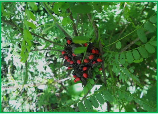
1.12.1. Abrus precatorius subsp. africanus Verdc. ... 32
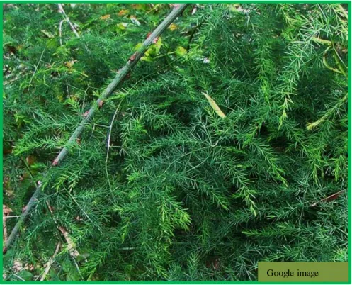
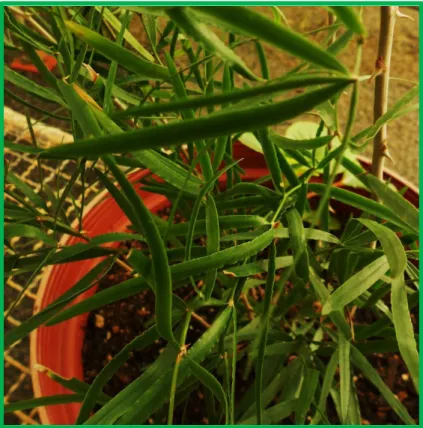
1.12.2. Asparagus africanus Lam. ... 33
1.12.3. Asparagus falcatus (L.) Oberm. ... 34
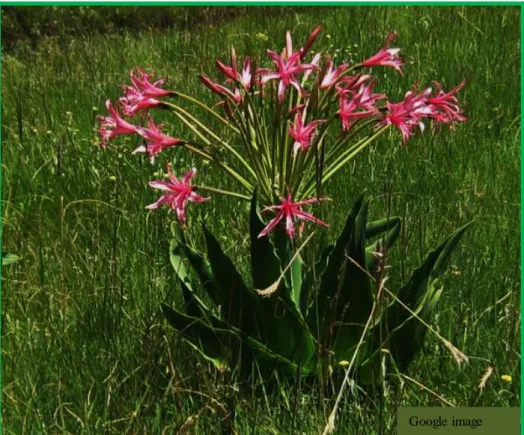
1.12.4. Brunsvigia grandiflora Lindl... 35
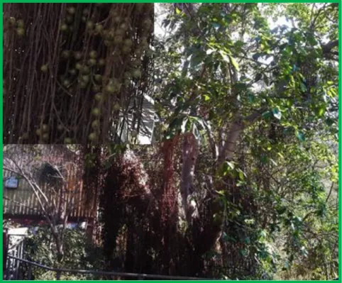
1.12.5. Ficus sur Forssk... 36
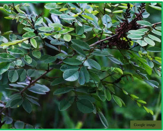
1.12.6. Indigofera arrecta Benth. ex Harv. & Sond. ... 37
1.12.7. Leonotis intermedia Lindl... 38
1.12.8. Pentanisia prunelloides Schinz ... 38
1.12.9. Polygala fruticosa P. J. Bergius ... 39
1.12.10. Terminalia phanerophlebia Engl. &Diels ... 40
Chapter 2: In vitro antimicrobial activity of extracts from plants used traditionally in South Africa to treat tuberculosis and related symptoms ... 42
2.1. Infectious diseases ... 42
2.1.1. Bacterial infections ... 43
2.2. Drug-resistance... 45
2.2.1. Multi drug-resistant tuberculosis ... 46
2.2.2. Extensive drug-resistant tuberculosis ... 47
2.3. Ethnopharmacological approach to drug resistance problem ... 48
2.4. Bacterial strains related to respiratory ailments that were selected in this study ... 49
2.4.1. Klebsiella pneumoniae... 49
xi
2.4.2. Staphylococcus aureus... 50
2.4.3. Mycobacterium aurum ... 50
2.4.4. Mycobacterium tuberculosis H37Ra ... 51
2.5. Materials and methods ... 52
2.5.1. Preparation of plant extracts ... 52
2.5.2. Bacterial strains and culture conditions ... 52
2.5.3. Procedure for microorganism long term storage ... 52
2.5.4. Subculturing for bioassays... 53
2.5.5. Antibacterial activity using microdilution ... 54
2.5.6. Antimycobacterial activity using the resazurin microplate assay... 54
2.6. Results and discussions ... 55
2.6.1. Plant extraction ... 61
2.6.2. Antimicrobial and antimycobacterial assay ... 68
2.7. Conclusions ... 78
Chapter 3: Inhibition of cyclooxygenase enzyme as an evaluation of anti-inflammatory property of selected plants ... 79
3.1. Introduction ... 79
3.2. Overview of inflammation response ... 80
3.3. Cyclooxygenase enzymes ... 81
3.4. Inflammation and tuberculosis ... 83
3.5. Anti- inflammatory agents ... 84
3.6. Materials and methods ... 86
3.6.1. Preparation of plant extracts ... 86
3.6.2. Substrate and enzyme preparation ... 86
3.6.3. Cyclooxygenase inhibitory activity ... 87
3.7. Results and discussions ... 88
3.7.1. Anti- inflammatory activity ... 88
xii
3.8. Conclusions ... 92
Chapter 4: Genotoxicity and cytotoxicity evaluation of biologically active extracts from plants used traditionally for treating tuberculosis and related symptoms in South Africa ... 93
4.1. Introduction ... 93
4.2. Mutagenicity... 94
4.3. Negative effect of mutagenicity in human beings... 94
4.4. Genotoxicity and cytotoxicity ... 95
4.4.1. Genotoxicity testing methods ... 96
4.4.2. Cytotoxicity testing methods ... 97
4.5. Materials and methods ... 100
4.5.1. Preparation of plant extracts for genotoxicity testing ... 100
4.5.2. In vitro genotoxicity evaluation of biologically active extracts using the Ames test ... 100
4.5.3. In vitro cytotoxicity testing of biologically active extracts using the MTT assay 101 4.6. Results and discussion... 102
4.6.1. Genotoxicity ... 102
4.6.2. Cytotoxicity ... 106
4.7. Conclusions ... 111
Chapter 5: Isolation and characterisation of antimicrobial compounds from Terminalia phanerophlebia Engl. & Diels leaf extracts ... 112
5.1. Introduction ... 112
5.2. Traditional uses and pharmacological studies of Terminalia phanerophlebia ... 113
5.3. Materials and methods ... 115
5.3.1. Bioassay guided fractionation of 80% methanol extracts of Terminalia phanerophlebia leaves ... 115
5.3.2. General ... 116
xiii
5.3.3. Plant material-collection and authentication... 116
5.3.4. Sample extraction ... 116
5.3.5. Solvent partitioning of the crude extracts ... 117
5.3.6. Isolation of compounds from Terminalia phanerophlebia ethyl acetate fraction 117 5.3.7. Structure elucidation of isolated compounds... 119
5.3.8. Antimicrobial assay procedure ... 120
5.4. Results and discussion... 120
5.4.1. Characterisation of compounds 1 and 2 ... 120
5.4.2. Antimicrobial assays results ... 121
5.5. Conclusions ... 147
Chapter 6: General conclusions ... 148
References ... 151
xiv
List of Figures
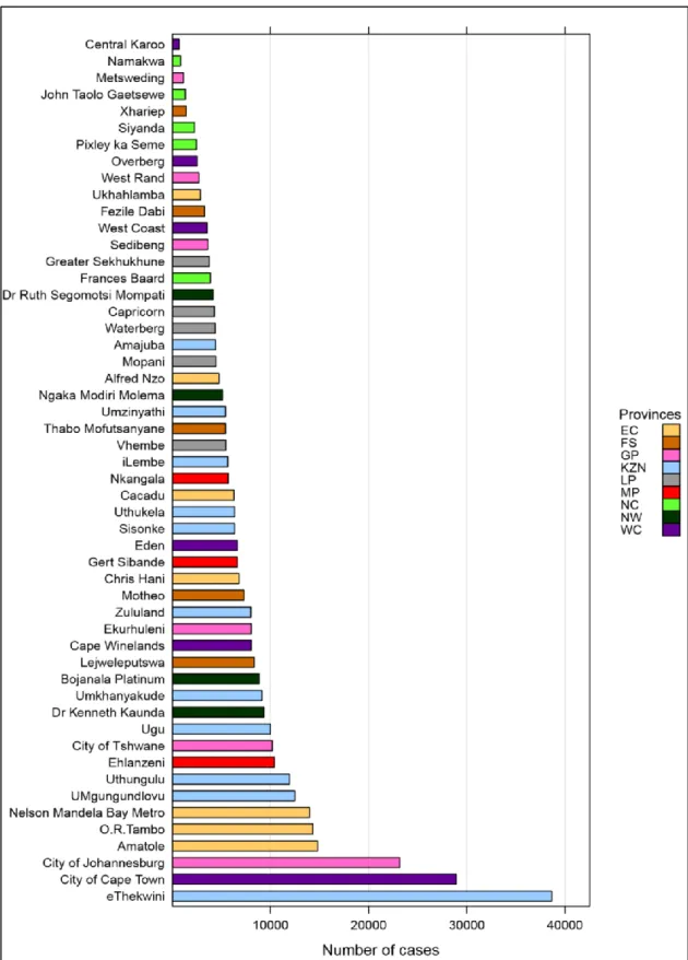
Figure 1.1: Number of tuberculosis cases reported by district in South Africa, 2008. EC, Eastern Cape; FS, Free State; GP, Gauteng province; KZN, KwaZulu-Natal; LP, Limpopo province; MP, Mpumalanga province; NC, Northern Cape; NW, North West, WC, Western
Cape (WHO, 2009b). ... 19
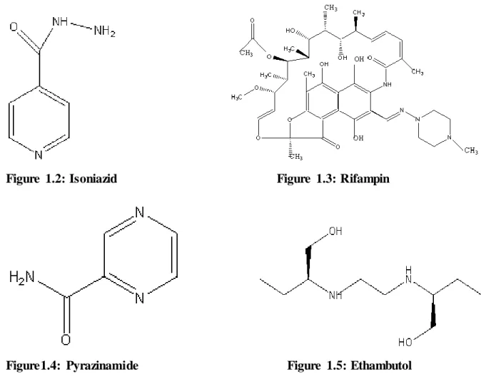
Figure 1.2: Isoniazid ... 21
Figure 1.3: Rifampin………21
Figure1.4: Pyrazinamide ... 21
Figure 1.5: Ethambutol……….21
Figure 1.6: Kanamycin... 22
Figure 1.7: Abrus precatorius subsp. africanus ... 32
Figure 1.8: Asparagus africanus (DEEDI, 2011) ... 33
Figure 1.9: Asparagus falcatus ... 34
Figure 1.10: Brunsvigia grandiflora (McMASTER et al., 2010) ... 35
Figure 1.11: Ficus sur. Inset shows the fruit of Ficus sur. ... 36
Figure 1.12: Indigofera arrecta (HYDE et al., 2014) ... 37
Figure 1.13: Leonotis intermedia ... 38
Figure 1.14: Pentanisia prunelloides. Inset shows the flowers of Pentanisia prunelloides. ... 39
Figure 1.15: Polygala fruticosa (VILJOEN and HITCHCOCK, 2002) ... 40
Figure 1.16: Terminalia phanerophlebia. Inset shows the fruit of Terminalia phanerophlebia ... 41
Figure 2.1: Percentage of global new tuberculosis cases with MDR tuberculosis in 2012 (WHO, 2013). ... 47
Figure 5.1: Fractionation scheme of the crude extracts from the leaves of Terminalia phanerophlebia ... 119
Figure 5.2(a): 1H NMR spectrum of Compound 1 ... 124
Figure 5.2(b):1H NMR spectrum of Compound 1 ... 125
Figure 5.3:13C NMR spectrum of Compound 1 ... 126
xv
Figure 5.4: DEPT NMR spectrum of Compound 1 ... 127
Figure 5.5: HSQC NMR spectrum of Compound 1 ... 128
Figure 5.6: HMBC NMR spectrum of Compound 1 ... 129
Figure 5.7(a): 1H NMR spectrum of Compound 2 ... 130
Figure 5.7(b): 1H NMR spectrum of Compound 2 ... 131
Figure 5.7(c): 1H NMR spectrum of Compound 2 ... 132
Figure 5.8(a): 13C NMR spectrum of Compound 2 ... 133
Figure 5.8(b): 13C NMR spectrum of Compound 2 ... 134
Figure 5.8(c): 13C NMR spectrum of Compound 2 ... 135
Figure 5.9: DEPT NMR spectrum of Compound 2 ... 136
Figure 5.10(a): COSY NMR spectrum of Compound 2 ... 137
Figure 5.10(b):COSY NMR spectrum of Compound 2 ... 138
Figure 5.11(a): HSQC NMR spectrum of Compound 2 ... 139
Figure 5.11(b): HSQC NMR spectrum of Compound 2 ... 140
Figure 5.11(c): HSQC NMR spectrum of Compound 2 ... 141
Figure 5.12 (a): HMBC NMR spectrum of Compound 2 ... 142
Figure 5.12(b): HMBC NMR spectrum of Compound 2 ... 143
Figure 5.13: Some definitive HMBC correlations of compound 2... 145
Figure 5.14: Structures of isolated compounds from Terminalia phanerophlebia... 145
xvi
List of Tables
Table 2.1: Medicinal plants used traditionally in South Africa to treat tuberculosis and related symptoms ... 57 Table 2.2: Percentage yields of extracts from plants used in this study ... 63 Table 2.3: Antibacterial (MIC values) effects of plants used traditionally as remedies in the treatment of tuberculosis and related symptoms in South Africa ... 72 Table 3.1: Anti-inflammatory activity (COX-2) of extracts from plants that are used for the treatment of tuberculosis and related symptoms in South Africa ... 91 Table 4.1: Number of revertant colonies of Salmonella typhimurium strains TA98 and TA100 induced by extracts of some plants used as remedies for the treatment of tuberculosis and related symptoms in South Africa... 104 Table 4.2: Average LC50, and selectivity index values of bioactive plant extracts ... 109 Table 5.1: Solvent systems used in column chromatography ... 118 Table 5.2: 1H (500 MHz) and 13C (125 MHz) NMR chemical shifts of compound 2 (MeOD) ... 144 Table 5.3: Antibacterial activity of the crude extracts, solvent fractions, column fractions from ethyl acetate sample and isolated compounds from the leaves of Terminalia phanerophlebia ... 146
xvii
List of Abbreviations
4-NQO 4-nitroquinoline-1-oxide
AA Arachidonic acid
AIDS Acquired immunodeficiency syndrome
ATCC American type culture collection
ATM African traditional medicine
BCG Bacillus calmette-guèrin
CAM Complementary and alternative medicine
CFU/ml Colony forming units/ml
COX Cyclooxygenase
CPE Cytopathic effects
DCM Dichloromethane
DMSO Dimethylsulfoxide
DNA Deoxyribonucleic acid
DOT Directly observed treatment short course
DPM Disintegration per minute
DPM background Disintegration per minute background
DPM solvent blank Disintegration per minute solvent blank
DPM extract Disintegration per minute extract
DW Dry weight
EtOAc Ethyl acetate
EtOH Ethanol
FCS Foetal calf serum
HCL Hydrochloric acid
HIV Human immunodeficiency virus
HPLC High performance liquid chromatography
ICC Interim coordinating committee
INH Isoniazid
LC50 Lowest concentration 50
LDH Lactate dehydrogenase
LPS Lipopolysaccharides
xviii
MDR tuberculosis Multi drug-resistant tuberculosis
MECs Members of executive council
MEM Minimal essential medium
MeOH Methanol
MH Mueller-Hinton
MHz MegaHertz
MIC Minimum inhibitory concentration
MRC Medical Research Council
MTT Mitochondrial reduction
NEMA National Management Act
NMR Nuclear magnetic resonance
NRF National Research Foundation
NSAIDs Non-steroidal anti-inflammatory drugs
NTM Non-tuberculous mycobacteria
NU Natal University
OADC Oleic acid, albumin, dextrose, and catalase
PBS Phosphate buffered saline
PGE2 Prostaglandin E2
PG12 Prostaglandin 12
PE Petroleum ether
REMA Resazurin microplate assay
TCM Traditional Chinese medicine
TLC Thin layer chromatography
TLR-4 Toll-like receptor-4
TM Traditional medicine
TRIS Tris(hydromethyl)aminomethane
UKZN University of KwaZulu-Natal
USA United States of America
WHO World Health Organisation
XDR tuberculosis Extensive drug-resistant tuberculosis
1
Abstract
Respiratory ailments are major human killers, especially in developing countries including South Africa. Tuberculosis is one of the most prevalent infectious respiratory tract disease posing a major threat to human healthcare worldwide. This disease is a highly contagious airborne bacterial disease that usually infects the lungs and sometimes other body parts.
Tuberculosis spreads easily in overcrowded conditions from one person with an active respiratory disease to another via droplets that are emitted when they sneeze or cough.
Approximately two million deaths that occur worldwide per annum are caused by tuberculosis and about 285,000 cases occur in South Africa. This is the seventh highest total number in the world. The emergence of drug-resistant tuberculosis and other pathogenic diseases over the past decades makes this disease a serious threat to human health worldwide.
Emerging drug-resistant tuberculosis strains and the long duration of treatment has established an urgent need to search for new effective agents. According to a 2012 report by the World Health Organisation (WHO), South Africa, China, India and Russia are the countries with the highest prevalence of multi drug-resistant (MDR) tuberculosis.
Most researchers in South Africa have focused on evaluating the antimycobacterial activity of medicinal plants against bacterial strains that cause tuberculosis, but there has not been sufficient focus on the related ailments. Therefore, one of the aims of the present study was the evaluation of the antimicrobial properties of the selected medicinal plants against Mycobacterium species and other bacterial strains related to respiratory infection. The floral diversity of South Africa has a potential for yielding new bioactive compounds, therefore pharmacological screening of plant extracts from this region is important. The aim of this study was the pharmacological evaluation of plants that are used traditionally in South Africa to treat tuberculosis and related symptoms against microorganisms that cause respiratory ailments, and the identification of compounds from antimicrobial active plant extracts.
Ten plants: Abrus precatorius subsp. africanus (leaves and seeds), Asparagus africanus (leaves), Asparagus falcatus (leaves), Brunsvigia grandiflora (bulb), Ficus sur (bark and roots), Indigofera arrecta (leaves and roots), Leonotis intermedia (leaves and stem), Pentanisia prunelloides (leaves and roots), Polygala fruticosa (whole plant), and Terminalia phanerophlebia (leaves, roots and twigs) were selected based on a survey of available
2
literature of medicinal plants used in South Africa for the treatment of tuberculosis and related symptoms. Ground plant material from different plant parts of the 10 plants were extracted sequentially with four solvents: petroleum ether (PE), dichloromethane (DCM), 80% ethanol (EtOH) as well as water, and a total of 68 extracts were produced. The plant extracts of the selected plants were evaluated for antibacterial activity against four microorganisms (Klebsiella pneumoniae, Staphylococcus aureus, Mycobacterium aurum A+
and Mycobacterium tuberculosis H37Ra) associated with respiratory infections using the microdilution assay. Cyclooxygenase-2 (COX-2) enzyme was used to evaluate the anti- inflammatory activity of the extracts. The Ames test and mitochondrial reduction (MTT) assays were used to establish toxicity of the extracts that showed noteworthy antimicrobial activity against the tested bacterial strains (Klebsiella pneumoniae, Staphylococcus aureus, Mycobacterium aurum A+ and Mycobacterium tuberculosis H37Ra). The extracts were tested for genotoxicity against Salmonella typhimurium (TA98 and TA100 strains) and cytotoxicity using monkey kidney Vero cells. Based on good antimicrobial activity observed, compounds were isolated from Terminalia phanerophlebia (leaves). Crude extracts obtained from 80% methanol (MeOH) of Terminalia phanerophlebia were successively extracted with hexane, DCM, ethyl acetate (EtOAc) and n-butanol. The fractions and isolated compounds obtained were tested for their antibacterial activity against Mycobacterium aurum A+, Mycobacterium tuberculosis H37Ra, Staphylococcus aureus and Klebsiella pneumoniae.
Structure elucidation was carried out using NMR (1D and 2D) spectroscopic methods.
This investigation revealed the pharmacological potential of the 10 plants used in South Africa for traditional treatment of tuberculosis and related symptoms: Abrus precatorius subsp. africanus (leaves and seeds), Asparagus africanus (leaves), Asparagus falcatus (leaves), Brunsvigia grandiflora (bulb), Ficus sur (bark and roots), Indigofera arrecta (leaves and roots), Leonotis intermedia (leaves and stem), Pentanisia prunelloides (leaves and roots), Polygala fruticosa (whole plant), and Terminalia phanerophlebia (leaves, roots and twigs).
The minimum inhibitory concentration (MIC) values of the tested plant extracts ranged from 0.098 to 12.5 mg/ml. Out of 68 extracts tested from different plant parts of the 10 plant species, 18 showed good antimicrobial activity against at least one or more of the microbial strains tested with MIC values ranging from 0.098 to 0.78 mg/ml. For anti-inflammatory results, only three extracts showed high inhibition (˃ 70%) of the COX-2 enzyme. In the Ames test using Salmonella typhimurium (TA98 and TA100 tester strains), all the extracts tested were non-genotoxic. However, in the MTT assay nine extracts demonstrated
3
cytotoxicity. Bioguided fractionation of the EtOAc fraction of Terminalia phanerophlebia (leaves) afforded two bioactive compounds: methyl gallate (methyl-3,4,5- trihydroxybenzoate) (1) and a phenylpropanoid glucoside; 1,6-di-O-coumaroyl glucopyranoside (2). These compounds are reported from Terminalia phanerophlebia for the first time. Both compounds showed good antimicrobial activity against all bacterial strains tested with MIC values ranging from 0.063 to 0.25 mg/ml. Inhibition of Mycobacterium tuberculosis by 1,6-di-O-coumaroyl glucopyranoside (2) at a MIC value of 0.063 mg/ml was noteworthy, as this bacterial strain is reported to be the leading cause of tuberculosis worldwide.
The good antimicrobial property of Abrus precatorius subsps. africanus, Asparagus africanus, Asparagus falcatus, Terminalia phanerophlebia, Indigofera arrecta, Ficus sur, Leonotis intermedia and Pentanisia prunelloides partially authenticate their traditional use in the treatment of respiratory diseases. However, these plants must be used with caution as some of their extracts showed cytotoxicity against Vero cells. The results observed in this study indicate that Abrus precatorius subsp. africanus, Asparagus africanus, Asparagus falcatus, Ficus sur, Pentanisia prunelloides and Terminalia phanerophlebia could be investigated further against drug-resistant tuberculosis strains. Good antimicrobial activity exhibited by the compounds isolated from Terminalia phanerophlebia (leaves) authenticate the traditional use of this plant in treating tuberculosis and its related symptoms. Compound (2), 1,6-di-O-coumaroyl glucopyranoside showed noteworthy activity against a Mycobacterium tuberculosis strain H37Ra (0.063 mg/ml), therefore this compound could potentially serve as a lead in tuberculosis drug discovery.
4
Chapter 1: Introduction and literature review
1.1. Introduction
Over the centuries, humans have depended on different parts of plants for basic needs such as food, clothing, and shelter (PHILLIPSON, 2001). Plants have been used as medicine since antiquity to cure various human diseases and ailments, they have also been used for other purposes such as production of fertilisers, flavours, and fragrances (HALBERSTEIN, 2005;
GURIB-FAKIM, 2011). Traditional medicine systems like the Chinese, Ayurvedic, Native American, and African ones have been in existence for centuries and they continue to provide mankind with new remedies (SALIM et al., 2008). This is evident from archaeological proof indicating that people regularly used medicinal plants in ancient times for curative and psychotherapeutic purposes (HALBERSTEIN, 2005). According to NEWMAN et al.
(2000) “the first annals on the therapeutic properties of medicinal plants dates back to 2600 BC”. The annals were written on clay tablets in cuneiform, from Mesopotamia (DIAS et al., 2012). The oils of Cedrus species and Cupressus sempervirens L., Glycyrrhiza glabra L., Commiphora species and Papaver somniferum L. are among the substances that were used, all of which still continue to be used today for the treatment of coughs, colds, parasitic infections and inflammation (GURIB-FAKIM, 2006). In developing countries people still use medicinal plants as their primary source of medicine and approximately 60-80% of the world’s population use plants for their healthcare needs (PALOMBO, 2006). Although there has been universal, cross-cultural utilisation of medicinal plants, scientific research validating their efficacy is being conducted on a worldwide scale (KONG et al., 2003). There are many research journals available, highlighting the biological activities and constituents of medicinal plants. Some of the therapeutic properties accredited to plants have been proven to be noteworthy, whereas others erroneous, yet the interest in plants as a source of potential chemotherapeutic agents continues (GURIB-FAKIM, 2011).
1.2. History of the medicinal use of plants by humans
The commencement of the use of medicinal plants by humans was instinctive, based on experience, as there was no information concerning which plant to use and how it could be used (PETROVSKA, 2012). As time passed, reasons for specific medicinal plant usage in
5
treating certain illnesses became available; therefore, the use of plants as medicine gradually abandoned the empiric basis and became founded on descriptive facts. Plants were used as the source of treatment and prophylaxis until the advent of chemical medicine in the 16 th century. However, the decreasing effectiveness of synthetic drugs make the use of natural remedies interesting once more.
According to CRABTREE (2013), the Sumerian clay slab from Nagpur (about 5000 years old) contained written proof of the use of plants for preparation of drugs. It contained 12 methods for drug preparation referring to over 250 plants. Camphor, the great yellow gentian, ginseng, jimson weed, cinnamon bark, and ephedra are some of 365 dried plant parts recorded in ‘Pen T’Sao’, a Chinese book on roots and grasses written by Emperor Shen Nung approximately 2500 BC (BENDER et al., 1966). Spices used today such as nutmeg, pepper, and clove originate from India, and are recorded in Indian holy books ‘Vedas’, as used for treatment of illnesses. A collection of 800 prescriptions referring to 700 plant species and drugs used for therapy such as pomegranate, castor oil plant, aloe, senna, garlic, onion, fig, willow, coriander, juniper, and common centaury are recorded on the Ebers Papyrus written approximately 1550 BC (BENDER et al., 1966).
According to information from the Bible and the holy Jewish book (The Talmud), aromatic plants such as myrtle and incense were used by humans during various rituals accompanying a treatment (PETROVSKA, 2012). Medicinal plants such as wormwood and common centaury were applied against fever; and garlic against intestinal parasites. Opium, henbane, deadly nightshade, and mandrake were used as narcotics; fragrant hellebore and haselwort as emetics. Sea onion, celery, parsley and asparagus were found in the works of the Hippocrates, classifying plants based on their physiological action (PETROVSKA, 2012).
Theophrastus was considered the founder of botanical science with his books “De Causis Plantarium”- Plant Etiology and “De Historia Plantarium”-Plant History (THANOS, 2005).
He produced a classification of more than 500 medicinal plants known at the time and amongst those plants there were cinnamon, iris rhizome, false hellebore, mint, pomegranate, cardamom, fragrant hellebore and monkshood (THANOS, 2005).
In early history, Dioscorides was the most prominent writer on plant drugs and is today known as the father of pharmacognosy, who studied medicinal plants wherever he travelled
6
while working as Nero's army military physician and pharmacognosist (PETROVSKA, 2012). Paracelsus was one of the advocates of chemically prepared drugs from plants and minerals. His beliefs were based on observations, and he simultaneously supported the
“Signatura doctrinae”-signature doctrine (PETROVSKA, 2012).
Although the early use of medicinal plants by ancient people was as simple pharmaceutical forms, infusions, decoctions and macerations, between the 16th and 18th centuries, the demand for compound drugs was increasing. The compound drugs contained medicinal plants along with drugs of an animal and/or plant source. The early 19th century saw the turning point in the use and knowledge of medicinal plants as there was the discovery, substantiation and isolation of alkaloids, glycosides, tannins, saponosides, etheric oils, vitamins and hormones (ZENK and JUENGER, 2007). Today, almost all pharmacopoeias worldwide describe plant drugs of medicinal value.
1.3. An overview of traditional medicine systems
Traditional medicine (TM), variously known as ethnomedicine, folk medicine, native healing, or complementary and alternative medicine (CAM) is an ancient and culture-bound method of healing that humans have used to cope and deal with various diseases that have threatened their existence and survival (ABDULLAHI, 2011). Several classifications have been attempted for defining and classifying TM (WACHTEL- GALOR and BENZIE, 2011).
However, the World Health Organisation (WHO) defined TM as the “sum total of the knowledge, skills, and practices based on the theories, beliefs, and experiences indigenous to different cultures, whether explicable or not, used in the maintenance of health, as well as in the prevention, diagnosis, improvement or treatment of physical and mental illness” (WHO, 2000).
Traditional medicine is used by a large proportion of the population either due to the system being accepted from a cultural and spiritual perspective or to the high cost of western pharmaceuticals (TAYLOR et al., 2001). There are many forms of TM systems that exist within cultures and in different regions (WACHTEL-GALOR and BENZIE, 2011).
Regarding raw material, production methods and quality control of products, in South Africa
7
and many other developing countries, there is currently no standardisation of TM (TAYLOR et al., 2001).
African and Latin American TM is marked strongly by an oral tradition of sharing information about medicinal plants even though training exists in various forms (from self- education to teaching by an expert practitioner) (UNESCO, 2013). On the other hand traditional Chinese medicine (TCM) has a formal structure and documentation is supported by the government (TAYLOR et al., 2001).
Traditional Chinese medicine is an ancient system (more than 3000 years) of medicine that is based on two separate theories about the natural laws that govern good health and longevity, namely ‘yin’ and ‘yang’, and the five elements (‘wu xing’) (WACHTEL-GALOR and BENZIE, 2011; YAO et al., 2013). Traditional Chinese medicine was incorporated into the national healthcare system by providing academic training and emphasising research and today it is taught in local medical schools and universities as part of a mixed curriculum (GURIB-FAKIM, 2006). In TCM, 60% of the curriculum is devoted to TM and the remaining 40% to modern medicine (WACHTEL-GALOR and BENZIE, 2011). Up to 40% of healthcare delivered in China is TCM and more than 90% of general hospitals have TM units (WACHTEL-GALOR and BENZIE, 2011).
Among the systems of medicine, African traditional medicine (ATM) is perhaps the most diverse. However, in the past the knowledge was usually transferred orally from one generation to the next, thus it is poorly recorded when compared to the TCM (MAHOMOODALLY, 2013). African traditional medicine in its diverse forms, is holistic involving both the body and the mind, with the healer typically diagnosing and treating the psychological basis of an illness before prescribing medicines to treat the symptoms (GURIB-FAKIM, 2006). The majority of the African population (80%) use TM for their primary healthcare needs (RICHTER, 2003). Some people in Africa utilise only TM, while others combine it with conventional drugs. Traditional medical practitioners (TMPs) are an important aspect of the health-care delivery system (ADEDEJI et al., 2013).
Africa is known to have a rich biological and cultural diversity with marked regional difference in healing practices, and is considered as the cradle of mankind (DA SILVA et al., 2011). There is an estimated 68000 plant species that are found in the African region of
8
which about 35000 are known to be endemic (ADEDEJI et al., 2013). More than 5000 plants are known to be used for curing various diseases and ailments, but only a few have been studied extensively (IWU, 1993).
In the recent past, there has been a growing interest in TM and its relevance to public health both in developed and developing countries. There is also a critical need to mainstream TM into public healthcare to achieve the objective of improved access to healthcare facilities (UNESCO, 2013). Traditional medicine practice is also spreading significantly in some developed countries as people become disillusioned by modern medicine and lose confidence in it. Almost half the population in industrialised countries use TM, either because they are convinced that this type of medicine is risk free and more natural (UNESCO, 2013).
Although many developing and developed countries recognise TM, not all of them make it an important part of the healthcare system. However, a few countries such as China and India have reached a level of offering TM as a healthcare option.
Due to the fact that TM lacks a scientific basis, although contributing significantly in healthcare it seldom cooperates with modern healthcare (TAYLOR et al., 2001). Other problems associated with TM are that its methods, and training are often kept secret, and does not keep pace with advances in science and technology (TAYLOR et al., 2001). The developed world initially showed rather little attention to indigenous traditional knowledge and provided minimal help to developing regions for preservation, collection and systemisation of the knowledge until the late 1980s (TAYLOR et al., 2001). However, recently there is an interest in natural products, this has led to an increasing respect for indigenous people and their customs and beliefs.
1.4. South Africa’s traditional plant based medicines
South Africa is estimated to have more than 24 000 plant species that occur within her borders, representing 10% of the world’s species. Thus, the South African region is considered to be a biodiversity hot spot (COETZEE et al., 1999). About 3000 plant species have been recorded by various cultural groups in South Africa as part of their materia medica (DE WET et al., 2013). Traditional medicine still forms an important part of the healthcare system of the South African population as is the case in most developing countries. It was
9
estimated that up to 60% of the South African population consult a traditional healer in addition to western trained doctors (TAYLOR et al., 2001). The South African population does not only use plants as medicine due to the rich biodiversity that exist in this region, but also because of coexisting diverse cultural groups with their associated beliefs and practices (PRETORIUS, 1999). Each culture in this region uses medicinal plants for preventing and curing various diseases and ailments. Traditional medicine treatment used in South Africa is holistic, it does not only deal with the physical aspect of the disease but also deals with psychosocial aspects (PRETORIUS, 1999).
A traditional healer is defined as a competent and community recognised person who provides healthcare using natural resources and has relevant knowledge about attitudes and beliefs regarding physical, mental and social well-being and the causation of disease (PRETORIUS, 1999; ELUJOBA et al., 2005). Although diverse cultural groups within South Africa subscribe to their own traditional systems of therapy, there are structural similarities that are evident. With different African cultures TM practitioners are classified into different categories as they do not perform the same functions. Among practitioners of TM are the diviner, they are known by different names in the different cultures of South Africa, for example, amagqirha in Xhosa, izangoma in Zulu, ngaka in Northern Sotho, selaoli in Southern Sotho and mungome in Venda and Tsonga (PRETORIUS, 1999; TRUTER, 2007). The popular press terms the diviner as isangoma, and the herbalist inyanga. However, nowadays the distinction between the diviner and the herbalist is not clear as a result of overlapping duties amongst the two. The prophet or faith healer that divines and heals within the framework of the African independent church is a more recent category of traditional healers. The interim coordinating committee (ICC) of traditional medical practitioners in South Africa has proposed to include traditional surgeons (iingcibi in Xhosa), and traditional midwives/birth attendants (ababelethisi in Zulu) in the proposed legislation. The healer:
population ratio in South Africa is estimated to be 1:200. There are approximately 200 000 traditional healers in South Africa (TAYLOR et al., 2001; TRUTER, 2007). However, this is likely to be an underestimation of people who provide medicinal plants-dependent services, as there are chances of ignoring people who do not identify themselves as full-time traditional healers, although they use medicinal plants to provide healthcare. Traditional healers are unlike medical doctors who only treat the disease, but also try to relink the social and emotional equilibrium of patients based on community rules and relationships. In many communities, traditional healers usually determine how to bring the sick person back into
10
harmony with the ancestors by acting as intercessors amongst the visible and invisible worlds; the living and the dead or ancestors, determining which spirits are working (ABDULLAHI, 2011). However, a significant turning point in the history of this old tradition and culture was marked by the influx of Europeans. Western medicine and culture introduction gave rise to a cultural-ideological clash which had previously created an unequal power relation that undermined and stigmatised the indigenous African way of medicine that included the use of medicinal plants (ABDULLAHI, 2011). However, that is no longer the case as medicinal plants are known to have great potential in human healthcare.
The majority of South Africans first call traditional healers when illness strikes (TRUTER, 2007; VAN NIEKERK, 2012). The reason for calling traditional healers first might be because they are easily accessible as they reside within the communities and that people believe in their healing abilities or in alleviating illnesses. Traditional healers are involved in activities such as tuberculosis control, nutritional, and social plant use programmes, and are part of student training programmes, thus playing a significant role in dealing with health problems in South Africa. Additionally, the acquired immunodeficiency syndrome (AIDS) foundation of South Africa, which supports human immunodeficiency virus (HIV) and AIDS education for traditional healers recognises that they are engaged in educating people and providing counselling (VAN NIEKERK, 2012).
Due to the fact that traditional healing techniques vary among geographic regions and cultures, the methods of healing need to be standardised. Standardisation of TM in South Africa has recently become a topic of discussion based on the fact that there is loss of oral information and there is a rapid pace of urbanisation in this country (TAYLOR et al., 2001).
National governments have made moves to encourage research into medicinal plants and the development of good policies, regulations and standards (trade or use of medicinal plants and training of traditional healers) by setting up research institutes and funding. In 1995, the South African government took the initiative for making ATM legitimate. The provincial governments were called by the national health minister and the provincial Members of Executive Council (MECs) for health to conduct public hearings on traditional healthcare availability (PRETORIUS, 1999). In 2011, a call was made by the president of the Republic of South Africa for standardisation of ATM. A follow up conference was held in South Africa in 2013 to determine how other countries such as China and Ghana standardised their traditional medicines and to use that as a guide to standardise the ATM in this region. Greater
11
interest in research of South Africa’s natural resources is being promoted by the country’s government through agencies such as the National Research Foundation (NRF) and Medical Research Council (MRC).
1.5. Secondary metabolites
A plant is similar to a factory, as it produces a vast and diverse range of organic compounds classified as primary and secondary metabolites (CROZIER et al., 2006). Primary metabolites are defined as compounds that have essential roles in processes such as photosynthesis, respiration and plant growth and development (CROTEAU et al., 2000;
CROZIER et al., 2006). Examples of primary metabolites include phytosterols, acyl lipids, nucleotides, amino acids and organic acids. On the other hand, secondary metabolites are structurally diverse compounds and vast numbers are distributed among plant species.
Examples of secondary metabolites include terpenes, phenolic compounds, and alkaloids, classified based on their biosynthetic origin (AGOSTINA-COSTA et al., 2012). Although ignored for some time, their role in plants is now attracting attention as some of these compounds have key roles in plant protection from microbial infection. Secondary metabolites are also of interest as they are viewed as potential sources of new natural drugs, antibiotics, and insecticides (CROZIER et al., 2006). A vast number of plant based natural products have been reported to provide a beneficial role in human health. This has inspired more research focusing on searching for new drugs and antibiotics (CROTEAU et al., 2000).
1.6. Drug discovery from medicinal plants
Natural products with therapeutic properties have been utilised as the main source of drugs for a long time (RATES, 2001). Between 1981 and 2002, about 28% of new chemical entities were natural product derivatives (BALUNAS and KINGHORN, 2005). Various infectious diseases and ailments are known to be treated by the use of herbal remedies.
Presently natural products and their derivatives represent more than 50% of all drugs in clinical use and 25% of that, originate from higher plants (MARIDASS and DE BRITTO, 2008). Out of 252 drugs considered by the WHO as basic and important, 25% are exclusively of plant origin (RATES, 2001). Examples of some 100 plant-based drugs that were introduced in United States of America (USA) drug market during 1950-1970 include
12
deserpidine, reseinnamine, reserpine, vinblastine and vincristine which are derived from higher plants. Other plant derived drugs such as artemisinin appeared worldwide from 1971- 1990 (VERMA and SINGH, 2008). Early drugs such as cocaine, codeine, digitoxin, morphine and quinine, some of which are still in use, were derived from medicinal plants (BALUNAS and KINGHORN, 2005).
In developing countries, about 33% of drugs that were produced in the early 1970s were derived from plants. This accounted for up to 40% of all prescriptions at that time (KONG et al., 2003). In 1980, more than US $1800 million was paid by consumers to the American economy in prescriptions containing plant-derived drugs (FARNSWORTH et al., 1985). In 1985 the annual production of traditional plant remedies contributed US $571 million dollars to the Chinese economy and sales of crude drugs in the country amounted to $1400 million (AKERELE, 1993). This shows the importance of plant-derived drugs in boosting the economy of developed and developing countries.
Ethnobotany and plants provide a rich resource of natural drugs for scientific development.
However, a very small percentage (about 15%) of plants have been investigated for their potential as sources of drugs and only about 5% for one or more biological activities (KONG et al., 2003; VERPOORTE et al., 2006). Even though there is a lot of research on medicinal plants that is published yearly, there are only a few plants that have been studied broadly for pharmacological properties (KONG et al., 2003). Due to these facts, medicinal plants obviously represent a great source of future new leads for the development of drugs.
Drugs discovered from plants benefit humans even today, although earlier, it was mostly a result of coincidence based on human practices (WANG et al., 2007). The isolation and characterisation of pharmacologically active compounds from medicinal plants continues and recently, drug discovery techniques have been applied to the standardisation of herbal medicines, to elucidate analytical marker compounds (BALUNAS and KINGHORN, 2005).
Natural products, particularly medicinal plants continue to be important sources of new drugs in spite of the current interest in molecular modelling, combinatorial chemistry, and other synthetic chemistry techniques by pharmaceutical companies (DIAS et al., 2012). These compounds can be used as leads for new drugs and new chemical entities other than being used directly as drugs (SALIM et al., 2008). However, in some situations these compounds
13
are not the first choice in treatment of severe life-threatening infections, but can be useful in many other ways (VAN WYK and WINK, 2004).
1.7. Respiratory tract infections
The respiratory system is made up of two parts classified as the upper and lower respiratory tract (FILE, 2002). Infectious diseases of the respiratory tract are common and they present a challenge in precise diagnosis and management, and are among the leading causes of mortality and morbidity worldwide (ELLIS, 1998; FILE, 2000). These infections occur among both the young and elderly with aged humans having a greater mortality from pneumonia, influenza, tuberculosis, infective endocarditis, urinary tract and intraabdominal infections (FILE, 2002). Depending on the part of the respiratory tract that is affected, infections can be caused by a huge variety of microorganisms. Most upper respiratory tract infections are caused by viral pathogens, they are not severe, and are self-limiting. In contrast, the lower respiratory tract infections are more likely to have a bacterial etiology, are more severe, and more often require antimicrobial intervention (FILE, 2002). Annually, about 7 million people die worldwide as a direct consequence of acute and chronic respiratory infections from the common cold to many life-threatening conditions such as tuberculosis (AKINGBADE et al., 2012). Infectious diseases of the respiratory tract are a widespread problem and will likely remain a problem for the future (ELLIS, 1998).
Globally, lower respiratory tract infections are a common reason for hospital outpatient visits and antimicrobial prescriptions (FILE, 2002). In 1998, the WHO reported that 3.7 million deaths were due to lower respiratory tract infections (CARROLL, 2002). Despite all the advances made by medical science, lower respiratory tract infections are still the most frequent among infectious causes of mortality worldwide (AKINGBADE et al., 2012).
Pulmonary tuberculosis is one of respiratory tract infections that arise from pathogens in the lower respiratory tract. According to WHO, tuberculosis is one of the most prevalent causes of death in Africa, responsible for about 1.5 million deaths in 2008 (BATES et al., 2013).
1.7.1. Overview of tuberculosis
14
Tuberculosis is an airborne bacterial disease that is highly contagious mostly infecting the lungs and sometimes other body parts like the central nervous system, adnexa, prostate seminal vesicles, bladder, ureter, adrenal glands, spleen, liver, peritoneum, bones, spine, psoas muscles, intestine, tonsils and middle ear (OKUNADE et al., 2004). The disease can be classified as pulmonary or extrapulmonary tuberculosis, and is usually caused by a human tubercle bacillus called Mycobacterium tuberculosis but occasionally by the bovine tubercle bacillus, Mycobacterium bovis or by Mycobacterium africanum (TRIPATHI et al., 2004).
Tuberculosis is one of the most prevalent infections of mankind having a major challenge in public health contributing considerably to illness and death worldwide (AHMAD, 2011). In the case of pulmonary tuberculosis, the bacterium usually destroys the soft tissue of the lungs, resulting in difficulty with breathing, and this cause cavities or holes in the lungs, and blood can be coughed up. If untreated it can cause death. The bacterium is very resistant to sunlight, antiseptics and disinfectants and can survive in dry sputum for weeks. Tuberculosis spreads easily in overcrowded conditions from one person to another via droplets from the throat and lungs of people with the active respiratory disease when they sneeze or cough (GREEN et al., 2010; TEKWU et al., 2012). Transmission of tuberculosis occurs after prolonged close contact with a person that tested smear positive for the bacteria. The symptoms of tuberculosis vary depending on the part of the body that is infected. However, the most common tuberculosis symptoms may include persistent cough (for three weeks or more), chest pains, tiredness and weakness of the body, loss of appetite and weight, night sweats even when it’s cold and coughing up blood or mucus, fever, easy fatigability, stiffness of the neck, headache, vomiting and skin rash (MOORE et al., 2009). Tuberculosis infection can be latent (showing no symptoms of tuberculosis infection while the bacteria remains alive and dormant) or active (have symptoms of tuberculosis infection). Tuberculosis infection is latent at the initial stage of infection and may progress to an active disease in later life particularly if a person’s immune system becomes weakened, for example in the case of HIV/AIDS or due to chemotherapy. The majority of tuberculosis infected people are unaware of their infection as they appear not to be sick. However, when the disease starts to be active they may die if they do not receive effective treatment immediately.
1.7.2. Historical viewpoint of tuberculosis
Historically, tuberculosis is one of the oldest, most prevalent and diverse health problems of mankind known since time immemorial (AHMAD, 2011; FYHRQUIST et al., 2014).
15
Scientists believe that the disease established itself in human populations about 8000 B.C. at the time of transition of humans from nomadic tribes to settled, agricultural based societies (DANIEL, 2006). Tuberculosis has been detected in prehistoric skeletons and Egyptian mummies, and reference was found in ancient Babylonian and Chinese writings (PAVAN et al., 2012). Many of the human remains found in Egypt show vertebrae erosion and fusion due to infection by tuberculosis. By the time the Europeans reached East Africa in the 19th century tuberculosis was already well established (DANIEL, 2006). During the emergence of Europe from the dark ages, a renewed interest in science coincided with an tuberculosis epidemiology that began in early 1600 and continued for the next century (BASEL, 1998).
Tuberculosis became known as the white death and was almost as feared as the black death also known as bubonic plague. In America, just like in Egypt, there is archeological evidence of early tuberculosis in Peruvian mummies. There is evidence that tuberculosis in America occurred prior to the arrival of the first European explorers (DANIEL, 2006). In ancient Greece, tuberculosis was called phthisis (consumption) and was noted by a Greek physician, Hippocrates (BASEL, 1998; DANIEL, 2006). In the beginning of the 20th century despite the rapid spread of tuberculosis in Europe, Nilotic North Africa, and America, in sub-Saharan Africa tuberculosis remained unknown, as has been reported by a number of medical observers (BLOOM, 1994). As noted by the army medical officers from Britain in places where immigration of Europeans had not occurred, tuberculosis was unknown. Livingstone and Lichtenstein also found no trace of tuberculosis in parts of South Africa in the first half of the 20th century (BLOOM, 1994).
In the Biblical books of Deuteronomy and Leviticus, tuberculosis is noted clearly using the ancient Hebrew word schachepheth (DANIEL, 2006). René Théophile Hyacenthe Laennec clearly identified the pathogenesis, described the physical signs of tuberculosis and also unified its concept whether pulmonary or nonpulmonary. By the time of Laennec, tuberculosis had surged across Europe, with London, Stockholm and Hamburg having a death rate approaching 800-1000 per 100000 annually (DANIEL, 2006). American cities also had similar death rates. During such tuberculosis incidences, the society responded by romanticising the disease with victims wan and pallid faces thought to be attractive (BASEL, 1998). Hermann Heinrich Robert Koch changed the history of tuberculosis when he produced evidence that the disease was caused by a specific microbe. Using a staining technique he visualised slender rods which he called tubercle bacilli. Wilhelm Conrad von Röntgen also
16
contributed to the diagnosis of tuberculosis by discovering X-rays that showed the development, course and severity of the disease (BASEL, 1998).
In the mid-19th century the mortality rate of tuberculosis began to decrease due to advanced knowledge about the disease and the use of vaccines and several antibiotics to control it (DANIEL, 2006). The decline of tuberculosis is still evident in Europe and America with the incident rate continuing to be low (DANIEL, 2006; WHO, 2013). However, Africa remains the only continent where tuberculosis rates are still increasing (KAMATOU et al., 2007).
1.7.3. Mycobacterium
Mycobacteria, belonging to the family Mycobacteriaceae are made up of Gram-positive, aerobic, non-motile and slightly curved or straight rod shaped bacteria that are characterised by a complex cell envelope containing a high percentage of lipids which includes mycolic acid (PARISH and STOKER, 1998; ROGALL et al., 1990; DOSTAL et al., 2003;
HASAN et al., 2013). The cell envelope isolates the cell from the environment and protects it from adverse conditions as well as making the bacteria relatively impermeable to antibiotics (PARISH and STOKER, 1998). Mycobacteria appear phenotypically most closely related to Nocardia, Rhodococcus and Corynebacterium (DOSTAL et al., 2003). It can infect most animal species, however most interest resides in the fact that it includes major human pathogens like Mycobacterium tuberculosis (the most disease infectious cause of mortality worldwide) and Mycobacterium leprae (PARISH and STOKER, 1998). Naturally and taxonomically mycobacteria falls into two major groups: the slow growers and fast growers, the former includes most of the human and animal pathogens whereas the latter consist of non-pathogenic species such as Mycobacterium aurum. The pathogenic species of mycobacteria that cause tuberculosis in humans and animals include Mycobacterium tuberculosis, Mycobacterium bovis, Mycobacterium canetti, Mycobacterium africanum, and Mycobacterium microti (McGAW et al., 2008a). The non-tuberculous group of mycobacteria that are mostly wide spread in the environment include Mycobacterium avium, Mycobacterium intracellulare, Mycobacterium paratuberculosis and Mycobacterium lepraemurium. The nonpathogenic strains of mycobacteria are encountered after surgery and as opportunistic infections in immunocompetent and immunocompromised individuals.
17
1.7.4. Global effect of tuberculosis
Tuberculosis has reemerged in recent years as a serious world health problem (CAMACHO- CORONA et al., 2007; MANN et al., 2008; McGAW et al., 2008a; SHUBLADZE et al., 2013). It is the most common cause of morbidity and mortality; more especially in HIV infections, and is becoming pandemic in some parts of the world (GANDHI et al., 2006).
There is estimation that one-third of the world’s population is infected with the tubercle bacillus (KAMATOU et al., 2007; BANSAL et al., 2009; BUENO et al., 2011; JENA et al., 2013). Approximately eight million new cases and two million deaths per annum are caused by tuberculosis (BUENO et al., 2011). In 2008, South-East Asia was declared by the WHO as a region having the largest number of new tuberculosis cases with 35% of newly reported cases globally. The sub-Saharan Africa had over 350 cases per 100000 members of the population killing both adults and children (WHO, 2010; RAMOS et al., 2013). Africa also has the worse death rate of tuberculosis patients. This is due to failing tuberculosis programs (GANDHI et al., 2006; MAKOMBE, 2006). Out of an estimated 1.7 million people that died from tuberculosis in 2009, the African region had the highest number (WHO, 2010). Tuberculosis remains a world health problem (WHO, 2009a; WHO, 2011).
Africa which is home to approximately 11% of the world’s population reports more than a quarter of the global tuberculos