1
A prospective audit of the use of diagnostic laparoscopy to establish the diagnosis of abdominal tuberculosis
By
J Islam
MBBS (Dhaka), DA (SA), FCS (SA)
Submitted for the requirement for the degree of
Master of Medical Science in Surgery
In the
Department of Surgery
Nelson R Mandela School of Medicine
University of Kwa-Zulu Natal
2011
2 Supervisor:
Professor SR Thomson MCh, FRCS (Eng & Ed)
Professor of Surgery, University of Cape Town
Co-supervisors:
DL Clarke
MBBCh, FCS (SA), MMed Sci (Natal), MBA (Natal) Principal Specialist, Department of Surgery
Grey’s Hospital, Pietermaritzburg
D Wilson MBChB, FCP (SA) Head, Department of Medicine Edendale Hospital, Pietermaritzburg
H Dawood
BSc, MBBCh, FCP (SA), LLM
Principal specialist, Department of Medicine Grey’s Hospital, Pietermaritzburg
3
Declaration
I, DR. JAHANGIRUL ISLAM declare that:
(i) The research reported in this dissertation, except where otherwise indicated, is my original work.
(ii) This dissertation has not been submitted for any degree or examination at any other university.
(iii) This dissertation does not contain other person’s data, pictures, graphs or other information, unless specifically acknowledged as being sourced from other persons.
(iv) This dissertation does not contain other person’s writing, unless specifically acknowledged as being sourced from other researchers. Where other written sources have been quoted, then:
a) their words have been re-written but the general information attributed to them has been referenced;
b) where their exact words have been used, their writing has been placed inside quotation marks, and referenced.
4 (v) Where I have reproduced a publication of which I am an author, co-author or editor, I have indicated in detail which part of the publication was actually written by myself alone and have fully referenced such publications.
(vi) This dissertation does not contain text, graphics or tables copied and pasted from the Internet, unless specifically acknowledged, and the source being detailed in the dissertation and in the References sections.
Signed: ________________________________ Date: ___________________
Name: Jahangirul Islam
Principal investigator
Signed: _______________________________ Date: ____________________
Name: Prof. SR Thomson
Supervisor
5
Dedication
To my loving wife, Ferdousi and wonderful sons, Fahmidul and Navidul.
Without their love, support and sacrifice, this work would not have been possible.
6
Acknowledgements
This work has been only possible with the selfless support of a number of individuals.
Fernando Ghimenton, a surgical mentor who was the inspiration of my surgical career and Victor Govindasamy, a senior colleague who was always very helpful in need. Damian Clarke, a colleague and co-supervisor, who inspired me with the idea of this work and Sandie Thomson, my supervisor who has given me a lot of his valuable time.
I am deeply indebted to all of these people.
Thank you.
7
Abstract
Introduction:
HIV epidemic is one of the major challenges to the South Africa’s socio-economic development. The incidence of tuberculosis is rising in sub-Saharan Africa, and in 2009 South Africa had the second highest incidence of tuberculosis in the world. Approximately 80% of incident tuberculosis cases in South Africa are HIV positive. In HIV positive individual, abdominal tuberculosis has been reported as the most common form of extra-pulmonary tuberculosis. HIV/AIDS has resulted in a resurgence of abdominal tuberculosis in South Africa. Making the diagnosis of abdominal tuberculosis is still difficult, though the condition is common. The role of laparoscopy in making the diagnosis is undefined.
Method:
All patients with clinically and radiologically suspected but histologically or microbiologically unconfirmed abdominal tuberculosis were referred to the investigating team and laparoscopy was performed to diagnose abdominal tuberculosis. Histology was performed on tissue biopsy specimens and TB culture on ascitic fluid and peripheral blood specimens.
8 Results:
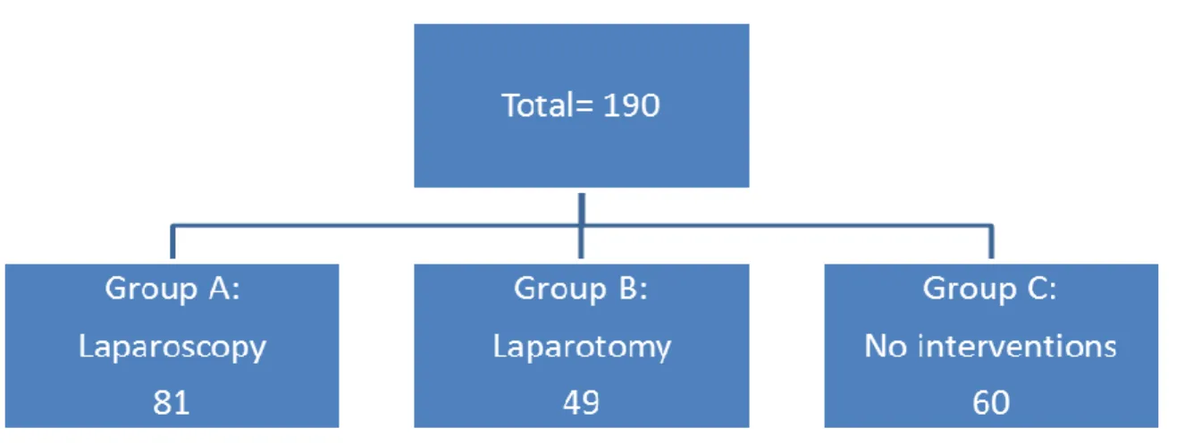
From January 2008 to June 2010 a total of 190 patients were referred to us. No surgical intervention was taken in 60 patients; all of them were HIV positive.
Twenty six of them died (43%) in the hospital during the evaluation period before the diagnostic laparoscopy, and the rest (57%) were unfit for anaesthesia. Forty nine patients required emergency laparotomy either for bowel obstruction or peritonitis and 39% of them died. Eighty one patients underwent diagnostic laparoscopy and 77% of them were HIV positive, in 16% the HIV status was unknown. Two percent had clinical ascites. Laparoscopic findings included intra- abdominal lymphadenopathy in 56, minimal ascitic fluid in 46, intra-abdominal mass in 17, and deposits on bowel wall, peritoneum or omentum in 20 patients.
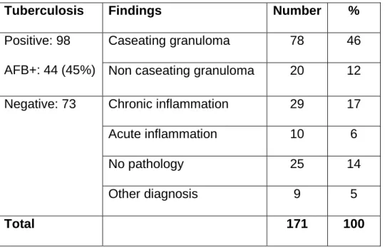
Fifty five patients (68%) had positive histology for tuberculosis. In 15 patients (19%) histology revealed non-specific inflammation, no pathology was found in one patient and no specimen was taken from one patient. Eighty percent of peritoneal deposits and 77% of lymph nodes were positive for tuberculosis, whereas 35% ascitic fluid culture was positive. In nine patients (11%) an alternative diagnosis was found (appendicitis, adenocarcinoma, lymphoma).
Conclusion:
Laparoscopy was feasible and showed a high yield to establish the diagnosis of abdominal tuberculosis and to provide an alternate diagnosis. Laparoscopy was useful to establish the gross features of abdominal tuberculosis and to provide
9 the adequate specimens for examinations. Very poor follow negated the evaluation of the clinical response to anti tuberculosis therapy.
10
Table of contents
Page
Title page ……… 1
Declaration ………. 3
Dedication ……….. 5
Acknowledgements ……….. 6
Abstract ……….. 7
Table of contents ……….. 10
Introduction ………... 15
1.1 Human Immunodeficiency Virus disease ………. 15
1.2 Tuberculosis (TB) ………. 16
1.3 HIV/ TB Co-infection ……… 16
1.4 Abdominal tuberculosis ……….. 18
1.5 Diagnosis of abdominal tuberculosis ……… 20
1.6 Traditional methods of TB diagnosis ……….. ..22
1.7 Alternate diagnostic adjuncts for TB ………. 22
11
1.8 Newer modalities to diagnose TB ………. 24
1.9 Imaging in the diagnosis of abdominal tuberculosis ………... 26
1.9.1 Ultrasonography (U/S) ………. 26
1.9.2 Computerized tomography (CT) scans ……….. 26
1.9.3 Upper and lower gastrointestinal series (Barium studies) 27 1.9.4 Endoscopy ……… 28
1.10 Laparotomy to diagnose abdominal tuberculosis ……… 28
1.11 History of laparoscopy in diagnosis of abdominal tuberculosis ..30
Hypothesis to be tested ……….. 34
Aims of the study ……….... 35
Methodology ………..…. 36
Laparoscopic technique ………..…. 40
Assessing diagnostic accuracy ………... 41
Statistical analysis ……….……….…... 42
Results ………..….. 43
2.1 Group A: Laparoscopy ………..… 44
2.1.1 Duration of symptoms and hospital stays ……….…. 44
12
2.1.2 Clinical presentation ……….… 44
2.1.3 Preoperative investigations ……… 46
2.1.3.1 Haemoglobin (Hb) % ……… 46
2.1.3.2 White Cells Counts (WCC) ……… 46
2.1.3.3 Neutrophils ……… 46
2.1.3.4 Lymphocytes ……… 46
2.1.3.5 Chest X-Ray (CXR) ………. 47
2.1.3.6 U/S abdomen ……… 47
2.1.3.7 CT scan abdomen ……… 47
2.1.3.8 ESR ……… 51
2.1.3.9 CRP ……… 52
2.1.3.10 Blood culture ……… 52
2.1.3.11 Endoscopies ………. 53
2.1.3.12 Contrast studies ……….. 53
Laparoscopic views ………. 54
3.1 Lymph nodes ……… 54
3.2 Ascitic fluid culture ………..…… 56
13
3.3 Omentum ……….… 57
3.4 Tubercles ……… 59
3.5 Peritoneum in the absence of tubercles ……… 60
3.6 Abdominal/ retroperitoneal mass ……… 61
3.7 Appendix ………. 61
3.8 Liver biopsy ………. 62
3.9 Cholecystectomy ……… 62
Complications ……… 71
Deaths ……….... 72
Follow up ……….….. 73
4 Group B: Laparotomy ………... 75
5 Group C: No surgical intervention ……… 76
Discussion ………..… 77
6.1 Clinical characteristics ………..… 78
6.2 Preoperative investigations ……….. 80
6.3 Imaging ……….. 80
6.4 ESR/ CRP ……….… 81
14
6.5 Blood culture ………..…….. 83
6.6 PCR ……… 83
6.7 Endoscopy ……….… 84
6.8 Laparoscopy ………. 84
6.9 Diagnosis other than tuberculosis ……….… 87
6.10 Non-specific chronic inflammations ………. 87
6.11 Perioperative complications/ deaths ………... 88
6.12 Laparotomy and Laparoscopy for TB abdomen …… 89
6.13 Follow up ………. 90
Limitations ………. 91
Conclusions ……….…. 93
References ……… 96
Appendices ……….. 109
List of tables ……… 122
List of figures ………..… 123
List of abbreviations ……… 124
15
Introduction
The current relevant epidemiology of Tuberculosis and Human Immunodeficiency Virus (HIV) and their interactions are outlined seriatim in the following sections.
They provide an essential background for the discussion on the diagnosis of abdominal tuberculosis in the HIV era.
1.1 Human Immunodeficiency Virus disease:
The HIV epidemic is one of the major challenges to South Africa’s socio- economic development. Worldwide, South Africa has the highest number of people living with HIV/AIDS (Acquired Immune Deficiency Syndrome), representing a quarter of the disease burden in sub-Saharan Africa and a sixth of the global disease burden.1 In 2009, an estimated 33.3 million people were living with HIV infection in the world; in South Africa alone it was 5.63 million and 1.8 million people died with AIDS in 2009.2 The national HIV prevalence in South Africa in general population for 2009 was 17.8%, which was 4th highest in the world and the highest percentage was in the province of KwaZulu-Natal, which was 25%.3 The highest provincial HIV prevalence among the ante-natal women aged 15-49 years was also recorded in KwaZulu-Natal in 2009 and it was 39.5%.3
16
1.2 Tuberculosis (TB):
Globally, there were an estimated 9.4 million new cases of tuberculosis in 2008, the incidence of tuberculosis is increasing by about 0.4% and approximately two million people die of it annually.4,5 The incidence of tuberculosis is rising in sub- Saharan Africa as well. South Africa had the seventh highest per capita incidence of tuberculosis. In 2003 and in 2009 it had the 2nd highest incidence in the world.6,7 In particular, the Western Cape province reported an incidence as high as 1000 cases per 100 000 population per annum.8 South Africa has one of the worst tuberculosis epidemics in the world, with high disease burden, incidence rates, and HIV co-infection rates, and growing epidemics of multidrug- resistant and extensively drug-resistant tuberculosis.1 A recent report noted that about 28% of medical admissions in Edendale Hospital, KwaZulu-Natal, were for active tuberculosis and 26.5% of these patients died in this hospital.4 The annual incidence of tuberculosis in KwaZulu-Natal was 1094 cases/100,000 in 2006.9 Tuberculosis has become the leading cause of death in South Africa by clinically determined death notification.10
1.3 HIV/ TB Co-infection:
The increased incidence in tuberculosis in the last decade is related to the HIV pandemic. Approximately 80% of incident tuberculosis cases in South Africa are HIV positive and these co-infected individuals are more likely to have smear- negative pulmonary or extrapulmonary tuberculosis, which can be very difficult to diagnose clinically.11 Deaths from tuberculosis among HIV positive people
17 account for 23% of the estimated two million deaths due to HIV/AIDS in 2007.12 Tuberculosis is the most common cause of death in people infected with HIV worldwide, and accounts for 11% of AIDS deaths.4 Tuberculosis and HIV co- infection act synergistically resulting in increased mortality. The incidence of tuberculosis is also increasing in the HIV negative population. HIV infection is a strong risk factor for developing both pulmonary and extrapulmonary tuberculosis. The incidence of extrapulmonary tuberculosis is 10% to 15% of total tuberculosis cases, and it could be as high as 50% to 70% in AIDS patients and often coexisting with pulmonary disease. Abdominal tuberculosis is the sixth most common site of extrapulmonary tuberculosis; in the range of 11% to 16%, and its incidence increases proportionally to the rising incidence of tuberculosis and HIV worldwide.13,14 In HIV positive individual abdominal tuberculosis has been reported as the most common form of extra-pulmonary disease with a rate of 74% in one series.15
Over the last three decades a number of audits of abdominal tuberculosis have been published from Southern Africa.16,17,18,19
However most of these audits are from pre-HIV era, and the relevance of these findings in the current high HIV disease prevalence is uncertain. It has been reported that hospital prevalence of HIV infection among adult surgical population is up to 39% in South Africa.20 The incidence of abdominal tuberculosis in the Western world is also increasing due to the presence of HIV and migration of population from the developing countries.13,21
18
1.4 Abdominal tuberculosis:
Abdominal tuberculosis may affect the gastrointestinal tract (gastrointestinal tuberculosis), peritoneum (tuberculous peritonitis), mesenteric and retroperitoneal lymph nodes (tuberculous adenitis) and solid organs e.g. liver, spleen, kidney, pancreas.22,23 Tuberculous peritonitis was the commonest form of abdominal tuberculosis before the HIV era and had contributed to 0.1% to 1.5% of total cases of tuberculosis.24 Peritoneal tuberculosis is classified conventionally into plastic (dry) and serous (wet) types. Mildly tender abdominal masses and doughy abdomen characterize the plastic type, ascites with or without signs of peritonitis characterize the serous type. Reports from early 1980’s stated that the disease involved the intestine in 37% of cases, tuberculous ascites and plastic peritonitis accounted for a further 57% of cases, while mesenteric lymphadenitis accounted for only 6% of cases.16 The recent large series by Clarke et al17 of patients with abdominal tuberculosis demonstrated that ascites is relatively uncommon and abdominal lymphadenopathy is a prominent feature and the authors also found that heterogenous complex retroperitoneal, abdominal masses were a common feature of abdominal tuberculosis.
Table 1 is taken from a recent report which summarizes and compares all the relevant audits of abdominal tuberculosis, and how it was diagnosed.17
19 Table 1: Comparison of the frequency of the various diagnostic criteria in the reported series.17
Parameter Novis25 Gunn26 Gilinsky16 Uygur-Bayramicli27 Clarke17
No. of patients 59 12 54 31 67
Period of data collection
1962–1971 1965–1969 1972–1981 1998–2001 2003–2005
AFB present in the lesion a
7 (12%) 3 (25%) 11 (20%) 5 (16%) 9 (13%)
Caseating granulomas a
14 (24%) 7 (58%) 9 (17%) 19 (61%) 11 (16%)
Culture a 1 (2%) 2 (17%) 2 (4%) NS Nil
Operative description
27 (46%) 4 (33%) 5 (9%) NS 14 (21%)
Evidence of TB elsewhere
10 (17%) NS 19 (35%) NS 2 (3%)
Response to treatment alone
Nil Nil 5 (9%) 9 (29%) 45 (67%)
AFB: acid-fast bacilli; NS: not stated.
a These criteria refer to the histologic and microbiologic findings in the resected lesion.
The comparison shows that HIV/AIDS has resulted in a resurgence of abdominal tuberculosis in South Africa. In addition the current clinical presentation of abdominal tuberculosis is different from the past. The disease frequently presents as an acute or semi urgent surgical referral with underlying chronic illness. Previously patients used to present with a primary surgical pathology and
20 HIV infection as a co-morbidity, but now increasingly patients present primarily with AIDS and AIDS related pathology.
1.5 Diagnosis of abdominal tuberculosis:
Establishing the diagnosis of abdominal tuberculosis has always been difficult because of vague and nonspecific clinical features, lack of efficient and sensitive diagnostic tools and the low yield of mycobacterium smear or culture. Delay in diagnosis and delay in initiation of therapy results in poorer outcome. It is very important to advocate a method that gives early diagnosis of this deadly disease, especially when existing treatment has been proven to be very effective and there is good evidence to suggest detrimental effects of delayed treatment.
Treatment delay has been proven to be the most significant factor of attributable to mortality from abdominal tuberculosis.28 Mortality rate can be as high as 60% if anti tuberculosis treatment is not started within 30 days of symptom onset.29 There has been a trend to make use of empirical trials of anti-tuberculosis therapy to establish the diagnosis. This is problematic as the definition of clinical recovery is vague and there are different diagnoses such as lymphoma or malignancy that can mimic abdominal tuberculosis.8,17,30 In addition commencing patients on unnecessary anti tuberculosis therapy exposes them to the risk of drug interactions and side effects without any benefit. Since the early 1980’s a diagnostic model has been widely used in South Africa to make the diagnosis of abdominal tuberculosis; the model included hard and soft criteria.16,25
21 The hard criteria include:
Microbiological or histological evidence of Mycobacterium tuberculosis
Granulomas with caseous necrosis
Successful culture of Mycobacterium tuberculosis from the tissue specimen
Evidence of tuberculosis at a distant site
Typical operative findings in conjunction with macroscopic caseation and caseating granulomas with or without acid-fast bacilli (AFB) on histology
Clinical diagnosis at autopsy
These hard criteria generally required an operative procedure with tissue resection or colonoscopic examination to provide histological or microbiological evidence of tuberculosis.
The soft criteria include:
Clinical features
Radiological features
Response to chemotherapy without recurrence.
22
1.6 Traditional methods of TB diagnosis:
If biopsy of palpable peripheral lymph node demonstrates caseating granuloma or the presence of Mycobacterium tuberculosis, the diagnosis is established and treatment can be started. This is hard evidence of tuberculosis. In the absence of easily accessible lesions we look for supportive evidence e.g. radiologic evidence or analysis of ascitic fluid. Abnormal chest X-rays, elevated erythrocyte sedimentation rate (ESR) and normocytic normochromic anaemia often accompany abdominal tuberculosis, but these tests are so non-specific that they have very little diagnostic importance.
1.7 Alternate diagnostic adjuncts for TB:
Serum C-reactive protein (CRP) is an acute phase reactant synthesized by the hepatocytes under the influence of interleukin-6 arising at sites of infection, inflammation and trauma. High levels of CRP are caused by infections, malignancies, chronic inflammatory diseases and trauma. But a CRP test cannot show where the inflammation is located or what is causing it. Serum concentrations of CRP increase within six hours of induction of an inflammatory process and due to its very short half live in the circulation, the initially elevated CRP levels return to normal value after resolution of the inflammatory process.
Though CRP values can never be diagnostic on their own and can only be interpreted at the bedside, in full knowledge of the other clinical and pathological results, it has been used both as diagnostic and prognostic tool for tuberculosis.31,32,33
23 Ascitic fluid can be aspirated only if there is clinically evident ascites. Most of the patients currently presenting with abdominal tuberculosis do not have ascites and they present with plastic (dry) type. Hence, there is no overt fluid to aspirate and evaluate to establish the diagnosis. If aspirated fluid is shown to be exudative in nature with lymphocytes predominant; it is only suggestive of tuberculosis, not confirmatory. Direct smear for Ziehl-Neelsen stain is unhelpful most of the time, with reported sensitivity ranging from 0% to 6%.24 The frequency of a positive culture for mycobacterium from small volumes of ascitic fluid has been less than 20% and it takes considerable time before results are available, although the positive rate can be improved by obtaining one litre of ascitic fluid concentrated by centrifugation.34
The role of ascitic fluid adenosine deaminase activity (ADA) has been studied to differentiate tuberculosis from other causes of ascites. ADA is an enzyme widely distributed in tissues and body fluids and the most important biologic activity is related to lymphoid tissues, because ADA is necessary for proliferation and differentiation of T-Lymphocytes. It has been suggested that an increasing ADA activity relates to the intensity of stimulation and the maturation state of the lymphocyte, due to the immune cellular response against Mycobacterium tuberculosis.13 Studies from a tuberculosis endemic area like South Africa have reported sensitivity and specificity exceeding 92% for this non-invasive test,35 whereas the study from the United States showed that the ascitic fluid ADA
24 activity has good accuracy but poor sensitivity and imperfect specificity, where the prevalence of tuberculosis is low and underlying cirrhosis is common.36
Gamma (γ) interferon, secreted by antigen-triggered CD4+ lymphocytes, is a key lymphokine that activates macrophages, increasing their bactericidal activity against Mycobacterium tuberculosis.34 Although the sensitivity and specificity of ascitic fluid ADA and γ-interferon activity are high in tuberculous peritonitis, their activities have been reported to be significantly lower in patients with low ascitic fluid protein concentration and in patients with AIDS, explained by a low lymphocyte activity due to the CD4+ lymphocyte depletion in HIV infection.13,34
Serum CA-125 level may be raised in tuberculous peritonitis, but the test is not specific because other conditions can give rise to the level e.g. carcinoma of the ovaries, though the decreasing CA-125 level is useful to evaluate the efficacy of therapy in TB peritonitis.37
1.8 Newer modalities to diagnose TB:
The yield of polymerase chain reaction (PCR) in the diagnosis of tuberculosis is high in tissues, but not in ascitic fluid; the specimens need to be fresh, the test is costly and above all the only way to get the tissue is either by laparoscopy/
laparotomy or colonoscopy.38 PCR could efficiently complement conventional bacteriological tools for the rapid diagnosis of tuberculosis but cannot replace them.39
25 A recently developed RD-1 gene-based assay for diagnosing tuberculosis infection shows promising results. Serological tests were performed to evaluate the interferon- γ producing T-cell response using peripheral blood mononuclear cells in patients with suspected abdominal tuberculosis and the results suggest that the tests are useful adjunct to the current tests for diagnosing abdominal tuberculosis.40 The test is called “Quantiferon Gold test” and is an enzyme-linked immunospot (ELISpot) assay which detects the release of interferon-gamma (IFN-g) in fresh heparinized whole blood from sensitized persons when it is incubated with mixtures of synthetic peptides simulating two proteins present in Mycobacterium tuberculosis. Though the sensitivity of detecting tuberculosis infection in persons with untreated culture-confirmed tuberculosis is approximately 80%,sensitivity for particular groups of tuberculosis patients (e.g., young children, immunocompromised patients with HIV infection and patients with immunosuppressive drugs) has not been determined.41 The other drawback of this test is: it has only role in diagnosing latent tuberculosis in immune competent patients and cannot distinguish active from latent disease.
26
1.9 Imaging in the diagnosis of abdominal tuberculosis:
1.9.1 Ultrasonography (U/S):
The traditional ultrasonic features of abdominal tuberculosis e.g. enlarged mesenteric lymph nodes of greater than 15 mm with hypoechoic or necrotic area, solid organ abscesses or hypoechoic lesions especially in the spleen, bowel wall thickening and ascites are well documented.42
Besides intra-abdominal fluid and lymphadenopathy, “club sandwich” or “sliced bread” and pseudokidney signs are highly suggestive of tuberculosis.43 “Club sandwich” or “sliced bread” sign is due to localized fluid between radially oriented bowel loops, due to local exudation from the inflamed bowel. Pseudokidney sign is the involvement of the ileo-caecal region which is pulled up to the subhepatic position. Though the sensitivity and specificity of U/S examination have been reported to be 45% and 96% respectively in one study,44 another study showed that though the sensitivity and specificity could not be established, U/S abdomen had clinical utility in the diagnosis and treatment follow up of abdominal tuberculosis in the resource constrain areas.45
1.9.2 Computerized tomography (CT) scans:
CT features of abdominal tuberculosis include thickening of the small bowel mucosa due to tuberculous infiltration, stranding and thickening of small bowel mesentery, omental and retroperitoneal lymph node involvement with central caseous necrosis and peripheral rim enhancement to give a typical halo appearance. Central necrosis with rim enhancement though not pathognomonic,
27 is a useful sign and readily seen in the current generation CT scanners.46 Ongoing necrotic breakdown results in large inflammatory retroperitoneal collections. CT reliably demonstrates the entire range of findings. Although peripheral rim enhancement is very characteristic of tuberculous lymphadenopathy, it is also noted in other processes such as lymphoma, metastatic malignancy, pyogenic infections and Whipple’s disease.47 All the patients with abdominal tuberculosis do not present with typical CT finding.48 Although no single CT feature is diagnostic of abdominal tuberculosis, CT findings interpreted in the light of clinical and laboratory data can be a valuable tool in the diagnosis of abdominal tuberculosis. The sensitivity and specificity of CT scan in the diagnosis of TB abdomen were 92% and 95% respectively in one study, though the sample size was small49 and the sensitivity was only 69% in another study.50
1.9.3 Upper and lower gastrointestinal series (Barium studies):
Tuberculosis can involve any region of the gastrointestinal tract, but in about 90%
of cases it affects the ileo-caecal valve, and the adjacent ileum and colon.47 Thickening of the ileo-caecal valve and/or wide gaping between the valve and narrowed terminal ileum is “Fleischner” or “inverted umbrella sign”, and localized stenosis opposite the ileo-caecal valve with rounded off smooth caecum and a dilated terminal ileum is called “purse string stenosis”. In advanced disease, the caecum becomes conical and shrunken resulting in a widely open ileo-caecal valve with fixed and narrowed terminal ileum due to fibrosis, which is called
28
“Stierlin’s sign, and the persistent narrow stream of barium indicating stenosis is called “String sign”. Although“Fleischner” or “inverted umbrella sign” and “purse string stenosis” are considered to be characteristic findings in ileo-caecal tuberculosis, “Stierlin’s sign” and “String sign” are not specific of intestinal tuberculosis and may also be found in Crohn’s disease.43,47,51
1.9.4 Endoscopy:
Upper endoscopy and colonoscopy with terminal ileoscopy are investigations of choice for the diagnosis of intestinal tuberculosis as it allows for direct visualization and tissue sampling for histology and culture, however it is limited by the accessibility of the small bowel.52,53 Diffuse involvement of the entire colon is rare and endoscopically lesions look very similar to ulcerative colitis and lesions mimicking carcinoma have also been described.43
1.10 Laparotomy to diagnose abdominal tuberculosis:
There is a drive to reduce the number of patients being treated for tuberculosis empirically due to the incomplete treatment or incorrect use of the drugs. This is especially true in the initial phase of anti-tuberculosis treatment as a cause of acquired multi-drug resistance (MDR) tuberculosis and severe hepatotoxicity occasionally causing acute liver failure and death.54 The mortality rate among patients with anti-tuberculosis treatment associated acute liver failure was high (67%) and 63% of patients in that study were prescribed anti-tuberculosis treatment empirically.55 Trials of TB treatment have been abandoned and the
29 current recommendation is that when the decision is taken for empiric TB treatment, then a full course should be given. A trial of TB treatment that is prematurely stopped in a patient who does have TB may contribute to drug resistance. We find it very difficult to establish a definitive diagnosis in patients with abdominal tuberculosis. This is because patients suffering from HIV disease and tuberculosis, a formal laparotomy which is required to obtain tissue for histological or microbiological analysis is associated with significant morbidity and potential high mortality.17,56 Furthermore the volume of patients with this problem would mean that resorting to laparotomy would place a great strain of resources. In the face of this high volume of chronically sick patients the general surgeons have resorted to commencing empirical trials of therapy and waiting for a response to therapy. There are problems with this approach. There is no standardization of what is meant by a positive response to treatment or what constitutes an adequate trial of therapy is also unclear; the weight gain, resolution of constitutional symptoms, improvement of haemoglobin percentage and reduction in CRP levels may be considered as indicators of response to therapy. Trial of therapy may cause delay in the diagnosis of other diseases which mimic abdominal tuberculosis e.g. Crohn’s disease, lymphoma, malignancy and other opportunistic infections.51,57,58,59,60
Until recently laparotomy was the only available diagnostic procedure to obtain adequate tissue to make a definitive diagnosis of abdominal tuberculosis. Many patients who present with signs and symptoms of an acute abdomen are
30 subjected to emergency laparotomy. Laparotomy in these patients may result in significant associated morbidity. It is important to avoid emergency surgery in patients without radiologic or clinical evidence of free perforation or disseminated peritonitis and in immunocompromised patients with significant co-morbidities.
Although elective laparotomy does not carry an unduly high mortality rate, in emergency laparotomy mortality rate can be as high as 60%.17 The most significant complications of laparotomy in patients with abdominal tuberculosis are anastomotic leak and fistula. It has been reported that enterolysis is the main cause of fistula formation and it is concluded that when a surgeon encounters tuberculosis at laparotomy, it would be advisable to avoid any enterolysis and to confine the procedure in taking specimens for microbiological and histopathological examinations, then the incision would be closed and the patient put on appropriate anti-TB medications.61
1.11 History of laparoscopy in diagnosis of abdominal tuberculosis:
Diagnostic peritoneoscopy with a direct optic scope was used in the 1960’s in patients with tuberculous peritonitis to obtain peritoneal biopsy for microbiological or histological examination. Peritoneal biopsy gives a better diagnostic value than ascitic fluid analysis alone.18
31 Blind percutaneous peritoneal biopsy used to be under taken with local anaesthesia with varying degrees of success. Peritoneal biopsy was first performed by Donohue in 1959 using Vim Silverman needle and later Abrams and Cope needles were used.62,63 There are several other small series of peritoneal biopsy in the 60’s, 70’s and 80’s.57,64,65 The procedure carries a high risk of complications like bowel perforation, bleeding, and even death.66,67,68 The main contraindication to blind percutaneous peritoneal biopsy is the absence of ascites. The role of blind percutaneous biopsy is limited in the cases of plastic (dry) type of abdominal tuberculosis which currently predominate.18,62,69
The difficulty in confirming the diagnosis, the considerable morbidity and mortality associated with formal laparotomy in patients with abdominal tuberculosis and HIV co-infection, and the reluctance to embark on poorly defined empirical therapy have generated interest in the use of laparoscopy to obtain specimens for histological and microbiological assessment.17,56,61 The advent of laparoscopy has allowed surgeons to visually inspect the abdominal cavity and take biopsies with minimal morbidity. Laparoscopy as an aid to diagnosis is well established in the management of both chronic and acute abdominal pain. There have been interests expressed in the use of laparoscopy to establish the diagnosis of abdominal tuberculosis.21,69,70,71,72,73
Table 2 shows the different laparoscopic series with their diagnostic yields.
32 Table 2: Different laparoscopic series with their yields.
Authors Series date No. of patients
Ascites (%)
HIV+ Diagnostic yield (%)
Mohamed70 2004-2008 13 100 not stated 85
Meshikhes73 1992-2008 20 100 not stated 75 Krishnan71 1999-2005 41 70 not stated 80
Al-Mulhim21 1995-2002 21 67 none 81
Rai72 1995-2001 25 32 not stated 92
Mimica69 1977-1987 32 75 not stated 85
The risk involved with laparoscopy is low but there is a definite incidence of complications which may be significant. Most of the complications associated with laparoscopy involve iatrogenic injury to major vessels or to hollow viscera.
With modern techniques to safely induce a pneumoperitoneum and to safely introduce operative ports the risk is less than 0.05%.66 Patients undergoing routine laparoscopic surgery face the same risk. The patient will also have to undergo a general anaesthetic. Once again the risks associated with modern general anaesthesia are extremely low. From retrospective studies it has been postulated that in patients suspected to have abdominal tuberculosis without evidence of extra-abdominal disease, early laparoscopy may be useful to establish a histological diagnosis with acceptably low morbidity.71 Although there are some publications of retrospective studies about the role of laparoscopy to establish the diagnosis of abdominal tuberculosis, there is no prospective
33 study.74,75,76 However, locally it was found that the use of laparoscopy in the diagnostic work up of suspected abdominal tuberculosis was poor and underutilized.17 Establishing a dedicated team that will aggressively pursue a diagnosis of abdominal tuberculosis with laparoscopy is an attractive concept at a busy hospital in South Africa with high incidence tuberculosis and HIV infection.
This will hopefully improve patient care and allow us to establish the diagnostic accuracy of laparoscopy for this condition.
34
Hypothesis to be tested
Laparoscopy is a minimally invasive procedure which will enable a definitive diagnosis of abdominal tuberculosis to be established in patients with clinically suspected but microbiologically or histologically unconfirmed abdominal tuberculosis. These patients would be eligible for empiric anti-tuberculosis treatment in terms of current standard of care.
35
Aims of the study
This study aimed to review several objectives in light of the HIV-tuberculosis co- infection pandemic in diagnosing abdominal tuberculosis:
1) To quantify and establish the role of diagnostic laparoscopy in the work up of patients with suspected abdominal tuberculosis.
2) To establish the gross laparoscopic features of abdominal tuberculosis.
3) To assess the capability of laparoscopy to provide adequate microbiological and histological specimens for analysis.
4) To audit the clinical response of patients with abdominal tuberculosis to appropriate therapy.
36
Methodology
A prospective clinical audit of the use of diagnostic laparoscopy in patients with suspected abdominal tuberculosis was conducted. The study has been approved by the Biomedical Research Ethics Committee (BREC) of the University of Kwa- Zulu Natal. The study was undertaken at Edendale Hospital in Pietermaritzburg.
All patients with clinically suspected but histologically or microbiologically unconfirmed abdominal tuberculosis were referred to the investigating team. The investigating team consisted of a general surgeon and an infectious disease physician. The patients then were jointly assessed by the investigating team. If a definitive diagnosis of extra abdominal tuberculosis could be achieved by another procedure e.g. sputum analysis, peripheral lymph node biopsy or fluid or lymph node aspiration; the patients were commenced on anti-tuberculosis treatment and excluded from this study cohort.
In order to maximize the diagnosis of extra abdominal tuberculosis, all study participants also had:
Induced sputum or tissue aspirate (if any) for tuberculosis culture
Microbiological blood culture
Pre-operative serum CRP level
The two clinicians reviewed abdominal U/S or CT scans reports if they were done. If the clinicians agreed that clinical and radiological features were suggestive of abdominal tuberculosis and the histological or microbiological
37 evidence of tuberculosis could not be obtained from any other site then the patients were offered a diagnostic laparoscopy.
Informed consents were taken from the patients. Moribund patients who are not expected to survive an anaesthetic were excluded from the study. All anaesthetic administered at Edendale Hospital are administered by trainees under direct consultant supervision. Pre-operative body weight, haemoglobin (Hb) % and CRP level were recorded in all patients especially to compare with post-operative response to treatment.
All the patients undergoing laparoscopy were started on standard anti- tuberculosis treatment without delay, whilst awaiting histology report. Positive diagnosis of abdominal tuberculosis was considered if there was: typical tuberculous granulomata containing Langhan’s giant cells with caseation or non caseating necrosis with the demonstration of AFB in the biopsied tissues or the isolation of Mycobacterium tuberculosis by culture from the ascitic fluid or biopsied tissues. This equates to “Hard criteria” and as such if one can reliably establish them then abdominal tuberculosis can be treated correctly. The surgeon prospectively followed up each patient enrolled in the study until the point at which we have confirmed a diagnosis of tuberculosis and evaluated the response of the treatment, which was at the end of eight weeks. If histological or microbiological confirmation of abdominal tuberculosis were obtained from laparoscopy then the patients were referred for follow- up to the tuberculosis
38 clinic at Edendale Hospital. The data were evaluated to see the diagnostic yield of laparoscopy in the form of macroscopic appearance and histological or microbiological results and the response to medications in the form of sensitivity and specificity.
All participants were to evaluated for tuberculosis treatment response by assessment at week 4 and week 8 following laparoscopy. Therapeutic response was considered if there were at least two or more of the following criteria present:
weight gain ≥5%, haemoglobin increase ≥1 gm%, 60% reduction in CRP, and at least half of the symptoms were much better or resolved, including assessment of adherence to tuberculosis treatment using the TB clinic “Green Card” were evaluated. All the patients had repeat abdominal U/S at follow up to compare the findings with the preoperative U/S reports.
Inclusions criteria:
In essence there were two broad groups of patients who were considered for laparoscopy:
1) Those with abdominal pain with mild peritonism and features compatible with HIV disease (or known to be HIV sero-positive) and a history of weight loss with drenching sweats for more than two weeks (dry type).
2) Those with ascites and features compatible with HIV disease (or known HIV sero-positive) and history of weight loss with drenching sweats for more than two weeks (wet type).
39 Exclusions criteria:
The patients were excluded from the study in the case of:
1) Moribund patients not fit for anaesthetic.
2) Patients with histologically or microbiologically confirmed TB abdomen from laparotomy for other reasons or ascitic fluid examinations
3) Informed consent not obtained 4) Minors less than 16 years of age
40
Laparoscopic technique
Laparoscopy was done under general anaesthesia in all patients. The first 10 mm trocar was introduced in the subumblical region under direct vision using visiport in most of the patients to avoid bowel injuries due to adhesions. Verese needle was used in only a few cases to insufflate the peritoneal cavity before inserting the trocar. A second 5 mm trocar was introduced under direct vision in the suprapubic region. A third 5mm or 10 mm trocar was introduced under direct vision according to the abnormalities found; most of the cases in the left iliac fossa. In most of the cases the abnormalities were found around the ileo-caecal region and were possible to biopsy with these three ports. Whole abdominal cavity was inspected: if there was any free fluid, it was aspirated and sent for microbiological examination, any abnormal looking tissues e.g. enlarged lymph nodes, mass, omentum, peritoneum or liver tissues were biopsied and sent for histological examinations. Due to a persistent logistic problem with the processing and delivery of tissue specimens for tuberculosis culture, only three specimens were deemed suitable for culture and hence no analysis of this technique was carried out. If the macroscopic feature was not suggestive of tuberculosis tru-cut biopsy of liver was done. Trocar sites were closed with non- absorbable sutures.
41
Assessing diagnostic accuracy
In assessing the diagnostic accuracy of a test or a group of tests using sensitivity and specificity as in this study, the gold standard definition of abdominal tuberculosis is key to defining the true positives. We appreciate that there are significant advances in diagnostic tests in pulmonary tuberculosis and in the diagnosis of disseminated tuberculosis, which have been discussed in the introduction. However the possibility of dual diagnosis of tuberculosis and another pathology led us not to focus purely on the diagnosis of tuberculosis but on the establishment of the diagnosis of abdominal tuberculosis. This was because we also wanted to establish if there was an alternate or metachronous diagnosis which would require specific therapy. Therefore our study focused on abdominal tissue and fluid sampling. We therefore chose the three-criteria stated in the statistical section as the gold standard.
42
Statistical analysis
Statistical evaluation of data entailed sensitivity (Se), specificity (Sp), positive predictive value (PPV) and negative predictive value (NPV) analysis. The “gold standard” for the diagnosis of abdominal tuberculosis was (1) presence of AFB in histological specimens, (2) presence of caseating granulomas in histological specimens and (3) TB culture.
In the case of quantitative data, means and 95% confidence interval (95% CI) were reported around sample estimates. MS Excel and Epicalc 2000 (Joe Gilman and Mark Myatt 1998, Brixton Books) were used to analyze the data.
43
Results
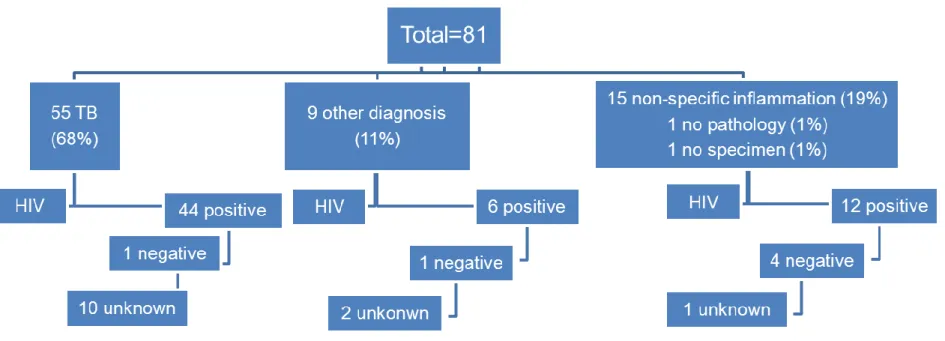
From January 2008 to June 2010 a total of 190 patients were referred to us with a provisional diagnosis of abdominal tuberculosis and no hard evidence of tuberculosis at a site other than the abdomen e.g. negative sputum for AFB, no positive culture from any aspirate. The mean age of the whole group was 33 years (95% CI 31- 34 years). 94 patients were male and 96 were female. The patients were divided into three groups: Group A- laparoscopy, Group B- laparotomy, Group C- no surgical intervention. Figure 1 shows the distribution of all the patients in the study.
Figure 1: Distribution of all the patients in the study.
44
2.1 Group A: Laparoscopy
Eighty one patients had diagnostic laparoscopy, 34 of them were male and 47 were female (male: female=1:1.38), mean age was 33 years (95% CI 31- 36 years). Only two patients were Indian origin and the rest were African. Sixty two patients were HIV positive (77%); only 10 of them had known CD4 counts and the mean counts were 138 (95% CI 57- 210) and 16 patients were on anti- retroviral therapies, six patients were HIV negative (7%) and 13 had an unknown HIV status (16%) because they declined HIV testing.
2.1.1 Duration of symptoms and hospital stays:
The mean duration of symptoms was 41 days (95% CI 31- 51 days) prior to the hospital admission and the mean duration of hospital stay was 11 days (95% CI 10- 13 days).
2.1.2 Clinical presentation:
The most common clinical features were abdominal pain, abdominal distension, lymphadenopathy, night sweats and weight loss. Preoperative body weight were recorded in the patients. The mean body weight was 55 kg (95% CI 53- 58 kg).
Table 3 is showing the clinical presentation of 81 patients.
45 Table 3: Clinical presentation of 81 patients.
Clinical features Numbers Percentage (%)*
Abdominal pain 81 100
Weight loss 69 85
Night sweats 61 75
Lymphadenopathy 52 64
Abdominal distension 22 27
Low grade fever 19 24
Vomiting 18 22
Previous lungs TB 18 22
Constipation 8 10
Diarrhoea 4 5
Ascites 2 2
*Percent rounded to the nearest integer
46
2.1.3 Preoperative investigations:
2.1.3.1 Haemoglobin (Hb) %:
Hb levels were checked in all the patients. The mean Hb was 9.69 gm% (95% CI 9.22- 10.14 gm%). Nineteen patients (23%) had Hb level less than 8 gm%.
2.1.3.2 White Cells Counts (WCC):
WCCs were checked in all the patients. The mean WCC was 8.62 x10⁹/L (95%
CI 7.56- 9.68 x 10⁹/L). Most of the patients (63%) had normal WCC (normal count is 4-11 x 109/L).
2.1.3.3 Neutrophils:
Neutrophils counts were checked in all the patients. The mean neutrophil count was 72.22% (95% CI 69.17- 75.26%). Only one patient (1%) had neutropenia, 33 patients (41%) had neutrophilia and 47 patients (58%) had normal neutrophil count (normal: 40-75%).
2.1.3.4 Lymphocytes:
Lymphocytes counts were checked in all the patients. The mean lymphocytes count was 17.6% (95% CI 15.36- 19.83%). There was no patient with lymphocytosis, 41 patients (51%) had lymphopenia and 40 patients (49%) had normal lymphocytes count (normal: 20-45%).
47 2.1.3.5 Chest X-Ray (CXR):
CXR were done in all patients but it was suggestive of tuberculosis in only ten patients (12%) though sputum cultures were negative in all of 10 patients. Most of the patients had non-productive cough with nonspecific pulmonary infiltrates.
Sputum cultures were performed only in patients with CXR suggestive of tuberculosis or in patients with associated cough and all cultures were negative for tuberculosis.
2.1.3.6 U/S abdomen:
U/S abdomen were done in 81 patients and all patients had evidence suggestive of abdominal tuberculosis, but histology/culture results were positive for tuberculosis in 55 patients (68%) and no specimen was taken from one patient.
Sonographic features were retroperitoneal lymphadenopathy (75), free fluid (11), complex ascites (17), thickened small bowel loops (14), retroperitoneal abscesses (3), and hypo-echoic/ necrotic echo pattern of the liver and spleen (3).
2.1.3.7 CT scan abdomen:
CT abdomen was done in 40 patients; 36 of them were suggestive of abdominal tuberculosis and four were not. Features suggestive of TB were mesenteric lymphadenopathy (38), thickened small bowel wall (12), hypodensity of liver and spleen (3), free fluid (17), and mesenteric stranding (10). Only 19 of them had positive (48%) histology for tuberculosis and 21 were negative (52%). The sensitivity and specificity of CT scan are 95% (95% CI 72- 100%) and 14% (95%
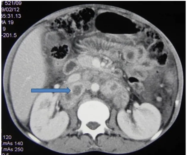
48 CI 4- 37%) respectively. The positive predictive value and negative predictive value of CT scan was 50% (95% CI 33- 67%) and 75% (95% CI 22- 99%) respectively. Figures 2-5 are showing CT scan findings in different patients.
Figure 2: CT scan showing enlarged retroperitoneal lymph nodes (arrow) with central area of necrosis; histology was positive for TB.
49 Figure 3: CT scan showing enlarged retroperitoneal lymph nodes (arrow), histology was negative for TB.
50 Figure 4: CT scan showing enlarged retroperitoneal lymph nodes (arrow), but no gland could be biopsied.
51 Figure 5: CT scan showing enlarged mesenteric lymph nodes (thin arrow) and thickened mesentery (bold arrow), histology showed metastatic adenocarcinoma.
2.1.3.8 ESR:
Thirty five patients had ESR done and the mean ESR was 89 (13-153) mm in 1st hour. Only three patients (9%) had normal ESR (less than 20) with 95% CI 2.24- 24.19 and 32 had high ESR (91%) with 95% CI 75.81- 97.76. Twenty two patients had positive histology/culture for tuberculosis (69%) of the 32 patients with high ESR. The sensitivity and specificity of ESR are 95% (95% CI 75- 100%) and 17% (95% CI 3- 49%) respectively. The positive predictive value and negative predictive value of ESR are 68% (95% CI 49- 83%) and 67% (95% CI 13- 98%) respectively.
52 2.1.3.9 CRP:
CRP was done in 24 patients and the mean CRP was 68 (5-128) mg/L. Fourteen patients had positive (58%) histology/ culture for tuberculosis, nine patients had negative histology (38%) and no specimen was taken from one patient (4%).
CRP was normal (less than 10 mg/L) in four patients (17%) with 95% CI 5.48- 38.19 and high in 20 patients (83%) with 95% CI 61.81- 94.52. Eleven patients had positive histology/ culture for tuberculosis (55%) of the 20 patients with high CRP. The sensitivity and specificity of CRP are 79% (95% CI 49- 94%) and 11%
(95% CI 1- 49%) respectively. The positive predictive value and negative predictive value of CRP are 58% (95% CI 34- 79%) and 25% (95% CI 1-78%) respectively.
2.1.3.10 Blood culture:
Blood cultures for tuberculosis were done in 51 patients. Thirteen (25%) of them had positive culture for tuberculosis and 38 (75%) were negative. The sensitivity and specificity of blood culture are 38% (95% CI 21- 58%) and 91% (95% CI 69- 98%) respectively. The positive predictive value and negative predictive value of blood culture are 85% (95% CI 54- 97%) and 53% (95% CI 36- 69%) respectively. Unfortunately it took 6-8 weeks to get the culture results back and it was not helpful in the diagnosis and management of the condition.
53 Table 4 is showing the diagnostic yields of preoperative investigations.
Table 4: Diagnostic yields of pre-operative investigations.
Investigations No. patients (%)
Number positive
(%) positive
Sensitivity (%)
Specificity (%)
PPV (%)
NPV (%)
ESR 35 (43) 22 69 95 17 68 67
CRP 24 (30) 14 58 79 11 58 25
CT scan 40 (49) 19 48 95 14 50 75
Blood culture 51 (63) 13 25 38 91 85 53
PPV= positive predictive value, NPV= negative predictive value
*Percent rounded to the nearest integer
The sensitivity, specificity, PPV and NPV calculations were based on the number who had the sample taken, and not on the total number (81) undergoing laparoscopy.
2.1.3.11 Endoscopies:
Upper gastrointestinal (GI) endoscopies were done in 10 (12%) patients and colonoscopies were done in nine (11%) patients, but none yielded any specific diagnosis.
2.1.3.12 Contrast studies:
Two patients (2%) had barium enema and three patients (4%) had upper GI series done, which were not significant.
54
Laparoscopic views
The typical appearance of abdominal tuberculosis is of ascites with multiple small deposits (tubercles) of about 0.5 cm in diameter adhering to the peritoneum and bowel walls and omentum, or there may be thin, filmy multiple white adhesions attaching the peritoneum to the bowel, omentum and liver or a deep abdominal mass from the involved mesenteric lymph nodes or thick oedematous omentum (omental cake).
3.1 Lymph nodes:
Mesenteric lymph nodes were biopsied from 56 patients (70%) and 43 had positive (77%) histology for tuberculosis. Thirteen lymph nodes (23%) were negative for tuberculosis and 24 patients (30%) did not have any lymph node, no specimen was taken from one patient. Amongst the 43 histology positive patients 35 (81%) had caseating granuloma and eight had non-caseating granuloma (19%), and 24 amongst all of them (56%) had acid fast bacilli (AFB) positive.
Amongst the 13 histology negative patients, 10 had nonspecific chronic inflammation with reactive lymph nodes (three had sinus histiocytosis), two had other diagnosis (adenocarcinoma and lymphoma) and one did not show any pathology.
The sensitivity and specificity of lymph nodes are 83% (95% CI 69- 91%) and 54% (95% CI 34- 72%) respectively. The positive predictive value and negative
55 predictive value of lymph nodes are 77% (95% CI 63- 87%) and 63% (95% CI 41- 80%) respectively. Figure 6 is showing mesenteric lymph node with pus.
Figure 6: Mesenteric lymph node with pus
56 3.2 Ascitic fluid culture:
Small amounts of ascitic fluid was aspirated and sent for culture from 46 patients (58%) and ascitic fluid was absent in other 34 patients (42%). Sixteen of them were positive (35%) for tuberculosis culture and 30 (65%) were negative. The correlation has been established between the ascitic fluid culture and histology results. It has been found that 13 patients had both fluid culture and histology positive, three patients had fluid culture positive but histology negative, 15 patients had fluid culture negative but histology positive, and 15 patients had both fluid culture and histology negative. The sensitivity and specificity of ascitic fluid culture are 46% (95% CI 28- 66%) and 83% (95% CI 58- 96%) respectively.
The positive predictive value and negative predictive value of ascitic fluid culture are 81% (95% CI 54- 95%) and 50% (95% CI 32- 68%) respectively.
Only two patients had drug resistant tuberculosis: one was resistant to Isoniazid (INH) and the other was resistant all drugs except INH and Rifampicin. Figure 7 shows straw coloured pelvic fluid.
57 Figure 7: Straw coloured pelvic fluid
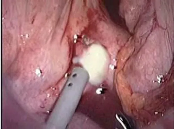
3.3 Omentum:
Forty one specimens (51%) were taken from omentum and sent for histological examination; 15 of them were positive (37%) for tuberculosis and 26 were negative (63%). Amongst the 15 positive patients 12 had caseating granuloma (80%) and three had non-caseating granuloma (20%), and only five amongst all of them (33%) had AFB positive. Amongst the 26 histology negative patients, 12 had nonspecific chronic inflammation, one had acute inflammation (the patient had bowel perforation), three had diagnosis other than tuberculosis (3- adenocarcinoma), and ten had no pathology found.
58 The sensitivity and specificity of omentum histology are 61% (95% CI 39- 80%) and 94% (95% CI 71- 100%) respectively. The positive predictive value and negative predictive value of omentum are 93% (95% CI 66- 100%) and 65%
(95% CI 44- 82%) respectively. Figure 8 is showing taking biopsy from the omentum.
Figure 8: Taking biopsy from the omentum.
59 3.4 Tubercles:
Peritoneal/ omental deposits (tubercles) were present and sent for histological examination in 20 patients (25%) and there was no deposit in the other 60 patients (75%); 16 of them were positive (80%) for tuberculosis and four (20%) were negative. Amongst the 16 positive patients 13 had caseating granuloma (81%) and three had non-caseating granuloma (19%), and only six amongst all of them (37%) had AFB positive. Amongst the four negative patients, two had diagnosis other than tuberculosis (2-adenocarcinoma) and two had nonspecific chronic inflammation.
No specimen was taken from one patient. The sensitivity and specificity of tubercles are 31% (95% CI 19- 45%) and 86% (95% CI 66- 95%) respectively.
The positive predictive value and negative predictive value of tubercles are 80%
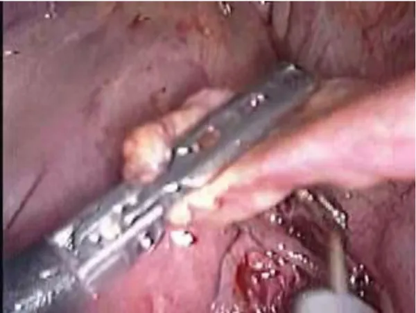
(95% CI 56- 93%) and 40% (95% CI 28- 53%) respectively. Figure 9 is showing multiple tubercles over the Ileo-caecal region.
60 Figure 9: Multiple tubercles over the Ileo-caecal region
3.5 Peritoneum in the absence of tubercles:
Twenty five specimens (31%) were taken from peritoneum and sent for histological examination; nine of them were positive (36%) for tuberculosis and 16 were negative (64%). Amongst the nine positive patients six had caseating granuloma (67%) and three had non-caseating granuloma (33%), and only one (11%) amongst all of them had AFB positive. Amongst the 16 negative patients, three had nonspecific chronic inflammation, one had acute inflammation and 12 had no pathology found.
61 The sensitivity and specificity of peritoneal tissue histology are 47% (95% CI 22- 73%) and 80% (95% CI 44- 96%) respectively. The positive predictive value and negative predictive value of peritoneum are 78% (95% CI 40- 96%) and 50%
(95% CI 26- 74%) respectively.
3.6 Abdominal/ retroperitoneal mass:
Intra-abdominal or retroperitoneal masses were present and biopsied from 17 patients (21%); histology of masses were positive for tuberculosis in 14 (82%) and negative in three patients (18%). Amongst the 14 positive patients 12 had caseating granuloma (86%) and two had non-caseating granuloma (14%), and eight amongst all of them (57%) had AFB positive.
No specimen was taken from one patient. The sensitivity and specificity of masses are 27% (95% CI 16- 41%) and 89% (95% CI 71-97%) respectively. The positive predictive value and negative predictive value masses are 82% (95% CI 56- 95%) and 40% (95% CI 28- 53%) respectively.
3.7 Appendix:
Five appendicectomies (6%) were done along with other tissues for histology and all of them were negative for tuberculosis. Three of them showed features of acute appendicitis with no other tissues positive for tuberculosis in these three patients, and final diagnosis was acute appendicitis. In one patient appendix specimen showed acute inflammation, but positive histology for tuberculosis in