SCREENING FOR BIOSURFACTANT PRODUCTION AMONGST AEROBIC ENDOSPORE FORMING BACTERIA ISOLATED FROM MFABENI PEATLAND
SEDIMENT CORE
BY
FOLASADE ABIMBOLA ADU
Submitted in fulfilment of the academic requirements for the degree of Master of Science in Microbiology
Discipline of Microbiology School of Life Sciences
College of Agriculture, Engineering and Sciences University of KwaZulu-Natal
Pietermaritzburg
South Africa
i
PREFACE
The research contained in this dissertation was completed by the candidate while based in the Discipline of Microbiology, School of Life Sciences of the College of Agriculture, Engineering and Science, University of KwaZulu-Natal, Pietermaritzburg Campus, South Africa.
The contents of this work have not been submitted in any form to another university and, except where the work of others is acknowledged in the text, the results reported are due to investigations by the candidate.
__________________________________
Signed: Dr. C.H. Hunter (Supervisor) Date:
ii
DECLARATION
I, Folasade Abimbola Adu, declare that:
i) the research reported in this dissertation, except where otherwise indicated or acknowledged, is my original work;
ii) this dissertation has not been submitted in full or in part for any degree or examination to any other university;
iii) this dissertation does not contain other person’s data, pictures, graphs, or other information, unless specifically acknowledged as being sourced from other persons;
iv) this dissertation does not contain other persons’ writing, unless specifically acknowledged as being sourced from other researchers. Where other written sources have been quoted, then:
a) their words have been re-written, but the general information attributed to them has been referenced;
b) where their exact words have been used, their writing has been placed inside quotation marks and referenced
v) this dissertation does not contain text, graphics, or tables copied and pasted from the internet, unless specifically acknowledged, and the source been detailed in the dissertation and in the references sections.
________________________
Signed: Folasade Abimbola Adu (Candidate) Date:
iii
ABSTRACT
Biosurfactants are surface-active agents that possess amphiphilic properties, which gives them the ability to reduce surface and interfacial tensions. They are produced by a wide range of microbes and are recognized for their industrial and environmental applications. Several classes of biosurfactant have been characterized, with lipopeptide production by members of the genus Bacillus being recognized as being significant. Endospore-formers within the order Bacillales can be considered to be a potential source of novel biosurfactants. A study was undertaken with the aim of screening for biosurfactant activity amongst aerobic endospore-forming bacteria (AEFB) isolated from Mfabeni peatland, St Lucia, KwaZulu-Natal, South Africa. This site has functioned continuously as a wetland since before the Holocene (>48 000 years) and represents an important ecosystem that has not been explored from a microbiological perspective. The isolates screened in this study were previously isolated from sections of a sediment core, which were radiocarbon dated from ca. 589 – 37,906 cal years BP. Eighty-two isolates were screened for biosurfactant activity using the hemolysis, drop collapse, and oil spreading assays. The oil- spreading assay was found to be the best method for assaying biosurfactant activity based on ease of use, sensitivity, and ability to give a clear difference between positive and negative results. Approximately 87% of isolates were judged to exhibit biosurfactant activity using this screening method. Isolates were further evaluated to determine the effect of pH (3.0 – 10.0), temperature (35o – 100oC) and salinity (0.5 – 15 %) on biosurfactant stability in cell-free culture supernatants (Tryptic soy broth). A surfactin producing isolate, Bacillus velezensis R16, was included in the assays for comparative purposes. Biosurfactant activity remained fairly thermostable in most instances over the temperature range tested. Under acidic conditions (pH 3 and 5.5) it was evident from the controls that the constituents of the TSB culture medium interfered with the oil spreading assay and no valid conclusions could be made. At pH 7 – 10 biosurfactant activity remained mostly consistent. Increasing salinity concentration had the most significant effect on biosurfactant activity leading to decreases in oil displacement activity for a number of the isolates. Eight isolates (viz., SAB19, SAB42, SAC15, SAC18, SAD5, SAD17, SAD18, and SAD23) exhibited promising biosurfactant activity over the different environmental parameters tested and were selected for further characterization and identification.
iv
Emulsification (E24) efficiency tests using sunflower seed oil and paraffin oil ranged from 19.5%
up to 61.85%. Using a Du-Nouy tensiometer it was established that isolates were able to reduce surface tension of culture medium from 57.3 mN/m to between 44.7 and 30.6 mN/m. Surfactant lipopeptides were extracted from isolates cultured in Landy medium, using acid precipitation followed by methanol extraction. Extracts were partially purified using thin layer chromatography (TLC) and hydrophobic fractions were characterized using liquid chromatography in conjunction with electrospray-ionization time-of-flight mass spectrometry.
The mass peaks detected by the UPLC-ESI-TOF MS were identified based on comparison to surfactin and iturin standards as well as a lipopeptide profile obtained from the B. velezensis R16 reference strain. All of the Isolates produced surfactin homologs as well as a hydrophobic compound (m/z 1326.1) that was putatively assigned as a precursor of the antibiotic Plantazolicin (PZN). A number of isolates also produced homologs of iturin/bacillomycin and/or fengycin lipopeptides. REP-PCR genomic fingerprinting allowed isolates to be differentiated at the strain level, with several groups of closely related strains being distinguished. Taxonomic classification revealed that the isolates could be separated into two genera namely Bacillus and Brevibacillus.
The Bacillus spp. isolates showed high levels of 16S rRNA gene sequence similarity (>99%) to members of the “B.amyloliquefaciens Operational Group” of related organisms; whereas the Brevibacillus isolates showed high levels of sequence similarity (>99%) to strains of Brev. brevis and Brev. formosus. Promising biosurfactant producers were isolated amongst the AEFB isolates screened in this study. However, novel biosurfactant was not identified.
v
ACKNOWLEDGEMENTS
My heartfelt gratitude to God for making it possible for me to reach this stage in my life and for providing all the help, support, encouragement, and direction I need through the following people:
My supervisor, Dr Charles Hunter, for his patience, kind words, accommodating heart and time in editing my thesis. Thank you for being the best supervisor. I really appreciate you.
Mrs. Celeste and Miss Fowlds for their prompt assistance anytime I need any equipment or reagent. Thank you.
Dr S. Yobo for assisting with the statistical analysis of my results. Thank you.
Heather Tredgold and Matthew Van Wyngaard for the daily advice, and assistance both for lab work and thesis almost round the clock. I am glad I met you guys.
Isaac Sanusi for his listening ears, and assistance in reviewing my thesis. Thank you for being my confidant and a brother from another mother. You are wonderful.
Mr. and Mrs. Adedoyin for the advice, encouragement, and prayers. Without you my starting this journey here might not have happened. I really thank you.
My Mum for all her prayers, sacrifices, and encouragement, which has helped me come this far.
Thank you Mummy. You are the world’s best.
Mr. and Mrs. Okunade and Oluwanifemi Adu for your support and encouragement. Thank you for being the best family members anyone could ever wish for. I really love you.
1 Table of Contents
PREFACE ... i
DECLARATION ... ii
ABSTRACT ... iii
ACKNOWLEDGEMENTS ... v
INTRODUCTION ... 9
CHAPTER ONE ... 11
LITERATURE REVIEW ... 11
1.1 INTRODUCTION ... 11
1.2 PRODUCERS OF BIOSURFACTANTS: - AEROBIC ENDOSPORE-FORMING BACTERIA (AEFB) ... 12
1.3 ECOLOGICAL ROLE OF BIOSURFACTANTS ... 13
1.3.1 BIOFILM FORMATION ... 13
1.3.2 MOTILITY ... 14
1.3.3 ANTAGONISM ... 14
1.4 TYPES OF BIOSURFACTANTS ... 15
1.4.1 LIPOPEPTIDES ... 15
1.4.1.1 SURFACTIN ... 16
1.4.1.2 ITURIN ... 17
1.4.1.3 FENGYCIN ... 18
1.5 FACTORS AFFECTING PRODUCTION OF BIOSURFACTANTS ... 18
1.5.1 ENVIRONMENTAL FACTORS ... 19
1.5.2 NUTRITIONAL FACTORS ... 19
1.6 APPLICATIONS OF BIOSURFACTANTS ... 20
1.6.1 BIOSURFACTANTS IN MEDICINE ... 20
1.6.2 BIOSURFACTANTS AND MICROBIAL CONSORTIA... 21
1.6.3 BIOSURFACTANTS IN AGRICULTURE ... 21
1.6.4 BIOSURFACTANTS IN FOOD INDUSTRY ... 22
1.7 PRODUCTION OF BIOSURFACTANTS ... 22
1.8 OPTIMIZATION OF BIOSURFACTANT PRODUCTION ... 23
1.9 METHODS FOR DETECTING BIOSURFACTANT PRODUCTION ... 24
1.9.1 OIL SPREADING ASSAY ... 24
1.9.2 DROP COLLAPSE ASSAY ... 25
2
1.9.3 EMULSIFICATION CAPACITY ASSAY ... 25
1.9.4 MICROPLATE ASSAY ... 25
1.9.5 DU-NOUY RING TENSIOMETER ASSAY ... 26
1.9.6 STALAGMOMETRIC ASSAY ... 26
1.9.7 HEMOLYSIS ... 27
1.9.8 CTAB AGAR PLATE ... 27
1.10 METHODS FOR IDENTIFYING AND CHARACTERIZING BIOSURFACTANTS ... 28
1.10.1 EXTRACTION OF LIPOPEPTIDE BIOSURFACTANTS ... 28
1.10.2 PURIFICATION OF BIOSURFACTANTS ... 28
1.10.2.1 THIN LAYER CHROMATOGRAPHY (TLC) ... 29
1.10.2.2 HPLC-UV/HPLC-MS ... 29
1.10.2.3 MATRIX ASSISTED LASER DESORPTION/IONIZATION-TIME OF FLIGHT (MALDI-TOF) MASS SPECTROMETRY ... 29
1.11 CONCLUSION ... 30
CHAPTER TWO ... 31
PRELIMINARY SCREENING OF BIOSURFACTANT ACTIVITY AMONGST AEROBIC ENDOSPORE-FORMING BACTERIA ISOLATED FROM MFABENI PEATLAND, SOUTH AFRICA ... 31
2.1 INTRODUCTION ... 31
2.2 MATERIALS AND METHODS ... 32
2.2.1 Bacterial isolates ... 32
2.2.2 Subculturing of AEFB ... 33
2.2.3 Screening for biosurfactant activity ... 34
2.2.3.1 Hemolysis test ... 34
2.2.3.2 Drop-collapse test ... 34
2.2.3.3 Oil-spreading test ... 35
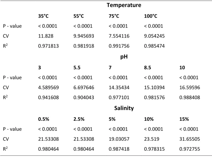
2.2.4 Determination of the effect of environmental parameters on biosurfactants produced ... 35
2.2.4.1 Effect of temperature on biosurfactant activity ... 35
2.2.4.2 Effect of pH on biosurfactant activity ... 35
2.2.4.3 Effect of salinity on biosurfactant activity ... 36
2.2.4.4 Statistical analysis ... 36
2.2.5 Emulsification index test ... 36
2.2.6 Surface tension measurement ... 37
2.3 RESULTS... 37
2.3.1 Preliminary screening for biosurfactant production amongst AEFB isolates ... 37
3
2.3.2 Effect of environmental parameters on the activity of biosurfactant produced by AEFB ... 39
2.3.2.1 Effect of temperature on biosurfactant activity ... 40
2.3.2.2 Effect of pH on biosurfactant activity ... 43
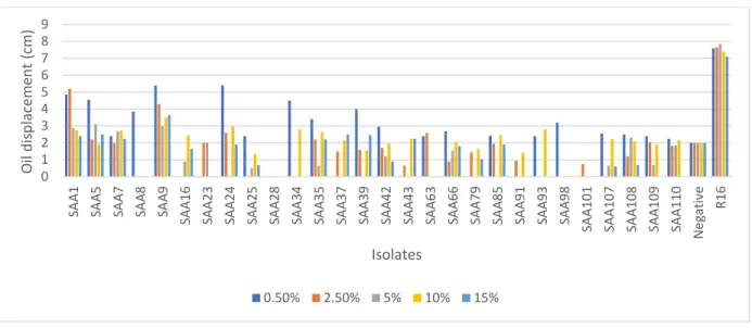
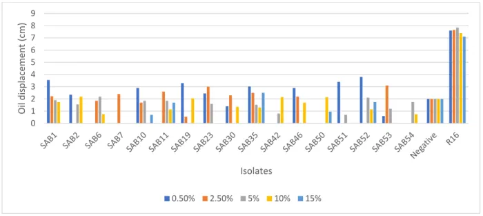
2.3.2.3 Effect of salinity on biosurfactant activity ... 46
2.3.3 Emulsification index (E24) assay ... 50
2.3.4 Surface tension measurement ... 52
2.4 Discussion ... 52
2.5 Conclusion ... 56
CHAPTER THREE ... 57
CHARACTERIZATION OF BIOSURFACTANT COMPOUNDS PRODUCED BY SELECTED AEROBIC ENDOSPORE- FORMING BACTERIA ISOLATES FROM MFABENI PEATLAND ... 57
3.1 INTRODUCTION ... 57
3.2 MATERIALS AND METHODS ... 59
3.2.1 Bacterial isolates ... 59
3.2.2 Biosurfactant production and acid precipitation ... 59
3.2.3 Thin layer chromatography (TLC) ... 60
3.2.4 Ultra Performance Liquid Chromatography (UPLC) in conjunction with mass spectrometry (MS) ... 61
3.2.5 Extraction of template DNA for Polymerase Chain Reaction (PCR) ... 61
3.2.6 Repetitive extragenic palindromic-Polymerase Chain Reaction (REP-PCR) ... 62
3.2.7 16S rRNA gene amplification ... 62
3.2.8 16S rRNA gene sequencing analysis ... 63
3.2.9 16S rRNA gene sequence phylogenetic analysis ... 63
3.3 Results ... 64
3.3.1 Acid precipitation and methanol extract ... 64
3.3.2 TLC separation and analysis ... 64
3.3.3 Mass Peaks detected by UPLC in conjunction with ESI-TOF MS ... 66
3.3.4 Determination of AEFB diversity using Rep-PCR ... 74
3.3.5 Determination of AEFB diversity using 16S PCR amplification ... 75
3.4 Discussion ... 78
3.5 Conclusion ... 82
CHAPTER FOUR ... 83
GENERAL OVERVIEW ... 83
4.1 Summary of findings ... 83
4
4.2 Future studies ... 86
REFERENCES ... 87
APPENDIX A: Measurement for the different buffer solutions used for pH adjustments ... 114
APPENDIX B: ANOVA analysis for the effect of temperature on biosurfactant activity ... 115
APPENDIX C: Certificate of analysis for surface tension measurement of selected AEFB isolates ... 118
Appendix D: Retention factor (Rf) values and mass peak assignment of lipopeptide compounds extracted from B. velezensis R16 determined using UPLC-ESI-TOF MS ... 119
APPENDIX E: UPLC ESI-TOF MS chromatograms and mass peaks commercial iturin and surfactin standards ... 120
APPENDIX F: 16S rRNA gene sequence PCR amplification of selected AEFB isolates... 122
5 LIST OF FIGURES
Figure 1.1: Cyclic structure of surfactin. It contains fatty acid chain with 13 to 15 carbon length
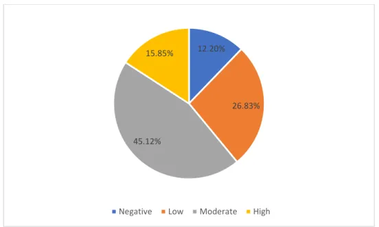
(Mulligan, 2009) ... 17 Figure 1.2: Cyclic structure of Iturin. It contains fatty acid chain with 14 to 17 carbon length (Meena and Kanwar, 2015) ... 18 Figure 1.3: Cyclic structure of fengycin. It contains fatty acid chain with 16 to 19 carbon length (Meena and Kanwar, 2015) ... 18 Figure 2.1: Pie chart illustrating the response of AEFB isolates to oil spreading assay. Zones of oil
displacement ranging from ≤ 20 mm represent a negative biosurfactant response, > 20 to < 40 mm low biosurfactant activity, ≥ 40 to < 60 mm moderate biosurfactant activity, and ≥ 60 to 85 mm high
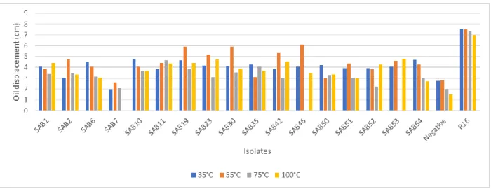
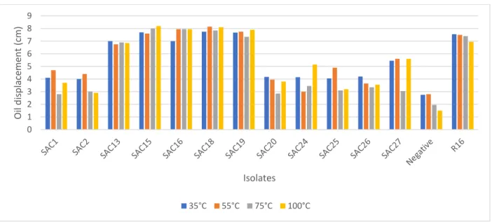
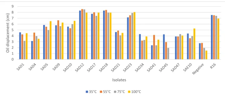
biosurfactant activity. ... 39 Figure 2.2: Effect of temperature on the biosurfactant activity of AEFB isolates cultured from Sample A (12 cm) of Mfabeni peatland determined using the oil spreading assay. Mean oil displacement values are presented (n = 2) ... 41 Figure 2.3: Effect of temperature on the biosurfactant activity of AEFB isolates cultured from Sample B (21 cm) of Mfabeni peatland determined using oil spreading assay. Mean oil displacement values are presented (n = 2) ... 42 Figure 2.4: Effect of temperature on the biosurfactant activity of AEFB isolates cultured from Sample C (89 cm) of Mfabeni peatland determined using oil spreading assay. Mean oil displacement values are presented (n = 2) ... 42 Figure 2.5: Effect of temperature on the biosurfactant activity of AEFB isolates cultured from Sample D and E (237 and 344 cm) of Mfabeni peatland determined using oil spreading assay. Mean oil
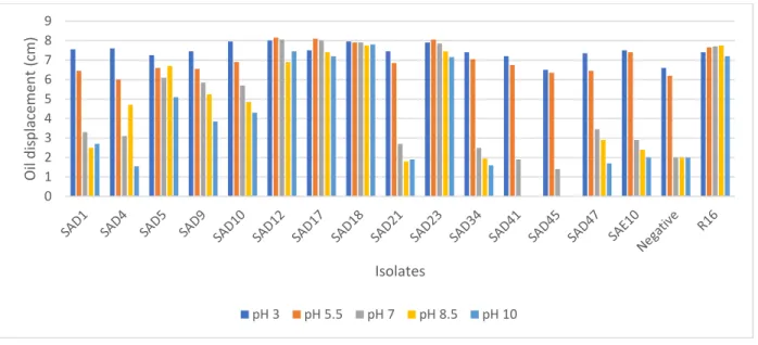
displacement values are presented (n = 2) ... 43 Figure 2.6: Effect of pH on the biosurfactant activity of AEFB isolates cultured from Sample A (12 cm) of Mfabeni peatland determined using oil spreading assay. Mean oil displacement values are presented (n
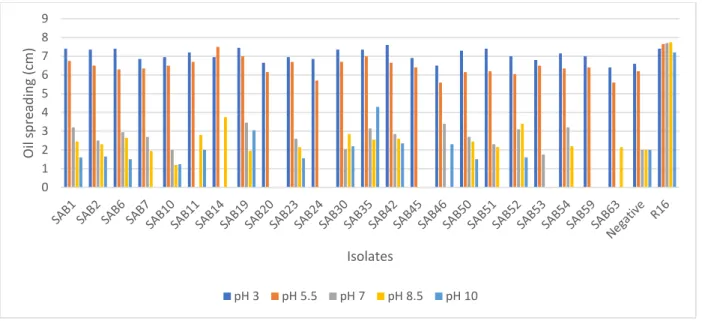
= 2) ... 44 Figure 2.7: Effect of pH on the biosurfactant activity of AEFB isolates cultured from Sample B (21 cm) of Mfabeni peatland determined using oil spreading assay. Mean oil displacement values are presented (n
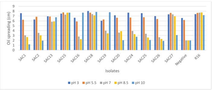
=2) ... 45 Figure 2.8: Effect of pH on the biosurfactant activity of AEFB isolates cultured from Sample C (89 cm) of Mfabeni peatland determined using oil spreading assay. Mean oil displacement values are presented (n
=2) ... 45 Figure 2.9: Effect of pH on the biosurfactant activity of AEFB isolates cultured from Sample D and E (237 and 344 cm) of Mfabeni peatland determined using oil spreading assay. Mean oil displacement values are presented (n =2) ... 46 Figure 2.10: Effect of salinity on the biosurfactant activity of AEFB isolates cultured from Sample A (12 cm) of Mfabeni peatland determined using oil spreading assay. Mean oil displacement values are
presented (n = 2) ... 47
6
Figure 2.11: Effect of salinity on the biosurfactant activity of AEFB isolates cultured from Sample B (21 cm) of Mfabeni peatland determined using oil spreading assay. Mean oil displacement values are
presented (n = 2) ... 48
Figure 2.12: Effect of salinity on the biosurfactant activity of AEFB isolates cultured from Sample C (89 cm) of Mfabeni peatland determined using oil spreading assay. Mean oil displacement values are presented (n = 2) ... 49
Figure 2.13: Effect of salinity on the biosurfactant activity of AEFB isolates cultured from Sample D and E (237 and 344 cm) of Mfabeni peatland determined using oil spreading assay. Mean oil displacement values are presented (n = 2) ... 50
Figure 2.14: The emulsification (E24) index observed for the uninoculated broth (negative) control (A), Isolate SAB19 (B) and the B. velezensis R16 control (C) using vegetable seed oil ... 51
Figure 3.1: Thin layer chromatography of methanol extracts demonstrating hydrophobic region associated with AEFB isolates: 1- B. velezensis R16 control; 2- SAB19; 3- SAB42; 4- SAC15; 5- SAC18; 6- SAD5; 7- SAD17; 8- SAD18; and, 9- SAD23. Compound bands were detected by spraying the surface with distilled water to detect hydrophobic regions. ... 66
Figure 3.2: UPLC-ESI-TOF MS chromatogram of crude extract from B. velezensis R16 control. Portions labelled A, B, and C are representatives of lipopeptide compounds. Peak A- bacillomycin, peak B- fengycin, and peak C- surfactin ... 67
Figure 3.3: Mass peak of peak A which eluted at time 21.15 with m/z 1031.5 represents a bacillomycin homolog ... 67
Figure 3.4: Mass peak of peak B which eluted at time 25.97 with m/z 1505.9 represents a fengycin homolog ... 68
Figure 3.5: Mass peak of peak C which eluted at time 28.99 with m/z 1036.7 represents a surfactin homolog ... 68
Figure 3.6: Chromatogram of the crude extract for isolate SAB19 obtained using UPLC ESI-TOF MS ... 68
Figure 3.7: Chromatogram of the crude extract for isolate SAB42 obtained using UPLC ESI-TOF MS ... 69
Figure 3.8: Chromatogram of the crude extract for isolate SAC15 obtained using UPLC ESI-TOF MS ... 69
Figure 3.9: Chromatogram of the crude extract for isolate SAC18 obtained using UPLC ESI-TOF MS ... 69
Figure 3.10: Chromatogram of the crude extract for isolate SAD5 obtained using UPLC ESI-TOF MS ... 70
Figure 3.11: Chromatogram of the crude extract for isolate SAD17 obtained using UPLC ESI-TOF MS .... 70
... 70
Figure 3.12: Chromatogram of the crude extract for isolate SAD18 obtained using UPLC ESI-TOF MS .... 70
Figure 3.13: Chromatogram of the crude extract for isolate SAD23 obtained using UPLC ESI-TOF MS .... 71
Figure 3.14: Chromatogram (A) and mass peak (B) of hydrophobic fraction (Rf 0.12 – 0.17) from TLC. ... 73
Figure 3.15: Agarose gel (1.5%) electrophoresis image comparing REP-PCR fingerprints of eight AEFB isolates from an ancient Mfabeni peatland sediment core. MWM: 1 kbp DNA ladder; R16- Positive control; Neg: DNA-free control. ... 75
7
Figure 3.16: Agarose gel (1.5%) electrophoresis image indicating 16S rRNA gene sequencing PCR
amplification products of eight AEFB isolates from an ancient Mfabeni peatland sediment core. ... 122 Figure 3.17: The evolutionary history was inferred using the neighbor joining method based on the Tamura-Nei substitution model (Saitou and Nei, 1987). Molecular phylogenetic analysis of AEFB isolates using neighbor joining method. The scale bar corresponds to 0.020 nucleotide substitutions per
sequence positions. Abbreviations: B= Bacillus; Brev. = Brevibacillus. ... 76 Figure 3.18: Molecular phylogenetic analysis of AEFB isolates by maximum likelihood method. The evolutionary history was inferred by using the maximum likelihood method based on the Kimura 2- parameter model (Kimura, 1980; Kumar et al., 2016). The scale bar corresponds to 0.050 nucleotide substitutions per sequence positions. Abbreviations: B= Bacillus; Brev. = Brevibacillus. ... 77
8 LIST OF TABLES
Table 2.1: AEFB isolates from Mfabeni peatland selected for biosurfactant screening ... 33 Table 2.2: Results showing biosurfactant activity amongst AEFB isolates ... 38 Table 2.3: Summary of ANOVA analysis of environmental parameters influencing biosurfactant activity40 Table 2.4: Results showing emulsification (E24) index of selected AEFB isolates ... 51 Table 3.1: Sequence of the primers used for REP-PCR and 16S rRNA reactions ... 63 Table 3.2: Rf values obtained for the hydrophobic region of the selected AEFB isolates visualized using TLC ... 65 Table 3.3: Detection of Lipopeptide compounds produced by AEFB isolates identified by UPLC-ESI-TOF MS analysis of methanol extracts from TLC crude extracts ... 72 Table 3.4: Unidentified peak associated with the hydrophobic region (Rf 0.12 – 0.17) from the scraped fraction of TLC plates ... 74
9
INTRODUCTION
Biosurfactants are surface-active compounds that are able to reduce surface and interfacial tensions (Desai and Banat, 1997; Mulligan, 2005). Their scope of application includes use as wetting agents, emulsifiers, foaming agents, detergents and dispersants. In recent years they have gained favor over synthetic surfactants because of their ecological acceptability due to reduced toxicity, higher biodegradability and stability over a range of environmental conditions (Desai and Banat, 1997; Lima et al., 2011). Increasingly, biosurfactants are being recognized for their industrial and environmental applications, which include bioremediation, decontamination of manufacturing wastes, clearing of oil spills, and microbial enhanced oil recovery (Joshi et al., 2012).
Biosurfactants are produced by a range of bacteria and fungi with several classes of biosurfactant compounds having been distinguished (Mata-Sandoval et al., 1999; Mata-Sandoval et al., 2001;
Chen et al., 2007a). Various classes or categories of biosurfactant are recognized, which include lipopeptides, mycolic acid, glycolipids, lipopolysaccharides, and phospholipids (Smyth et al., 2010). The interest in biosurfactants has resulted in a growing number of studies that are focused on screening for novel biosurfactant compound producers, screening for strains with enhanced biosurfactant production capabilities, and, screening for biosurfactant compounds that can function over a wide range of environmental conditions.
Wetlands and peatlands are ecosystems which support a diverse array of aerobic and anaerobic microbial communities that play important roles in the recycling of organic matter. Mfabeni Peatland, located within the St. Lucia Wetland Park, KwaZulu-Natal is regarded as one of the most significant peatland ecoregions in South Africa (Grundling et al., 2013). The diversity and functioning of microbes within this ecosystem is largely unexplored and represents an untapped source of microbial diversity with potential biotechnological applications (Naidoo, 2017). Aerobic endospore-forming bacteria (AEFB) are of special interest due to their association with the production of industrially significant enzymes and bioactive chemicals such as antibiotics, biopesticides and biosurfactants (Couto et al., 2015). Peatlands are associated with an accumulation of partially degraded plant material that builds up over time as the result of anoxic
10
conditions that arise under waterlogged conditions. Under these conditions endospores from AEFB may become trapped within layers of accumulating organic material. Peatland sediment therefore, has the potential to serve as an archival record of AEFB diversity from this environment (Naidoo, 2017).
A study was undertaken with the aim of screening for biosurfactant activity amongst a phylogenetically diverse collection of aerobic endospore-forming bacteria (AEFB) isolated previously from sections of a sediment core taken from Mfabeni peatland. These samples were taken from radiocarbon dated sections of the core which were dated from ca. 589 to 37,906 cal years BP.
The objectives of the study were:
i) To screen for biosurfactant production amongst aerobic endospore forming bacteria from a subset of isolates obtained from a Mfabeni peatland sediment core;
ii) To determine the effect of environmental parameters namely, temperature, pH, and salt concentration on the activity and efficacy of biosurfactants produced by AEFB;
iii) To extract, partially purify and characterize biosurfactant compound(s) produced by selected AEFB using acid precipitation followed by methanol extraction, Thin Layer Chromatography (TLC) and reverse phase Ultra Performance Liquid Chromatography (UPLC) used in conjunction with Electrospray Ionization-Mass Spectrometry (ESI-MS);
iv) To differentiate and classify selected AEFB isolates using REP-PCR genomic fingerprinting and 16S rRNA gene sequence analysis.
This dissertation has been divided into four chapters. Chapter one is a literature review that provides an overview of biosurfactants, specifically focusing on those produced by Bacillus spp..
Chapters two and three cover specific aspects of the research undertaken in the study and are each presented in the format of an independent scientific paper. Chapter four provides a summary of the major findings and highlights their significance.
11
CHAPTER ONE LITERATURE REVIEW 1.1 INTRODUCTION
Microbial surfactants, or biosurfactants, are surface active, amphiphilic, low molecular weight compounds produced by a wide range of different microorganisms (Banat, 1995; Padmavathi and Pandian, 2014). Biosurfactants act by lessening the interfacial and surface tensions between particles in two liquids or between a liquid and a solid (Banat et al., 2000). Lipopeptides, fatty acids, polysaccharides, glycolipids, phospholipids, lipoproteins, neutral lipids, and polymerics are examples of biosurfactants that have been distinguished (Neu, 1996; Muthusamy et al., 2008).
Biosurfactants are favored over synthetic surfactants because of their unique properties such as reduced toxicity, higher biodegradability, ecological acceptability, and stability under adverse conditions (Desai and Banat, 1997; Lima et al., 2011).
Biosurfactants are increasingly regarded as having commercial value due to a range of potential applications such as; acting as wetting agents, emulsifiers, foaming agents, detergents, and dispersants. They are applicable in a wide range of industries including cosmetics, petroleum, food processing, agricultural, and pharmaceutical industries (Banat et al., 2000). They are also suitable for use in oil recovery and in the management and remediation of contaminated sites (Banat, 1995; Al-Sulaimani et al., 2011). They have also been investigated for various biomedical applications as well; for example, as antiviral, antifungal, antibacterial, and anti-adhesive agents against a range of drug resistant pathogens (Gudiña et al., 2010; Luna et al., 2011).
Aerobic endospore-forming bacteria (AEFB) are Gram positive, rod-shaped organisms, suited to culturing on a large scale and are mostly non-pathogenic (Fritze, 2004; Neves et al., 2007). They are amongst the diverse group of microorganisms that produce biosurfactants (Al-Bahry et al., 2013; Couto et al., 2015). This group of bacteria include members of the genus Bacillus and are regarded as a promising group of bacteria because of their biotechnological potential (Mandic- Mulec and Prosser, 2011; Al-Bahry et al., 2013; Couto et al., 2015).
12
Lipopeptides are one of the most widely studied classes of biosurfactant associated with AEFB.
Structurally, they are made up of a lipid tail joined to a short linear or cyclic oligopeptide. In addition to their surfactant properties, they are known for their antimicrobial, antitumor, cytotoxic, and immunosuppressant properties (Cameotra and Makkar, 2004; Donadio et al., 2007; Gross and Loper, 2009; Pirri et al., 2009). Surfactin, a lipopeptide synthesized by Bacillus subtilis, is regarded as one of the most effective biosurfactant discovered to date (Arima et al., 1968; Gudiña et al., 2013). Other examples of lipopeptides produced by B. subtilis include fengycin, mycosubtilin, iturin, and bacillomycin (Vater et al., 2002).
Considering the advantages that microbial surfactants have over artificial surfactants, there is a need to screen and identify novel biosurfactant producers, and also identify AEFB strains that have enhanced biosurfactant production capabilities.
1.2 PRODUCERS OF BIOSURFACTANTS: - AEROBIC ENDOSPORE-FORMING BACTERIA (AEFB)
The genus Bacillus was established in 1872 by Cohn and is amongst the first set of bacteria to be described. They have been isolated from various environments, which includes soil, sea water, dust, and even ocean basin cores (Singh et al., 2007; Wilson et al., 2008; Aislabie et al., 2009;
Teixeira et al., 2010; Vollú et al., 2014). The ability of AEFB to produce spores makes isolation, cultivation, and maintaining them in the laboratory relatively easy. Dormant endospores are readily dispersed, this allows them to be distributed widely and colonize a wide range of habitats (Wipat and Harwood, 1999).
AEFB have adapted to a broad range of environmental conditions and are able to inhabit diverse habitats (Nicholson et al., 2000). For example, members of the Halobacillus, Thermobacillus, and Psychrobacillus genera have been isolated from halophilic, thermophilic, and psychrophilic areas respectively (Brandes et al., 2011). AEFB have also been isolated from various environments like salt marshes, marine sediment, thermal acid waters, marine sponges, glaciers, wetlands, volcanic soil, and geothermal vents (Margesin and Miteva, 2011; Phelan et al., 2012; Sonalkar et al., 2014;
Aanniz et al., 2015). Many of these strains have traits like proteolytic, amylolytic, and
13
antimicrobial activities which are of biotechnological interest (Phelan et al., 2012; Aanniz et al., 2015).
AEFB are of special biotechnological interest because of their ability to produce industrially significant enzymes and bioactive chemicals, their ability to degrade a range of pollutants, as well as their use as biopesticides. Biosurfactants are among the most significant bioactive chemicals synthesized by AEFB (Couto et al., 2015). Biosurfactants are normally produced as secondary metabolites during late exponential growth or stationary phase (Mahdy et al., 2012). They may be retained intracellularly or excreted extracellularly into the surrounding medium (Kazim et al., 2017). The biosurfactants produced assists in making insoluble substrates available to the microorganism through solubilization and desorption (Viramontes-Ramos et al., 2010; Mahdy et al., 2012). They also enhance the surface area of hydrophobic surfaces and adjust the joining or removal of microorganisms to surfaces (Saravanan and Vijayakumar, 2015).
Additionally, biosurfactants have been linked to a range of functions that include environmental remediation, and biological control (Ron and Rosenberg, 2001; Mandic-Mulec and Prosser, 2011).
Among the biosurfactants produced by the AEFB, surfactin and lichenysin are two well-studied lipopeptide surfactants made by B. subtilis and B. licheniformis respectively (Sekhon et al., 2012).
These compounds are able to function under extreme temperature (e.g. 4°C to 100°C) and pH (e.g. 3 to 10) conditions; these properties have allowed them to be used in a range of diverse applications (Rodrigues et al., 2006; Jacques, 2011; Kaloorazi and Choobari, 2013).
1.3 ECOLOGICAL ROLE OF BIOSURFACTANTS
Biosurfactants have been reported to have a range of ecological roles, which may be specific to the bacteria producing the surfactant (Raaijmakers et al., 2010). Some of these roles are
discussed in the following sections.
1.3.1 BIOFILM FORMATION
Biofilms are dense collections of bacterial cells that form on surfaces as a result of cell division and multiplication. The bacterial cells release proteins that form slime materials which keeps the biofilm together (Stewart and Franklin, 2008). Within biofilms, bacteria are shielded from harsh
14
environmental conditions such as exposure to antibiotics. Lipopeptide biosurfactants synthesized by Bacillus species have been associated with biofilm formation and attachment to surfaces. It has been suggested that biosurfactants enhance the circulation of oxygen and food by maintaining liquid-filled channels in biofilms (Davey et al., 2003; Klausen et al., 2003).
Hofemeister et al. (2004) demonstrated that B. subtilis strain AI/3 needed surfactin to be able to develop pellicles, and biofilms.
1.3.2 MOTILITY
Twitching, swarming, and swimming are the main ways by which bacteria are able to move on surfaces (Henrichsen, 1972). During the swarming and swimming mode of movement, bacteria merge their flagella, and are thrust forward as a result of the rotation of the flagella (Harshey, 2003). Bacterial cells locomote in clusters for swarming but singly in the case of swimming (Raaijmakers et al., 2010). Biosurfactants can influence cell movement due to their ability to modify the thickness of surface layers (Lindow and Brandl, 2003; Raaijmakers et al., 2010). It is believed that lipopeptides participate in cell aggregation and in the coordination of their movement (Raaijmakers et al., 2010). They also assist their producers in moving to locations on plant surface that are rich in nutrient by acting as wettability agents ( Lindow and Brandl, 2003;
Nielsen et al., 2005).
1.3.3 ANTAGONISM
It has been observed that microorganisms which produce lipopeptide biosurfactants have better competitive benefits compared to other microorganisms (Raaijmakers et al., 2010). These biosurfactants inhibit growth and cause lysis of a wide array of microorganisms such as oomycetes, viruses, fungi, and bacteria (Raaijmakers et al., 2010). Surfactin has been found to deactivate viruses through the disruption of the viral constituents. It also inhibits various animal and human pathogens by forming pores in their membranes causing cellular disruption (Vollenbroich et al., 1997; Huang et al., 2006). Lipopeptides are also effective against fungi; for instance, fengycin is known for its antifungal activity against Botrytis cinerea and Fusarium graminearum (Romero et al., 2007; Wang et al., 2007) while iturin is effective against Rhizoctonia solani and Penicillium roqueforti (Yu et al., 2002; Chitarra et al., 2003).
15
1.4 TYPES OF BIOSURFACTANTS
Biosurfactants have been classified based on the kind of charge present on each moiety which includes; positively charged cationic biosurfactants, negatively charged anionic surfactants, amphoteric surfactants, and non-ionic biosurfactants (Ginkel, 1989; Rahman and Gakpe, 2008).
Biosurfactants have also been categorized based on their molecular weight and chemical composition. These include low molecular weight and high molecular weight biosurfactant compounds. The low molecular weight compounds are generally active at reducing interfacial and surface tensions; the most widely studied of which are the lipopeptides and glycolipids (Rahman and Gakpe, 2008). Examples of lipopeptides include surfactin, iturin, fengycin, and bacillomycin, while the glycolipids comprise the sophorolipids, rhamnolipids, and trehalolipids (Ron and Rosenberg, 2001). High molecular weight compounds, such as bioemulsan, are the most efficient stabilizing agents and are effective in steadying oil in water colloids (Dastgheib et al., 2008; Salihu et al., 2009). In this literature review, the focus is on lipopeptides because they are one of the most widely studied classes of biosurfactant associated with Bacillus species.
1.4.1 LIPOPEPTIDES
Lipopeptides are a well-known group of biosurfactants synthesized by Bacillus species (Perfumo et al., 2010). They exhibit wide-ranging activities which include antimicrobial, antifungal, and antitumoral characteristics (Donadio et al., 2007; Perfumo et al., 2010). Lipopeptides have the ability to form pores in cell membranes, which causes membrane imbalance and death in sensitive organisms (Bender et al., 1999; Baltz, 2009).
Lipopeptides are made up of a cyclic oligopeptide moiety attached to a lipid tail (Stein, 2005;
Perfumo et al., 2010; Raaijmakers et al., 2010). These lipopeptides have been divided into four classes namely; surfactins, iturins, fengycins, and kurstakins (Hathout et al., 2000; Ongena and Jacques, 2008). Surfactins consists of surfactin, esperin, pumilacidin, and lichenysin, whereas the Iturin family consists of mycosubtilin, iturin A, AL, and C, and bacillomycin D, F, L, and LC. The fengycin family consists of fengycin A and B, and plipastatin A and B (Ongena and Jacques, 2008).
A fourth class- the kurstakins were originally isolated from strains of B. thuringiensis and little is
16
known about their ecological significance or applicability (Hathout et al., 2000; Béchet et al., 2012).
1.4.1.1SURFACTIN
Surfactin is a cyclic lipoheptapeptide connected to a hydrophobic β-hydroxy fatty acid side chain (Figure 1.1) (Seydlová et al., 2011). Based on the different amino acid sequence, surfactin has been categorized into A, B, and C groups (Rodrigues et al., 2006; Korenblum et al., 2012).
Normally, several surfactin isoforms with fatty acid side chains of varying carbon chain length (13 to 15 carbon) can be found together within a mixture of various peptide variants in cells (Tang et al., 2007). The structure and composition of the fatty acid side chain and the amino acids in surfactin molecule depends on both the culture conditions and the producer strain (Seydlová et al., 2011). Surfactins are known to possess antiviral, antifungal, antimicrobial, antibacterial, and antitumor abilities which makes them of interest for use in medical, and environmental applications (Kim et al., 1998; Ahimou et al., 2000; Nitschke and Costa, 2007; Mulligan, 2009;
Banat et al., 2010).
Surfactants, such as surfactin can be used as detergents and soaps (Heerklotz et al., 2004; Dufour et al., 2005). Several mechanisms have been proposed to describe the molecular mechanism involved in the disruptive action of surfactants (Deleu et al., 2003). In the first instance, surfactin is thought to act as a cation-carrier that is able to carry cations through an organic barrier. The fatty acid portion of surfactin penetrates a phospholipid bilayer to interact with the acyl chain of phospholipid. The heptapeptide headgroup aligns with the polar head region of the phospholipids. When a cation, such as calcium ion forms a complex with the surfactin molecule, it causes a ‘flip-flop’ response whereby the surfactin reorientates itself across the lipid bilayer (Heerklotz et al., 2004). The second explanation is that surfactin form pores, or cationic channels across a phospholipid membrane. This then leads to an osmotic imbalance, which causes disruption of the lipid membrane (Deleu et al., 2003). The third hypothesis describes the detergent effect of surfactin being a result of its fatty acid chain inserting and causing disruption or disorganization of the phospholipid bilayer (le Maire et al., 2000). This effects membrane
17
permeability and can lead to membrane solubilization. The formation of mixed micelles can also occur (Kragh-Hansen et al., 1998).
Surfactins are synthesized by a surfactin synthetase enzyme, which is a non-ribosomal peptide synthetase (Hue et al., 2001). Surfactin is considered to be a potent biosurfactant. At a concentration of 20 µM, it is able to lower the surface tension of water from 72 mN/m to 27 mN/m (Yeh et al., 2005). Water tension is lowered by the surfactant molecule occupying the intermolecular space between water molecules; and reducing the forces of attraction between these molecules. This creates a more fluid solution, which increases the wetting ability of the water (Dufour et al., 2005).
Figure 1.1: Cyclic structure of surfactin. It contains fatty acid chain with 13 to 15 carbon length (Mulligan, 2009)
1.4.1.2ITURIN
Iturin is a cyclic heptapeptide connected to a β-amino fatty acid chain of varied length, ranging from 14 to 17 carbon (Figure 1.2) (Tsuge et al., 2001; Meena and Kanwar, 2015). It is produced by a number of Bacillus spp. that fall within the B. subtilis complex of closely related taxa (Tsuge et al., 2001). They are potent antifungal agents which makes them potential candidates for use as biopesticides (Vater et al., 2002; Romero et al., 2007; Pecci et al., 2010). The cytoplasmic membrane of the fungi is penetrated by the hydrophobic part of iturin which causes pore formation and leads to osmotic disruption of the cell cytoplasm (Stein, 2005).
18
Figure 1.2: Cyclic structure of Iturin. It contains fatty acid chain with 14 to 17 carbon length (Meena and Kanwar, 2015)
1.4.1.3 FENGYCIN
Fengycin is a cyclic lipodecapeptide which is connected to a β-hydroxy fatty acid of varying chain length (16 to 19 carbons) (Steller and Vater, 2000; Wei et al., 2010). Fengycin is an antifungal agent and is particularly effective against filamentous fungi (Steller and Vater, 2000; Deleu et al., 2008). Two groups of fengycin have been distinguished, Fengycin A and Fengycin B. Fengycin A possesses the amino acid alanine (Ala) at position 6 of the oligopeptide structure while Fengycin B has valine (Val) at this position (Steller and Vater, 2000; Meena and Kanwar, 2015).
Figure 1.3: Cyclic structure of fengycin. It contains fatty acid chain with 16 to 19 carbon length (Meena and Kanwar, 2015)
1.5 FACTORS AFFECTING PRODUCTION OF BIOSURFACTANTS
Environmental, and nutritional factors are the main factors which affect the production of biosurfactants (Rahman and Gakpe, 2008). Not only do these factors determine the quantity of biosurfactant produced but they can also influence the type produced (Salihu et al., 2009).
19
1.5.1 ENVIRONMENTAL FACTORS
Environmental factors like pH, temperature, salinity, oxygen availability, and growth conditions impact production of biosurfactant (Desai and Banat, 1997; Rahman et al., 2002; Ilori et al., 2005;
Raza et al., 2007). For instance, oxygen transfer was found to be one of the major limiting factors for scaling up production of surfactin in B. subtilis (Sheppard and Cooper, 1990). The presence or absence of glucose as a carbon source in a growth medium can also determine the availability and amount of biosurfactant produced (Walter et al., 2010). Bacillus species have been reported to be able to produce biosurfactants at temperatures up to 100°C, pH up to 10, and at salinity concentrations up to 10% (Desai and Banat, 1997; Echigo et al., 2005; Márquez et al., 2011; Md, 2012; Varadavenkatesan and Murty, 2013; Liang et al., 2017).
For application in microbial enhanced oil recovery (MEOR), surfactant producing bacteria must have the capacity to withstand inhospitable conditions such as low oxygen, high temperature, salinity, and pressure (Couto et al., 2015). Alternatively, they should produce stable biosurfactant compounds that can function under a range of adverse conditions. Biosurfactant producers which meet these criteria have the potential to be used in enhancing bioremediation and in the clean-up of oil spills (Sekhon et al., 2012).
1.5.2 NUTRITIONAL FACTORS
Several carbon substrates have been used for biosurfactant production such as glycerol, glucose, and crude oil (Desai and Banat, 1997; Ilori et al., 2005; Rahman and Gakpe, 2008). The kind, and amount of biosurfactant produced depends largely on the concentration of the carbon source used (Zajic and Donaldson, 1985; Rahman and Gakpe, 2008; Raza et al., 2007). For example, Pseudomonas aeruginosa produced higher yields of biosurfactant (100 – 165 mg g-1) when supplied with long chain alcohols, corn oil, and lard compared to succinate acid and glucose, which produced lower yields (12 – 36 mg g-1) (Mata-Sandoval et al., 2001). This isolate was also able to produce rhamnolipid from a range of carbon substrates, which include C11 and C12 alkanes, olive oil, glycerol, succinate, glucose, and fructose (Rahman and Gakpe, 2008). In a study carried out by Saharan et al. (2011), biosurfactant production by B. subtilis MTCC 2423 was
20
enhanced after supplementing culture media with beef extract, sucrose, sodium pyruvate, and glucose as carbon source.
1.6 APPLICATIONS OF BIOSURFACTANTS
Biosurfactants are useful to man and have found application in a wide range of industries due to their emulsification, foaming, surface tension reduction, lubrication, and moisture retention properties (Desai and Banat, 1997; Barros et al., 2008; Banat et al., 2010; Damasceno et al., 2012).
Currently, the need for surfactants has largely been met by synthetic surfactants which in many instances are not only harmful but are also often non-biodegradable; these limitations have heightened the demand for natural surfactants.
1.6.1 BIOSURFACTANTS IN MEDICINE
The establishment of bacterial biofilms on surfaces are potentially important sources of nosocomial infections (Singh and Cameotra, 2004). Significantly, bacteria found in biofilms can exhibit increased tolerance to antibiotics; they are also effective in avoiding host defense mechanism. Factors such as swarming, motility, and biofilm formation are important determinants of the ability of a bacterium to colonize surfaces. Biosurfactants such as surfactin have anti-adhesive properties, which hinders the binding of pathogenic microorganisms to surfaces or infection sites. Surfactin has been shown to reduce the amount of biofilm formed by strains of Escherichia coli, Salmonella enterica, and Proteus mirabilis (Seydlová and Svobodová, 2008). In controlled laboratory studies, coating catheters by running them through a solution of surfactin was found to reduce biofilm formation. The coating of catheters with surfactin has been proposed as a means to limit biofilm development by potential pathogens (Seydlová and Svobodová, 2008).
Surfactin has also been identified as an anti-inflammatory agent because of its ability to hinder inflammation in eukaryotic cells, which occurs as a result of the interaction between these cells and lipopolysaccharide (Seydlová and Svobodová, 2008; Kim et al., 2006). However, intravascular use of surfactin is not recommended due to its non-specific cytotoxic effect on cell membranes
21
(Seydlová and Svobodová, 2008). Concentrations below 25 µM were not found to be disruptive to cell membrane (Seydlová and Svobodová, 2008).
1.6.2 BIOSURFACTANTS AND MICROBIAL CONSORTIA
It has been shown that the use of microbial consortia can improve biodegradation efficacy compared to monocultures (Kadali et al., 2012). This has been attributed to mutually beneficial interactions, which can have a positive influence on growth and survivability (Sampath et al., 2012). For example, the addition of rhamnolipid biosurfactants has been shown to improve the biodegradation efficiency of slow degrading consortia. Conversely, biosurfactants can have an opposite effect whereby they cause a significant decrease in the biodegradation rate associated with fast degrading consortia (Owsianiak et al., 2009). This phenomenon is attributed to differences in substrate uptake modes. In instances where hydrocarbon uptake occurred directly from the aqueous phase, the addition of surfactants was thought to increase solubilization of hydrocarbons and improve the biodegradation. Conversely, in instance where high initial rates of biodegradation of hydrocarbons occurred at the interface boundary with a biofilm, the introduction of surfactants could potentially restrict the contact between microorganisms and substrates, thereby negatively effecting biodegradation rate (Owsianiak et al., 2009).
1.6.3 BIOSURFACTANTS IN AGRICULTURE
Several biosurfactants such as iturin A, fengycin, and surfactin demonstrate antimicrobial activity against fungal and bacterial plant pathogens and can be regarded as promising biocontrol agents suited to sustainable agricultural practices (Sachdev et al., 2013). For instance; surfactants produced by Bacillus spp. have been reported to show biocontrol actions against several plant pathogens, which include Dickeya and Pectobacterium spp. which cause soft rots, Colletotrichum gloeosporioides which causes anthracnose on papaya leaves and Fusarium spp. which cause damping off of vegetable seedlings (Kim et al., 2010; Velho et al., 2011; Eddouaouda et al., 2012;
Krzyzanowska et al., 2012).
Biosurfactants can also be applied to improve antagonistic action of microorganisms and their associated microbial products (Jazzar and Hammad, 2003; Kim et al., 2004). For example, weed species have been eliminated by spraying the plants with a combination of biosurfactants (0.2%)
22
and Myrothecium verrucaria (2.0 x 10⁷ conidia ml-1 at 300 L ha-1) (Boyette et al., 2002; Hoagland et al., 2007). Thus, these microbial surfactants and/or their producers are potential eco-friendly alternatives for environmentally hazardous chemical insecticides and pesticides used in agriculture (Sachdev et al., 2013).
1.6.4 BIOSURFACTANTS IN FOOD INDUSTRY
In the food industry, emulsification is of great importance in influencing the solubilization of odor producing compounds, as well as the texture and consistency of food products (Radhakrishnan et al., 2011). Emulsifiers control clustered globules and keeps activated systems steady so as to stabilize emulsions (Nitschke and Costa, 2007; Patino et al., 2008). An emulsion is a varied system comprising of an immiscible liquid that is distributed into another liquid, in droplet form (Nitschke and Costa, 2007). Biosurfactants are able to improve the consistency of such systems by decreasing the interfacial tension, which in turn decreases the surface energy between different phases (Nitschke and Costa, 2007; Muthusamy et al., 2008). These surfactants can also alter rheological properties and influence the shelf life of products (Nitschke and Silva, 2018).
The emulsification index also known as E24 is a fast and qualitative method used to ascertain the emulsifying properties of a biosurfactant (Desai and Banat, 1997). In a recent study done by Mnif et al. (2012), the addition of a lipopeptide biosurfactant (0.075% concentration) was found to improve the volume and structure of bread crumbs when compared to soya lecithin. The biosurfactant also decreased staling and multiplication of microbes as well as improve texture of the bread after eight days (Mnif et al., 2012). Addition of 0.1% lipopeptide has also been reported to improve the adhesive and cohesive texture of cookie dough (Zouari et al., 2016).
1.7 PRODUCTION OF BIOSURFACTANTS
Although biosurfactants have many industrial benefits and attributes compared to their chemical counterparts, producing these on an industrial scale has not been attempted fully because of high costs (Deleu and Paquot, 2004). The use of renewable, low-cost waste materials such as molasses, cassava wastewater, grape pomace, starch rich wastes and wastes from oil refineries have been investigated as potential solutions to this challenge (Makkar and Cameotra, 2002;
Nitschke and Pastore, 2006; Rivera et al., 2007; Sobrinho et al., 2008; Saharan et al., 2011).
23
Dubey and Juwarkar (2001) were able to produce biosurfactant from Pseudomonas aeruginosa using a synthetic medium supplemented with hexadecane and glucose. Other low-cost materials, which are also easily assessible include vegetable oils and their wastes e.g. soybean and sunflower, marine oils, and tallow (Pekin et al., 2005). These oils are efficient in promoting the production of biosurfactant during microbial growth (Rahman et al., 2002; Bednarski et al., 2004).
Potatoes are a major source of starch like substances and have been used for biosurfactant production (Saharan et al., 2011). They also provide a good source of vitamins, sulphur, and nitrogen for growth (Saharan et al., 2011). For example, B. subtilis 21332 is able to produce surfactin when cultured on potato wastewater whether it was either supplemented with trace element or not (Thompson et al., 2000; Noah et al., 2005). Carbohydrate rich effluents from cassava industries have also been shown to induce surfactin production in B. subtilis (Nitschke and Pastore, 2006).
1.8 OPTIMIZATION OF BIOSURFACTANT PRODUCTION
The optimization of biosurfactant production is an area of study that has received growing attention in recent year (Sarubbo et al., 2001; Sahoo et al., 2011; Liu et al., 2012; Kazim et al., 2017; Moshtagh et al., 2018). The type, quantity, and quality of biosurfactant produced is dependent on a range of cultural conditions, which include; temperature, agitation, pH, aeration, nitrogen source, metal ion concentration, as well as the type of carbon sources used (Saharan et al., 2011). The process conditions for biosurfactant production also need to be improved in order to increase yields to make production commercially viable (Dubey and Juwarkar, 2001; Hewald et al., 2005; Saharan et al., 2011). For example, addition of iron (2.0 mM) and manganese (0.2 mM) to a growth medium was found to enhance biosurfactant production (up to 4.8 g/l) by B.
subtilis (Gudiña et al., 2015).
Changing one variable at a time while keeping the others constant has been used as a traditional way of optimizing bioprocesses (Saharan et al., 2011). More recently, response surface methodology (RSM), a statistical optimization tool has been developed for process optimization (Saharan et al., 2011). This tool explores the relationships between selected explanatory variants
24
and one or several response variables (Saharan et al., 2011). RSM has been used to optimize inoculum, pitching rates, substrate concentration as well as environmental conditions in order to improve surfactin production by B. subtilis (Sen and Swaminathan, 2004). It has also been used for the enhancement of biosurfactant production by Pseudomonas aeruginosa AT10, Lactococcus lactis, Streptococcus thermophilus, and B. licheniformis (Abalos et al., 2002; Rodrigues et al., 2006). Optimization methods assist industries in selecting and formulating the best mixture of inexpensive substrates for media production and in employing the most beneficial environmental conditions for enhanced production of biosurfactant (Saharan et al., 2011).
1.9 METHODS FOR DETECTING BIOSURFACTANT PRODUCTION
A number of screening methods have been developed to detect biosurfactant production amongst microorganisms (Walter et al., 2010). Assays such as the Du-Nouy ring tensiometer assay, axisymmetric drop shape assay, and pendant drop shape assay measure the surface and interfacial tensions of biosurfactants directly; whereas the drop collapse, oil spreading, emulsification capacity, hydrophobic interaction chromatography (HIC) and microplate assays give an indirect measurement of surfactant production (Walter et al., 2010). Hemolysis and cetyltrimethylammonium bromide (CTAB) assays are regarded as specialized screening methods that are used to screen for specific types of surfactant and they are not appropriate for a general biosurfactant screening approaches (Walter et al., 2010).
1.9.1 OIL SPREADING ASSAY
The oil spreading assay, also known as oil displacement test, relies on the displacement of oil by the biosurfactant (Morikawa et al., 2000). A fixed volume of oil is added to distilled water in a petri dish and an aliquot of cell-free supernatant containing the suspected biosurfactant is then added to the middle of the oil droplet (Morikawa et al., 2000; Walter et al., 2010). Displacement of the oil as indicated by a zone of clearing is considered to be a positive indication of the presence of surfactant compounds (Morikawa et al., 2000; Walter et al., 2010). The diameter of the displacement corresponds to the level of biosurfactant activity (Morikawa et al., 2000; Walter et al., 2010). The oil spreading method is simple, fast, requires a small amount of sample and does not require specialized equipment (Płaza et al., 2006). It is also sufficiently sensitive to
25
detect biosurfactant activity at low concentrations. Several studies have used the oil spreading technique as a reliable means to screen for biosurfactant compound production amongst various microorganisms (Youssef et al., 2004; Płaza et al., 2006).
1.9.2 DROP COLLAPSE ASSAY
The drop collapse assay relies on changes in interfacial tension that occurs to a drop of liquid when exposed to surfactant compounds (Jain et al., 1991). A drop of surfactant containing supernatant is added onto a surface already coated with oil. If the supernatant contains surfactant, the drop collapses, increasing in diameter due to a reduction in interfacial tension between the drop and the surface. In the absence of a surfactant, the drop remains stable. This method is simple and fast and does not require large amount of sample.
1.9.3 EMULSIFICATION CAPACITY ASSAY
The emulsification capacity assay, also known as the E24 test, relies on the ability of a surfactant molecule to form a stable emulsion (Cooper and Goldenberg, 1987). This assay is carried out to determine the emulsification property of surfactant compounds. It involves the formation of micelles where hydrophobic liquids become dispersed within hydrophilic liquids (e.g. water), thereby forming an emulsion of two substances, which are normally immiscible (Banat, 1995).
Cell-free supernatant is added to oil and vortexed for 2 minutes, after which the mixture is left to stand for 24 h and then the height of the emulsion layer is measured (Cooper and Goldenberg, 1987; Walter et al., 2010). The E24 test determines emulsification activity and correlates to the surfactant concentration. It is calculated by determining the ratio of the height of emulsion layer and the total height of liquid (Cooper and Goldenberg, 1987). The equation used is shown below:
𝐸24 =ℎ𝑒𝑖𝑔ℎ𝑡 𝑜𝑓 𝑒𝑚𝑢𝑙𝑠𝑖𝑜𝑛 (ℎ𝑒𝑚𝑢𝑙𝑠𝑖𝑜𝑛)
𝑡𝑜𝑡𝑎𝑙 ℎ𝑒𝑖𝑔ℎ𝑡 𝑜𝑓 𝑙𝑖𝑞𝑢𝑖𝑑 (ℎ𝑡𝑜𝑡𝑎𝑙) 100%
1.9.4 MICROPLATE ASSAY
The microplate assay was developed as a qualitative assay that is used to detect the presence of surfactant compounds and relies on the property of biosurfactants being able to cause visual
26
distortion in cell-free supernatants (Vaux and Cottingham, 2007; Walter et al., 2010)). Cell-free supernatant samples are added to a 96- micro-well plate and then placed on a sheet of grid paper.
Each well is viewed from above to determine whether there is biosurfactant present which results in a concave surface, distorting the image of the grid. The microplate test is simple, fast, sensitive, and allows for an instantaneous detection of surface active compounds (Chen et al., 2007b). The method uses small amount of sample, making it suitable for automated high throughput screening (Chen et al., 2007b).
1.9.5 DU-NOUY RING TENSIOMETER ASSAY
The Du-Nouy ring method quantifies the force needed to separate a platinum wire loop from a liquid surface interface (Tadros, 2005). The force exerted is directly proportional to the interfacial tension and is quantified using an automated tensiometer (Walter et al., 2010). The Du-Nouy Ring test has been used extensively to screen microbes for biosurfactant production (Cooper and Goldenberg, 1987; Bodour and Miller-Maier, 1998; Rahman et al., 2002; Płaza et al., 2006). A culture is regarded as a promising surfactant producer if it is able to reduce the surface tension of a liquid medium to 40 mN/m or less (Cooper and Goldenberg, 1987). Willumsen and Karlson (1996) defined a good biosurfactant producer as a culture that can reduce the surface tension of a growth medium by ≥20 mN/m compared to a distilled water control. Although this method is accurate and relatively easy to use, it requires specialized equipment and is limited in that it is unable to measure different samples simultaneously (Walter et al., 2010). Another restriction of this method is that relatively large sample volumes are needed for analysis; these samples may also need to be diluted due to a limited concentration range that can be analyzed (Bodour and Miller-Maier, 1998).
1.9.6 STALAGMOMETRIC ASSAY
The stalagmometric assay is an assay that uses a Traube stalagmometer to measure the surface tension of a liquid (Dilmohamud et al., 2005). A Traube stalagmometer is an instrument that allows for the consistent formation of uniform volumes of liquid droplets and subjects them to gravitational force within a modified capillary tube. The number of drops that fall per unit of volume is compared to a control liquid (i.e. water) of known density and surface tension, in order
27
to establish the sample density. This information is then used to calculate the surface tension of the sample according to the following equation:
Where
𝜎
L is surface tension of the liquid to be examined,𝜎𝑤
is the surface tension of the water, NL is the number of liquid drops from the sample, 𝑁𝑤 is the number of water drops, P𝐿 is density of the sample, and 𝑃w is density of water (Dilmohamud et al., 2005). The disadvantage of this test is that only consecutive measurement can be carried out (Walter et al., 2010).1.9.7 HEMOLYSIS
Some biosurfactants are able to lyse erythrocytes and this principle is used to detect surfactant using the hemolysis assay (Mulligan et al., 1984). Cell cultures of bacteria are grown on sheep blood agar for 48 hours. A positive result, which is indicated by zone of clearance around the growth is observed for strains that can lyse blood cells. The hemolysis assay is usually used as an initial screening for isolates that can produce biosurfactants (Schulz et al., 1991; Youssef et al., 2004; Płaza et al., 2006). One of the restrictions of this method is that it is not precise, as there are enzymes that can also lyse blood cells. Another limitation is that diffusion of the biosurfactant may be restricted due to physicochemical constraints of the compound; which can hinder the formation of clear zones (Jain et al., 1991). Some studies have demonstrated that certain biosurfactants do not exhibit any hemolytic activity at all and that the method lacks specificity (Schulz et al., 1991; Youssef et al., 2004; Plaza et al., 2006). Hence, this method is prone to giving false negative and false positive results. Mulligan et al. (1984) suggested that the blood agar method should only be used as a preliminary screening method and, ideally, should be backed up by other techniques based on surface activity measurements.
1.9.8 CTAB AGAR PLATE
The cetyltrimethylammonium bromide (CTAB) agar plate method is a convenient screening method used for detecting anionic biosurfactants (Siegmund and Wagner, 1991). The organisms
28
under investigation are grown on a CTAB agar plate comprising of methylene blue and cetyltrimethylammonium bromide. Microorganisms that produce anionic biosurfactants, displace the cationic CTAB which results in dark blue zones forming around the colonies (Walter et al., 2010). This assay has been adopted to evaluate biosurfactant production under various culture conditions such as different temperatures or substrates that are added directly to the agar plate (Walter et al., 2010). A major disadvantage of the CTAB assay is that it is potentially harmful to some microorganism and could hinder growth (Walter et al., 2010). Siegmund and Wagner (1991) have suggested that CTAB could be substituted by another cationic surfactant in order to overcome these limitations.
1.10 METHODS FOR IDENTIFYING AND CHARACTERIZING BIOSURFACTANTS
Various techniques and methods have been developed over the years to identify and characterize biosurfactant compounds ranging from simple colorimetric assays to more complex sequencing and mass spectrometry approaches (Smyth et al., 2010). Based on size and structural complexity, low molecular weight biosurfactants tend to be easier to characterize compared to biopolymers.
Mass spectrometry is used extensively to characterize novel biosurfactant compounds. After isolating and purifying the surfactant compounds, a mass spectrometer can then be used to determine the molecular mass of the biosurfactant. Other techniques such as peptide sequence determination and fatty acid analysis can then follow (Smyth et al., 2010).
1.10.1 EXTRACTION OF LIPOPEPTIDE BIOSURFACTANTS
Biosurfactants need to be extracted so as to obtain a crude extract (Smyth et al., 2010). This is done by centrifuging the cell culture followed by acid precipitation, after which the crude biosurfactant is extracted using a suitable solvent (Vater et al., 2002).
1.10.2 PURIFICATION OF BIOSURFACTANTS
Crude extracts containing biosurfactants need to be further purified because some impurities might have been extracted along with the targeted biosurfactant. Various approaches or
29
methods have been used for purifying lipopeptides. These i