THE SECRETORY APPARATUS OF
CERATOTHECA TRILOBA (PEDALIACEAE):
MORPHOLOGY AND CHEMICAL COMPOSITION OF THE SECRETION
Taariq Karim
A research report submitted to the Faculty of Science and Agriculture, University of KwaZulu- Natal, in fulfilment of the requirements for the degree of Master of Science in Biological Sciences (Plant Science).
January 2012
DECLARATION
I, Taariq Karim, declare that this dissertation is my own work. It is being submitted in fulfillment of the requirements for the degree of Master of Science in Biological Sciences (Plant Sciences) at the University of KwaZulu-Natal. It has not been submitted before for any degree or examination at this or any other university.
...
Taariq Karim
...day of...2012
DEDICATION
This dissertation is dedicated to my parents who have supported me and always motivated me to do my very best.
ABSTRACT
Ceratotheca triloba (Bernh.) E. Mey. ex Hook. f., commonly known as the wild foxglove, is a fast growing annual that is indigenous to southern Africa. The surface of the plant is covered with fine, hair-like trichomes, which exude sticky, aromatic substances. The plant is traditionally used to treat various abdominal ailments, insect infestation of the skin and leaf extracts are administered to induce abortion. In order to provide a scientific basis for the medicinal properties of C. triloba, this study was initiated to characterise the morphology and ultrastructure of the foliar trichomes and to determine the chemical composition of the secretion. Phytochemical tests and a range of microscopic techniques including stereo microscopy, scanning electron microscopy, transmission electron microscopy, histochemistry and fluorescence microscopy were used. Leaf surface imaging with stereo microscopy and SEM indicated the presence of two morphologically distinct glandular trichomes, capitate and peltate. The capitate trichomes are tall, consisting of a single basal cell, 2-4 stalk cells, a neck cell and a head that is made up of four secretory cells. Capitate trichomes of up to 14 stalk cells were observed on the abaxial side of flower petals. The peltate trichomes consisted of a basal cell, a single stalk cell and a multicellular head. The peltate trichome head generally consisted of four cells, but occasionally were seven or eight-celled. Fully developed trichomes were found to be concentrated on emergent leaves and probably serve protective function. The density of trichomes decreased as the leaf expanded. TEM showed that the secretory cells of peltate trichomes contained centralised nuclei and numerous peripheral vacuoles. Numerous mitochondria and ER cisternea were found throughout the cytoplasm. Osmiophilic plastids which were found near nuclei and golgi apparatus were in close proximity to the peripheral vacuoles. Similar observations were made for the stalk cells of both trichomes. The head cell of capitate trichomes however, contained large amounts of osmiophilic substances, ER cisternae and mitochondria. The head cells of peltate trichomes store the secretory product in vacuoles and secrete them upon cell rupture or cell dissolution. The secretory product of capitate trichomes is secreted through the cell membrane via an eccrine mode of secretion. Peltate and capitate trichomes appear to be secreting similar compounds, but the composition of each compound in the secretory material may vary between the trichomes. Histochemical and phytochemical tests reveal that the secretions are comprised of mucilage, phenolic compounds, lipids, flavonoids, tannins, saponins
and fixed oils which may contribute to the medicinal properties of C. triloba. The observations made in this study provide useful information for additional research in the Pedaliaceae, and specifically in C. triloba. Future studies should isolate the active compounds for antimicrobial and antioxidant testing. Cytotoxicity testing should also be undertaken to test safety and efficacy of the active compounds.
CONFERENCE CONTRIBUTIONS FROM THIS DISSERTATION
Karim, T., Naidoo, Y., Naidoo, G., 2009. Foliar trichomes of Ceratotheca triloba (Pedaliaceae):
The morphology and chemical composition of the secretion. 4th Global Summit on Medicinal and Aromatic Plants, Kuching, Sarawak, Malaysia.
ACKNOWLEDGEMENTS
I would like to thank my supervisor, Dr. Yougasphree Naidoo, for her support, expertise and patience throughout this research. Her commitment will always be remembered and appreciated.
I would also like to thank my co-supervisor, Prof. G. Naidoo for his guidance and expertise throughout this research and for the award of a NRF grantholder‟s bursary.
I would like to thank the following people:
The staff of The Electron Microscope Units at the University of KwaZulu-Natal, Westville and Pietermaritzburg campuses.
C.T. Sadashiva for assistance with phytochemistry.
N. Kasim for assistance in preparing phytochemical and histochemical reagents.
Dr. S.H.A Ibrahim for assistance with transmission electron microscopy.
Finally, a special thank you is extended to my family and friends for their moral support. They were a source of motivation and encouragement when this project seemed an impossible task.
TABLE OF CONTENTS
Page
DECLARATION ii
DEDICATION iii
ABSTRACT iv
CONFERENCE CONTRIBUTIONS FROM THIS DESSERTATION vi
ACKNOWLEDGEMENTS vii
TABLE OF CONTENTS viii
LIST OF TABLES ix
LIST OF FIGURES x
LIST OF ABBREVAITIONS xii
CHAPTER 1: INTRODUCTION 1
CHAPTER 2: LITERATURE REVIEW 5
CHAPTER 3: MATERIALS AND METHODS 18
CHAPTER 4: RESULTS 26
CHAPTER 5: DISCUSSION 63
CHAPTER 6: CONCLUSION 76
REFERENCES 78
LIST OF TABLES
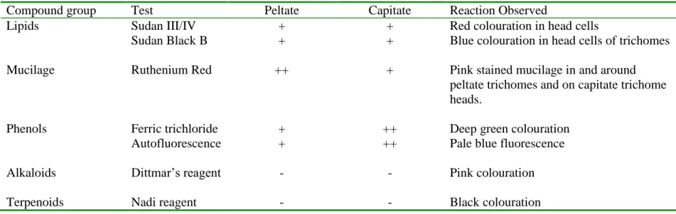
Page Table 1: Results of histochemical and fluorescence tests for the identification of
compounds in the secretory product.
51
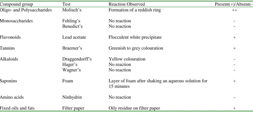
Table 2: Results of phytochemical tests of crude methanolic extracts of powdered leaves of C. triloba.
62
LIST OF FIGURES
Page Figure 1: Ceratotheca triloba (Bernh.) E. Mey. ex Hook. f. growing along a
roadside at the University of Kwa Zulu-Natal, Westville Campus.
3
Figure 2: Glandular trichomes of the Lamiaceae 10 Figure 3: Mucilage hairs of Sesamum indicum and Ceratotheca melanosperma. 17 Figure 4: Capitate trichomes observed on leaves of C. triloba. 29 Figure 5: Peltate trichome on leaves of C. triloba. 31 Figure 6: Development of peltate trichomes on leaves of C. triloba. 33 Figure 7: SEM showing a decrease in density of trichomes as leaves mature. 35 Figure 8: Trichome frequency on the adaxial surface of leaves at three stages of
development, emergent, young and mature.
37
Figure 9: Trichome frequency on the abaxial surface of leaves at three stages of leaf development, emergent, young and mature.
37
Figure 10: Surface imaging of trichomes on flower petals of C. triloba. 39 Figure 11: Light micrographs of transverse sections of leaves of C. triloba stained
with Sudan Black.
42
Figure 12: Light micrographs of transverse sections of leaves of C. triloba stained with Sudan III and IV.
44
Figure 13: Light micrographs of transverse sections of leaves of C. triloba stained with Ruthenium Red.
46
Figure 14: Autofluorescence of transverse sections of leaves of C. triloba. 48 Figure 15: Light micrographs of transverse sections of leaves of C. triloba stained
with ferric trichloride.
50
Figure 16: Transmission electron micrographs of a capitate trichome. 55
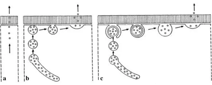
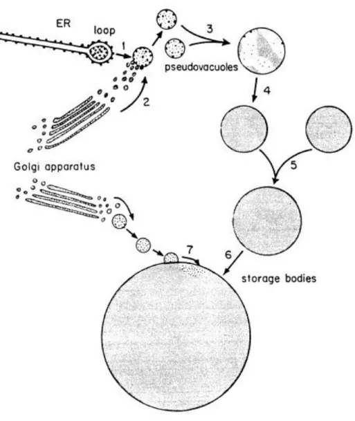
Figure 17: Transmission electron micrographs of head cells of peltate trichomes. 57 Figure 18: TEM of cell wall region of a peltate trichome. 59 Figure 19: TEM showing vesicle formation in peltate trichomes. 61 Figure 20: Illustration showing different pathways of secretion in secretory cells. 72 Figure 21: Illustration showing likely origin of protein-carbohydrate mucilage in
mango fruit ducts.
73
LIST OF ABBREVIATIONS
Bc Basal cell
C Capitate trichome
Ch Chloroplast
Cp Cellular pedestal
CPS Capitate trichome in the post secretory phase
Cw Cell wall
DC Developing capitate trichome DP Developing peltate trichome
ER Endoplasmic reticulum
ESEM Environmental scanning electron microscopy/micrograph
Gb Golgi body
L Lipid drop
LM Light microscopy/micrograph
M Mitochondria
Mc Mucilage
Me Multimembranous structure
N Nucleus
Nc Neck cell
Nm Nuclear membrane
Nu Nucleolus
O Oil drop
P Peltate trichome
Pd Plastids
Pn Phenolic Compounds
PPS Peltate trichome in the post secretory phase
SC Subcuticular chamber
Sc Secretory cell
Sec Secretory material
SEM Scanning electron microscopy/micrograph
Sh Secretory head
St Stalk cell
TEM Transmission electron microscopy/micrograph
V Vacuole
Vc Vesicle
Vn Vein
WHO World Health organisation
CHAPTER 1: INTRODUCTION
1.1 ETHNOBOTANICAL MEDICINE
Traditional medicine can be defined as any non-western form of medication or medical practice.
It is a very broad term as it incorporates the use of animal, plant and mineral based products for the diagnosis and treatment of ailments. Therefore, to describe medicinal practices involving the use of plants and plant products only, the term ethnomedicine or ethnobotanical medicine will be used instead.
The use of plants for medicine can be dated back in fossil records some 60 000 years ago to the Middle Paleolithic age and the development of ethnomedicinal practices can be traced back through documents described in ancient Ayurvedic and Chinese literature. The World Health Organisation (WHO) estimates that 85% of the world population still rely on some sort of plant product for medicine (Obi et al., 2006). Even in developed countries, the use of drugs containing plant products is also common (Fabricant and Farnsworth, 2001). In Germany the consumption of plants for medicinal purposes has risen by 20%. In the USA, 25% of all drugs dispensed by pharmacies contained plant active compounds (Farnsworth et al., 1985). Despite this, pharmaceutical companies in the USA have not shown much interest in ethnomedicinal research.
Developing countries, on the other hand, have invested in ethnomedicinal research which is beneficial as they have a rich source of information on traditional medicinal plant species which are used by the local people (Fabricant and Farnsworth, 2001).
An approach to researching plants for medicinal practices is by utilizing the information acquired from traditional healers. The information obtained from ancient ethnomedicinal literature has been useful in modern medicinal research. A survey carried out by Fabricant and Farnsworth (2001) to determine if ethnomedicinal information had any use in modern day medicinal research showed that 122 plant-derived pure compounds were identified as established drugs used in modern medicine. Of these, 80% are still being used to treat the original ailments as described in ethnomedicinal literature. It was also found that these compounds were isolated from only 94 plants. The number of flowering plant species occurring globally is estimated to be 250 000,
leaving many plants to be studied and disease treating compounds still to be discovered (Ayensu and DeFilipps, 1978).
1.2 DESCRIPTION OF Ceratotheca triloba
Ceratotheca triloba (Bernh.) E. Mey. ex Hook. f., commonly known as the wild foxglove, is a fast growing annual that is indigenous to southern Africa (Fig. 1). The plant is also known as the South African foxglove because it is the only species of Ceratotheca in South Africa. The plant thrives in disturbed habitats and is commonly observed in dry grasslands along roadsides.
Plants of C. triloba may be bushy or may grow as a single stem that can reach a height of 2 m, depending on the soil moisture. The entire plant is covered in fine hair-like structures which exude sticky, aromatic substances. The plants display either pink or white flowers that grow in pairs along the uppermost part of stems. The petals have five lobes with the lowermost being longer than the rest and displaying streaks of thin lines leading into the flower. The soft leaves expand to about 50 mm in length and divide into three lobes at the base with bluntly serrated margins. Seeds of the plant are protected by a capsule that displays three horns at the tip.
In South Africa, leaf extracts of C. triloba are used by traditional healers to treat diarrhoea, nausea and painful abdominal and menstrual cramps, whereas in Zimbabwe, the same leaf extracts are administered to induce abortion. Furthermore, in Botswana, the entire plant of C.
triloba is used to treat insect infestations of the skin and pediculosis. Despite its potential medicinal value, no significant scientific research has been carried out on this plant.
Figure. 1. Ceratotheca triloba (Bernh.) E. Mey. ex Hook. f. growing along a roadside at the University of Kwa Zulu-Natal, Westville Campus. a) whole plant, b) mature three lobed leaf, and c) pink flower with five lobes; the lower most lobe has streaks of lines running into the corolla tube.
a
c
b
1.3 RATIONALE OF THIS STUDY
Trichomes have been studied previously because of their functional usefulness in nature and the economic value of the secretory product in industry (Valkama et al., 2003). Trichomes are highly variable structures that provide a mechanical and chemical barrier against herbivores and the secretory product that they produce has been used in the food, fragrance and pharmaceutical industry (Fabricant and Farnsworth, 2001; Valkama et al., 2003). Trichome morphological traits have also been key characteristics in plant taxonomic studies. Therefore the morphology of trichomes, chemical nature of the secretory product, and how the secretory product is produced are vital aspects that need to be addressed.
1.4 AIMS AND OBJECTIVES
The genus Ceratotheca has been neglected in terms of trichome research. Not much is known about the trichomes or the chemical properties of the secretions they produce. Since C. triloba has been used by traditional healers for a variety of ailments, this study was undertaken with the following aim and objectives:
Aim
To provide a scientific basis for the medicinal properties of C. triloba.
Objectives
a. characterise the foliar trichomes of C. triloba according to morphology and distribution b. determine the chemical composition of the secretory product
c. provide a description of the secretory process
d. provide a phytochemical assessment of crude leaf extracts
CHAPTER 2: LITERATURE REVIEW
Secretory tissues in plants can be defined as multicellular structures or specialised single cells that eliminate specific substances to the external environment. These structures secrete specialised compounds which may aid in pollination, defence against herbivory and pathogens, and regulation of water and ions. In all cases, secretory structures serve as part of the metabolic pathway of plants, and in plant interrelations with the external environment. These structures include salt secreting glands, laticifers, nectaries, mucilage secreting cells, and trichomes (Fahn, 1979). There is great variation in structure and function of secretory tissues between species of plants. In addition, development and composition of the secretion can also vary. This review will focus on plant trichomes which are the most diverse group of secretory structures. The botanical literature has listed more than 300 morphologically distinct types of trichomes which have been used in plant classification (Kelsey et al., 1984).
Trichomes, derived from the Greek word trikhoma which means „growth of hair‟, are fine hair- like outgrowths found on the aerial regions of most angiosperms, some gymnosperms and bryophytes (Johnson, 1975). They are highly variable structures that are in contact with the external environment and therefore function in response to various biotic and abiotic stimuli (Tooker et al., 2010). Trichomes serve as the first line of defence against plant natural enemies and they may attract and guide pollinators (Young et al., 1984; Wagner, 1991). They also reflect UV radiation, and assist in drought tolerance by reducing leaf temperature and preventing excessive water loss (Levin, 1973; Wagner, 1991; Werker, 2000). In addition to variation in structure and function, differences may also occur in trichome development (Werker, 2000;
Serna and Martin, 2006).
2.1. EVOLUTION OF PLANT TRICHOMES
Co-evolution between species is widely accepted as one of the fundamental mechanisms that produced the Earth‟s diversity (Ehrlich and Raven, 1964). The process of co-evolution is a closely linked interaction between species. Interactions such as pollination, predation and mutualism between species may bring about genetic and morphological changes in one species
triggered by changes in the other (Ehrlich and Raven, 1964; Janzen, 1980). According to the co- evolutionary hypotheses, characteristics of plants that offer protection such as spines, thorns and trichomes evolved due to selection pressures imposed by natural enemies on plants (Juniper and Southwood, 1986). Therefore, plants evolved trichomes as a defence strategy against herbivores and plant pathogens.
Arabidopsis thaliana grown in an environment free of herbivores and pathogens displayed a decrease in production of two known characteristics that offer protection against natural enemies i.e. glucosinolate concentration and trichome density (Mauricio and Rausher, 1997).
Glucosinolate is a secondary compound that is toxic to herbivores and pathogens (Mauricio and Rausher, 1997; Rosa and Rodrigues, 1999; Tierens et al., 2001) and trichome density provides defence against plant‟s natural enemies (Ambrosio et al., 2008). These results indicate that selection pressure enforced by plant natural enemies causes an increase in defence characteristics, which is supportive of the co-evolution hypothesis.
In contrast to co-evolution, the sequential evolution hypothesis considers the resistant features of trichomes as incidental and points to abiotic selections pressures, such as temperature and UV radiation, as the underlying cause of trichome evolution (Mauricio and Rausher, 1997). Unlike the co-evolution hypothesis, evidence to support the sequential evolution hypothesis is circumstantial.
2.2. TRICHOME CLASSIFICATION
Trichomes are highly diverse structures that vary in function, location, density, distribution, ability to secrete and timing of secretion. These differences can occur between species of plants, between plants within a single species and between organs of an individual plant. Classification of trichomes using one of the above characteristics is difficult because of overlap between species of plants and neither characteristic on its own is sufficient (Werker, 2000). The function of a trichome may vary according to its location on the plant. Trichomes located on vegetative organs may serve to repel herbivory, whereas trichomes located on reproductive organs may
2.2.1 Morphological variation
a) Non-glandular trichomes
Non-glandular trichomes are generally classified according to their morphology. They range from unicellular to multicellular structures that can be uniseraite, biseriate or multiseriate, branched or unbranched (Werker et al., 1994). The tips of non-glandular trichomes may be tapering or blunt (Werker, 2000). Thickness of cell walls may vary within and between trichomes and the hardening material within the walls may vary resulting in varying strength (Werker, 2000). Trichome length may differ between different plants, between organs of the same plants or within a single organ (Economou-Amilli et al., 1982).
Occasionally more than one type of non-glandular trichome may be found on leaves as in the case of Salvia blepharophylla and Coridothymus capitatus (Economou-Amilli et al., 1982; Bisio et al., 1999). In S. blepharophylla, the three types were found: 1. multicellular, long trichomes with tapering tips, located along the leaf margins; 2. multicellular, uniseriate, short trichomes (usually 4-5 celled) with swollen bases located on the midrib and veins; and 3. unicellular papillae with enlarged bases and tapering tips, which were common on the abaxial surface of leaves. The reason for this variation in trichome morphology, however, is not known.
b) Glandular trichomes
Glandular trichomes include a variety of glands that synthesise, accumulate and secrete essential oils (Croteau, 1987; Gershenzon et al., 1989). In order to classify these trichomes, the chemical composition of the secretion and the secretory process are considered in relation to trichome location and function.
The structure of glandular trichomes range from unicellular to multicellular and these can be uniseriate or multiseriate (Werker, 2000). The uniseriate, multicellular glandular trichome comprises of a base of one to a few cells, a stalk of one to many cells and a secretory head made up of one to many secretory cells (Fahn, 1988). Occasionally, a neck cell is located between the secretory head cells and stalk cells, and is morphologically different from the two.
In species of Lamiaceae, glandular trichomes are generally characterised in two major groups, the capitate and the peltate trichomes, named according to the shape of their secretory heads (Fahn, 1988). Capitate (Latin – capitis meaning “of a head”) trichomes usually have a globose secretory head made up of 1-4 cells, subtended by a stalk of one to many cells. They have been termed “short term trichomes” because they develop and release their secretion early in leaf development, thereby providing protection against herbivory even before any form of mechanical disturbance occurs (Werker, 2000). Peltate (Latin – peltata meaning “shield shaped”) trichome secretory heads are usually flattened and round made up of 4-18 cells subtended by a single stalk cell located at the centre of the secretory head. These have been defined as “long term trichomes” since they accumulate oil that is only release upon mechanical disruption (Werker, 2000). Similar forms of trichomes have been observed in other families such as the Cannabaceae (Dayanandan and Kaufman, 1976), Fabaceae (Zoric et al., 2009), Curcurbitaceae (Kolb and Müller, 2004) and Pedaliaceae (Naidoo et al., 2006).
Variation in trichome structure may occur between species within a family or genus. Mentha peperita and Leonotis leonurus of the family Lamiaceae possess both capitate and peltate trichomes (Fahn, 1988; Ascensão et al., 1995, Fig. 2). Both contain peltate trichomes consisting of a single basal cell, a stalk cell and a secretory head made up of eight secretory cells arranged in a circle (Fahn, 1988; Ascensão et al., 1995). However, other species of the same family such as Satureja thymbra, have peltate trichomes of similar structure but the head contains more cells arranged in two concentric circles (Bosabalidis, 1990).
Differences in trichome structure may also be found within a single genus as in the case of Salvia blepharophylla (Bisio et al., 1999) and S. aurea (Serrato-Valenti et al., 1997), which contain peltate trichomes with secretory heads of four cells arranged in a quadrant, and eight cells arranged in a circle respectively. The capitate trichomes of S. aurea and S. blepharophylla are however, similar (Serrato-Valenti et al., 1997; Bisio et al., 1999) comprising of a single stalk cell and a bicellular secretory head. In contrast, those found on leaves and flowers of S. scleria and S.
dominica are highly diverse, comprising of six different forms differing in structure, size, location, and manner of secretion (Werker et al., 1985a). In addition, the short stalk capitate
al., 1999) also differ with respect to the number of head cells, with the former possessing a bicellular head and the latter a unicellular one.
Variation may also extend to trichomes located on the same organ of a plant and between groups of cells within a single trichome. Meyberg et al., (1991), reported that capitate trichomes found on leaves of Nicotiana tobaccum secreted different products even though they appeared to be morphologically analogous. Some trichomes secreted a resinous compound while the others secreted oil droplets. The exact functions of the different groups of cells that make up a trichome, and between cells of the same group are not clear. Ultrastructural studies indicated that trichomes with multicellular secretory heads may contain cells that secrete different substances than their neighbouring head cells (e.g. Artemisia annua, Duke and Paul, 1993) whereas other studies have highlighted the possibility that some head cells may not even be active in secretion (e.g. Mallotus philipinensis, Roth, 1977). Ultrastructural studies have also shown differences in cells of the stalk with the lowermost cells being highly vacuolated and the uppermost cells having dense cytoplasm (Turner et al., 2000).
Figure 2. Glandular trichomes of Lamiaceae. a) Peltate trichome of Mentha peperita with oil drop (O) in subcuticular chamber (SC). The side walls of the stalk are highly cutinised (arrow) to prevent apoplastic backflow. b) Capitate trichome with a single stalk cell and unicellular
O
SC
a
b
c) Integrated forms of glandular and non-glandular trichomes
In most cases, trichomes can be clearly differentiated into glandular and non-glandular.
However, there exist trichomes that cannot be characterised explicitly into either group. In the case of certain species of Lamiaceae, unbranched glandular trichomes are clearly differentiated from branched non-glandular trichomes. However, there are instances in which compound glandular trichomes are branched having many non-glandular branches with one glandular branch (e.g. Phlomis, Azizian and Cutler, 1982; Rosmarinus officinalis, Werker et al., 1985c;
Hyptis, Rudall, 1980). In addition, Ascensão et al., (1995) reported that the stalk walls of capitate trichomes in Leonotus leonurus displayed cuticular warts which are a characteristic of the non- glandular trichomes of that species. It was concluded by Ascensão et al., (1995) that these capitate trichomes represented an intermediate form between glandular and non-glandular trichomes. Fahn (1988) suggested that these findings may also provide some answers with regards to the evolution of these structures.
2.2.2 Variation in the process of secretion
In Lamiaceae, the peltate trichomes accumulate the secretory product in a subculticular chamber, which forms when the cuticle detaches from the cell wall of the secretory cells (Werker et al., 1985a, b). These peltate trichomes are assumed to release their contents when the elevated cuticle is damaged, usually by plant herbivores. Ceratotheca sesamoides of the family Pedaliaceae has peltate trichomes that lack a subcuticular chamber. They secrete mucilage when the secretory cell ruptures (Abels, 1975) or dissolves (Ihlenfeldt, 2001), resulting in cell death.
Capitate trichomes may also accumulate oils in a subcuticular chamber and release them either by cuticular rupture or by diffusion through micropores in the cuticle. The secretory product of the “short stalked capitate trichome” of Plectranthus madagascariensis accumulates in a small subcuticular space and is probably released by diffusion through micropores, whereas “the long stalked capitate trichome” releases its secretory product via cuticular rupture (Ascensão et al., 1998). Similar variations were also reported for other species of Lamiaceae (e.g. Ocimum basilicum, Werker et al., 1993; Plectranthus annua, Ascensão et al., 1999) The “capitate Type Ι hairs” of S. sclarea however lack a subcuticular chamber and release their contents through
micropores in the cell wall and cuticle (Werker et al., 1985a). Accumulation of secretory product in the cell lumen and direct diffusion through the cell wall were also observed for capitate trichomes in S. aurea (Serrato-Valenti et al., 1997) and O. basilicum (Werker et al., 1993). Kolb and Müller (2004) observed that the capitate trichomes found on leaves of Cucurbita pepo var.
styriaca released their secretion when they interacted with the non-glandular trichomes and bristles which indicates that secretion in this species is dependant on trichome density.
2.2.3 Trichome function
a) Protection
Plants may require protection from a variety of abiotic (extreme temperature, intense light and water loss) and biotic (herbivores, pathogens and alleopathy from plant competitors) factors.
Trichomes provide protection against all of the above threats, especially when they densely cover plant organs. The effect of trichome density on herbivore resistance has been noted in Thithonia diversifolia (Ambrosio et al., 2008).
In the case of abiotic factors, a direct correlation has been observed between environmental stresses and the density of trichomes found on the plant organ (Clausen et al., 1940). Johnson, 1975 reported that Potentilla (Rosaceae) planted in sunny sites contained more pubescent trichomes than those grown in shady sites. Warming (1909) has already emphasised the point that a greater number of trichomes will appear in plants that are subjected to a harsher moisture regime. He also noted the differences in trichome cover between plants of the same species which are found in xeric and mesic habitats. He observed that the plants which are found in xeric habitats contained more trichomes. In addition, the resin secreted by glandular trichomes, in Betula pendula found at high altitudes, serves as a moisture repellent, protecting the plant against the harsh winter and spring (Lapinjoki et al., 1991). Non-glandular trichomes may serve as structural defences against plant herbivores. Gilbert (1971) observed a highly effective defence strategy of Passiflora adenopoda against the heliconiine butterfly larvae. The hook-like trichomes killed the larvae by entrapping them, followed by inducing many puncture wounds in the larval integument.
Glandular trichomes provide a variety of defence strategies by secreting specialised compounds that may either trap or poison herbivores, or provide an antimicrobial effect against pathogens.
These compounds include terpenoids (Dawson et al., 1966; Bakker et al., 1972; Turner et al., 2000), flavonoids (Kelsey et al., 1984), tannis (Feeny, 1968) alkaloids (Rodriguez et al., 1984) and phenolics (Yu et al., 1992). Non-volatile compounds such as alkaloids, tannins and phenols may act as a deterrent to herbivory. In a study carried out by Harley and Thorsteinson (1967), several alkaloids (including nornicotine, lobeline, tomatine and hordinine) that were added to the diet of the two striped grasshopper, Melanoplus bivittatus, either repelled the insects or killed them. Similarly, phenolic compounds such as the coumarins, which are produced by Melilotus species, have an inhibitory effect on the vegetable weevil (Matsumoto, 1962). Some glandular trichomes secrete mucilage that interacts with phenols to trap herbivorous insects. Tingey and Gibson (1978) reported a defence strategy of Solanum polyadenium and S. berthaultii against the aphid Empoasca fabea. It was reported that the mouthparts and tarsi were encased by a sticky substance which hardened, resulting in starvation and death of the aphid. This is probably due to the release of phenolic compounds which are oxidised to quinines by polyphenol oxidase. The resulting quinines polymerise and trap insects (Yu et al., 1992).
Tannins interact with proteins to form tannin-protein complexes, which reduce the total protein that can be ingested by herbivores. Since these complexes reduce the dietery content in plants and most angiosperms contain tannins, it is highly possible that insects use tannins as a negative feeding cue. In addition, chemoreceptors found on legs and mouthparts of insects detect possible fatal chemicals on the elevated head cells of trichomes (Dethier, 1963). This prevents feeding as the insect realises the host is unsuitable for consumption. In this way trichomes serve as a deterrent to herbivores by advertising the presence of highly toxic chemicals.
b) Attraction
Much of the research characterising trichomes has focused on the vegetative organs with the reproductive organs being neglected. Trichomes on flowers serve conflicting roles in deterring natural enemies and attracting pollinators (possibly the natural enemies of other plants). In some species of Lamiaceae, large glandular trichomes were observed on stamens, under the anther lobes (Cannabis sativa, Mahlberg et al., 1984; Leonotus leonurus, Ascensão et al., 1995).
Whether these trichomes secrete compounds that attract pollinators or whether they deter herbivores, or both, is not known.
Many authors have observed differences in trichome structure and distribution between vegetative and reproductive organs. This may be indicative of differences in function. In Cannabis sativa, stalked capitate trichomes were observed on floral organs, whereas the vegetative organs displayed various other trichomes (Mahlberg et al., 1984). In many species, the calyx, which covers the flower during development, is covered in glandular and non- glandular trichomes similar to those found on the vegetative organs. These trichomes are probably for protection as described previously. The trichomes on the calyx of Origanum vulgare (Werker et al., 1985b) are different from those on vegetative organs with respect to the ratio of the various types of glandular trichomes and the proportion of the chemical constituents they secrete, thus indicating that trichomes on the calyx may also serve as attractants. In Salvia dominica and S. sclarea, both the reproductive and vegetative organs are covered in capitate and peltate trichomes. The morphology of peltate trichomes is uniform between the two organs whereas the capitate trichomes differ in structure, size and mode of secretion. The main components of the essential oil, neryl acetate and linalyl acetate, also differed between vegetative and reproductive organs (Werker et al., 1985a).
Variations in trichome occurrence and distribution were also observed within a flower. In some species of Lamiaceae such as Origanum vulgare (Werker et al., 1985b) and Leonotus leonurus (Ascensão et al., 1995), peltate trichomes were the only types observed on the outside of corolla tubes whereas the inside of the tubes displayed a variety of other glandular trichomes. Variation in trichome structure and distribution between and within organs may indicate differences in function. However, evidence to support this is inferred and more work is required to clarify these issues.
2.3 TRICHOME DEVELOPMENT
Most of the information regarding trichome development is drawn from studies on mutants of Arabidopsis thaliana. These studies show that the first cells (protodermal cells) to differentiate from the epidermis develop into trichomes. Trichomes are therefore generated in early leaf development and the rate of differentiation is rapid (Duke and Paul, 1993; Werker, 1993). This often provides protection to the leaf primordia which are more susceptible to herbivory. In Inula viscosa, fully developed trichomes were observed on leaves not more than 2 mm long (Werker and Fahn, 1981). Whether all the trichomes produced on leaf primordia are predetermined or develop as the leaf ages is still not clear.
The various processes in trichome development are discussed in the following section.
Development of other protuberances, such as emergences and „false hairs‟ will not be considered as they are not true trichomes.
2.3.1 Development of single cells
The epidermal cell, with a dense cytoplasm and a large nucleus, divides periclinally to form a protrusion above the epidermis. These cells can then undergo further periclinal divisions to form a stalk and anticlinal divisions to form secretory head cells (Werker, 2000). Uphof (1962) characterised the periclinal development of a multicellular, uniseriate trichome according to direction of division of the existing stalk cells. Acropetal division occurs when the uppermost stalk cell divides to form a new cell, whereas basipetal division occurs when the lowermost stalk cell undergoes division. In Pelargonium scabrum, capitate stalked trichomes result from a development of the protodermal cell which divides periclinally to form a single stalk cell (Oosthuizen and Coetzee, 1983). Further stalk cells develop from acropetal division. The peltate trichome in species of Lamiaceae undergoes two periclinal, acropetal divisions to form a stalk cell and a single secretory cell which undergoes further anticlinal division (e.g. Origanum dictamnus, Bosabaladis and Tsekos, 1982a; Leonotus leonurus, Ascensão et al., 1995).
A common misconception with biseriate and multiseriate trichomes is that they all develop from more than one cell. In most biseriate trichomes, especially those of the Asteraceae, initial division occurs from a single protodermal cell which undergoes an anticlinal division to form two cells along the leaf epidermis (e.g. Holocarpha virgata, Carlquist, 1958; Artemisia campestris, Ascensão and Pais, 1987; Stevia rabaudiana, Montiero et al., 2001). These cells then divide periclinally to form a fully developed biseriate trichome. There are variations to this form of development. In Helichrysum aureonitens, the first division is periclinal but the anticlinal division occurs soon after (Afolayan and Meyer, 1995).
In most trichomes, in addition to differentiation of the protodermal cell, other epidermal cells may differentiate around the protodermal cell. These generally form a ring of differentiated cells normally referred to as a cellular pedestal, which provides support and anchors the trichome in the epidermis (Werker, 2000). Some cellular pedestals may even grow above the epidermis thereby elevating the trichome.
2.3.2 Development from multiple cells
Initially only one protodermal cell divides anticlinally to form four cells, which undergo numerous periclinal divisions to form a developed trichome. In addition to the single protodermal cell, many epidermal cells also differentiate and divide together to form the developing trichome, and in most cases add to the structure as in Helicteres hirsute (Uphof, 1962).
2.4 THE FAMILY PEDALIACEAE
The Pedaliaceae is a small family, consisting of 17 genera and 80 species. It is a predominantly African family with the exception of a few species, such as Pedalium murex, which is found in Yemen (Cronquist, 1981; Wood and Haig-Thomas, 1997). Sesamum is the largest genera consisting of 20 species, most of which are well documented for their oily seeds. Taxonomically,
the genera Sesamum and Ceratotheca are the closest, with the horns on seed capsules of Ceratotheca the only characteristic separating the two (Bruce, 1953).
Much of the research on this family focused on the oil producing seeds of Sesamum. In terms of research on trichomes, the Pedaliaceae has been somewhat neglected. Solereder (1908) described the mucilage hairs found on leaves of Sesamum indicum and Ceratotheca melanosperma. These trichomes were peltate, consisting of a head made up of four cells arranged in a cross (Figure 3).
A capitate trichome was also described as having a multicellular stalk and a small spherical head of four cells which were shaped like small palisade cells. Similar observations were made for C.
sesamoides. Naidoo et al., (2006) described glandular trichomes found on leaves of Harpagophytum procumbens consisting of a multicellular stalk and head made up of four cells that were arranged in a quadrant, which secreted viscous material.
Figure 3. Mucilage hairs of Sesamum indicum and Ceratotheca melanosperma. (from Solereder, 1908)
a b
CHAPTER 3: MATERIALS AND METHODS
3.1 COLLECTION AND PROPAGATION OF PLANT MATERIAL
Intact plants of Ceratotheca triloba were collected from the University of KwaZulu-Natal (UKZN), Westville campus and Umhlanga Rocks (290 43‟ 33.6” S, 310 4‟ 12” E) and transported to the greenhouse for propagation in pots. Voucher specimens were prepared and kept at the School of Biological and Conservation Sciences Ward Herbarium (Voucher specimen no.
KARIM - 1).
3.2 SAMPLING
For light microscopy and scanning and transmission electron microscopy, samples were taken from mature plants in the field from the tip, mid and basal regions of leaves. Three stages of leaf development were sampled in this study i.e. the emergent (width < 2 mm, length < 9 mm) young (width 2 < 18 mm, length 9 < 26 mm) and mature (width > 18 mm, length > 26 mm) leaves.
3.3 PREPARATION FOR SCANNING ELECTRON MICROSCOPY (SEM)
Three techniques were used for SEM studies: freeze-drying, chemical fixation and environmental SEM.
3.3.1 Freeze-drying
Leaf segments were prepared by quenching in liquid nitrogen slush and freeze drying for 96 h in an Edwards Modulyo freeze dryer at -40 0C to -60 0C at a vacuum of 10-1 Torr. After securing samples on brass stubs with carbon conductive tape, they were sputter coated with gold in an atmosphere of argon with a Polaron Sputter coater unit SC500. The leaf segments were viewed with a Leo 1450 SEM at 5 kV and a working distance of 15 mm.
3.3.2 Chemical fixation
Segments of leaf blades and petals were prepared for chemical fixation for SEM. These samples underwent a primary fixation in a solution of 2.5 % glutaraldehyde (4 0C) buffered with 0.1 M phosphate buffer (pH 7.2) for 2-24 h followed by a post fixation at room temperature in 0.5%
osmium tetroxide (OsO4) for 1 h. Samples were then dehydrated in a graded alcohol series of 30
%, 50 %, 75 % and 100 % ethanol. Critical-point-dried samples were then secured on brass stubs with carbon conductive tape and sputter coated with gold and viewed as described previously.
3.3.3 Environmental scanning electron microscopy (ESEM)
Segments of fresh leaves and petals were placed on brass stubs with carbon conductive tape and viewed under a Philips XL 30 ESEM at 15 kV and a working distance of approximately 13 mm.
3.4 PREPARATION FOR TRANSMISSION ELECTRON MICROSCOPY (TEM)
Two standard methods (differing in embedding time and the embedding medium) of sample preparation were used for TEM.
3.4.1 Method 1
Leaf segments (< 0.5 mm2) were prepared by prefixing in a solution of 2.5% glutaraldehyde (4
0C) buffered with 0.1 M phosphate buffer (pH 7.2) for 2-24 h followed by a post fixation at room temperature in 0.5% osmium tetroxide (OsO4) for 1 h. The samples were washed three times for 5 min before and after post fixation in phosphate buffer followed by dehydration in a graded acetone series (i.e twice for 5 min each in 30 %, 50 %, 75 % and 100 % acetone). The leaf segments were allowed to stand in equal parts Spurr‟s (1969) resin and acetone for 4 h followed by Spurr‟s resin only for 24 h. Samples were then polymerized in whole resin at 70 0C for 8 h.
Ultrathin sections were obtained with a Reichhert Ultracut E microtome using glass knives and collected on uncoated 200 square mesh copper grids. The sections were stained with 2 % aqueous uranyl acetate (10 min) followed by Reynold‟s (1963) lead citrate (10 min). Sections were viewed with a Jeol 1010 TEM.
3.4.2 Method 2
Leaf segments (< 0.5 mm2) were prepared by prefixing in a solution of 3 % glutaraldehyde (4 0C) buffered with 0.05 M cacodylate buffer for 8 h followed by post fixation at room temperature in 2 % OsO4 for 2 – 4 h. The samples were washed twice for 30 min before and after post fixation in cacodylate buffer (0.05 M), followed by dehydration in a graded alcohol series of 10 min each in 10 %, 30 %, 50 % and 70 % ethanol. The samples were allowed to stand in 70 % ethanol overnight at 4 0C and further dehydrated for 10 min each in 80 % and 90 % ethanol followed by three 10 min changes in 100 % ethanol. The leaf segments were then washed twice in propylene oxide for 30 min and taken through increasing concentrations of Epon resin, diluted with propylene oxide (i.e. 25 % - 2 h, 50 % - 2 h, 75 % - 12 h and 100 % - 24 h). Leaf material was then embedded in 100 % resin at 70 0C for 48 h. Ultrathin sections were obtained with a Reichhert Ultracut E microtome using glass knives and collected on uncoated 200- square mesh copper grids. The sections were stained with 2 % aqueous uranyl acetate (10 min) followed by Reynold‟s (1963) lead citrate (10 min). Sections were viewed with a Jeol 1010 TEM.
3.5 LIGHT MICROSCOPY (LM)
For light microscopy, thin, hand cut sections and sections embedded in wax or Spurr‟s (1969) resin were used.
3.5.1 Wax embedded material
Excised leaf samples (~ 3 mm2) were initially placed in formaldehyde (70 %) for 48 h.
Thereafter, the leaf samples were dehydrated with eight incubations in a graded ethanol series (3 x 70 % - 30 min, 80 % - 2 h, 90 % - 4 h, 100 % - 90 min, 100 % - overnight, 100 % - 2 h). The samples were then placed in a graded series of xylene: ethanol mixtures (25:75, 50:50, 75:25) for an hour each before being placed in two xylene incubations for 15 min each. Paraplast Plus®
wax pellets (McCormick Scientific) were added four times at 3 h intervals, followed by a final addition of wax before sectioning.
Wax sections were de-waxed by exposing them to three 2 min baths of xylene (100 %), three 2 min baths of ethanol (100 %), a single 2 min bath in 70 % ethanol and a final 2 min wash in tap water.
3.5.2 Spurr embedded material
Leaf material was prepared in Spurr‟s (1969) resin (according to the standard protocol 3.4.1) and semi-thin sections were placed on pre-cleaned glass slides. Monitor sections (1 µm) were stained with 1 % Toluidine Blue - O (Feder and O‟Brien, 1968)
3.6 FLUORECSENCE MICROSCOPY
3.6.1 Cellulose cell walls
Excised leaf segments were placed in a non-toxic, 0.01 % solution of Cellufluor for 5 h, followed by washing in tap water for 30 min. Thereafter, the leaf segments were hand sectioned transversely and the sections mounted in 70 % glycerine on pre-cleaned slides before viewing with ultraviolet light. Untreated sections, viewed under UV light, were used as controls.
3.6.2 Phenols and lignin
Untreated freehand cross sections of leaves were viewed with UV light for the detection of phenols (Mabry et al., 1970) and lignins. Sections for fluorescence microscopy were viewed and imaged with a Nikon Eclipse 80i Microscope fitted with an Ultra Violet (UV) filter assembly (excitation wavelength 330 – 380 nm).
3.7 HISTOCHEMISTRY
3.7.1 Pectin and mucilage (Johansen, 1940)
Freehand cross-sections of fresh leaves were stained with ruthenium red for 10 min for detection of mucilage in the secretion and pectins in the cell walls.
3.7.2 Phenolic compounds (Johansen, 1940)
For the detection of phenolic compounds, transverse sections of fresh leaves and wax embedded material were stained with 10 % ferric trichloride. A stock solution of ferric trichloride was prepared by diluting 10 g ferric trichloride pellets in 100 ml distilled water, followed by filtration through Whatman No.1 filter paper.
3.7.3 Lipids, cutin and suberin (Pearse, 1968)
For the detection of lipids, hand sections and wax embedded sections of leaf material were stained with aqueous solution of Sudan III and IV for 15 min. The stain was washed off with 70
% ethanol and the sections were mounted in 70 % glycerol.
Sudan Black B was also used to detect lipids. Sections were stained in an alcoholic solution of Sudan Black B for 30 min in a moist environment. The stained sections were washed with 70 % ethanol and mounted in 70 % glycerine. Controls leaves were pre-treated with methanol and chloroform (1:1) (Lison, 1960)
3.7.4 Alkaloids (Furr and Mahlberg, 1981)
Sections of wax embedded leaf material were stained with Dittmar reagent (1 g potassium iodide, 1 g sodium nitrite, 30 ml HCl and 30 ml H2O) for the detection of alkaloids. Leaf sections that were pre-treated with tartaric acid were used as controls (Johansen, 1940)
3.7.5 Terpenoids (David and Carde, 1964)
Leaf sections, embedded in wax, were treated with Nadi reagent (David and Carde, 1964) for the detection of terpenoids. Stock solutions of solution A (1 % α-naphthol in 95 % ethanol) and solution B (1 % N, N-26 dimethyl-p-phenylenediamine HCl in water) were prepared and a mixture of equal volumes of each were used for staining. Sections stained with Nadi reagent were incubated in the dark for 60 min followed by a wash with sodium phosphate buffer for 2 min. Leaf sections that were pre-treated with 2 N HCl were used as controls (Mace et al., 1974)
All sections for histochemistry were examined with a Nikon Eclipse 80i Microscope using bright field illumination. Images were viewed and saved with NIS Elements – D (version 3.0) software.
3.8 PRELIMINARY PHYTOCHEMICAL TESTS
3.8.1 Preparation of crude leaf extract (European Pharmacopiea, 1975)
Fresh leaves were dried at room temperature for one week and then ground down to a powder.
Extracts were prepared by continuous extraction method a soxhlet apparatus and methanol as the organic solvent. The extracted solution was filtered, concentrated with a rotary evaporator, and stored at 4 0C for further use.
3.8.2 Phytochemical screening of leaf extracts
Preliminary phytochemical tests were carried out for the following phytocompounds:
a) Monosaccharides Fehling’s solution
For the detection of monosaccharides, extracts (3 ml) were mixed with an equal volume of Fehling‟s solution. Copper sulphate solution (34.66 g copper sulphate dissolved in 500 ml distilled water) and alkaline tartrate solution (173 g potassium sodium tartrate and 50 g NaOH dissolved in 500 ml water) were prepared. Both solutions were mixed with equal volumes before use. A brown colour reaction was indicative of monosaccharides.
Benedict’s solution
Extracts were also mixed with Benedict‟s solution for the detection of monosaccharides.
Benedict‟s solution is made up of two solutions: solution 1 (173 g sodium citrate and 100 g sodium carbonate in 800 ml distilled water while heating) and solution 2 (17.3 g copper sulphate in 100 ml distilled water). Solution 2 is added to solution 1 and made up to 1 L. The development of a reddish-brown precipitate is indicative of monosaccharides.
b) Oligosaccharides and polysaccharides
Plant methanolic extracts (3 ml) were added to water and filtered. The filtrate was then subjected to Molisch‟s reagent (15 g α-naphthol dissolved in 100 ml absolute ethanol). The formation of reddish-brown rings indicated the presence of di- and polysaccharides.
c) Flavonoids
Methanolic extracts (5 ml) were mixed with a solution of lead acetate (2 ml). Flocculent white precipitate indicated the presence of flavonoids.
d) Tannins
Breamer‟s test was performed for the detection of tannins. Plant methanolic extract (3 ml) was mixed with a 10 % alcoholic ferric trichloride solution. A dark blue or greenish grey colouration of the solution indicated the presence of tannins.
e) Alkaloids
Draggendorf’s reagent
A drop of extract was spotted on a silica gel plate, followed by spraying with Draggendorf‟s reagent. An orange colouration of the spot indicated the presence of alkaloids.
Stock solutions were prepared as follows: Solution A – bismuth nitrate (0.17 g) dissolved in glacial acetic acid (2 ml) and distilled water (8 ml), and Solution B – potassium iodide (4 g) dissolved in glacial acetic acid (10 ml) and distilled water (20 ml). Both solutions were mixed in equal volumes and diluted to 100 ml.
Hager’s reagent
Methanolic extracts were treated with Hager‟s reagent for the detection of alkaloids. Stock solution of Hager‟s reagent was prepared by dissolving 1 g of picric acid in 100 ml distilled water. The development of a yellow precipitate indicated the presence of alkaloids.
Wagner’s reagent
Methanolic extracts were treated with Wagner‟s reagent (2 g iodine and 6 g potassium iodide dissolved in 100 ml distilled water). A reddish-brown precipitate was a positive result for alkaloids.
f) Saponins
The foam test was carried out for the detection of saponins. Methanolic extract (2 ml) was mixed with 20 ml distilled water and shaken. A 1 cm layer of foam, occurring at the top of the mixture, indicated the presence of saponins.
g) Amino acids
Methanolic extract (1 ml) was mixed with distilled water and ninhydrin solution (1 ml) was added. A blue colour change indicated the presence of amino acids.
h) Fats and fixed oils
A drop of extract was placed between two pieces of filter paper. Occurrence of oil stains indicated the presence of oils.
3.9 IMAGE ANALYSES – TRICHOME COUNTS AND MEASUREMENTS
Images obtained from SEM were analysed with iTEM software (Soft Imaging System GmBH, Münster, Germany). Trichomes were counted in quadrants for each treatment (differentiated according to region on the adaxial and abaxial surfaces of leaves and the stage of development) with a total number of 20 images per treatment. The length of the stalk of 100 capitate trichomes and the diameter of the secretory head were measured randomly from developing and mature peltate trichomes.
3.10 STATISTICAL ANALYSES
All statistical analyses were performed with the SPSS statistical software package for windows (Rel. 15.0.0. 2006. Chicago: SPSS Inc.). The frequency (count mm-2) of trichomes was compared between treatments with a one-way analyses of variance (ANOVA) using the Tukeys Multiple Comparisons Test to compare differences among treatments. The diameter of peltate trichomes between developing and mature trichomes was compared with an independent sample t-test. A value of p < 0.05 was considered statistically significant.
CHAPTER 4: RESULTS
4.1 TRICHOMES
4.1.1 Trichome distribution
The leaves of C. triloba possess two morphologically distinct trichomes, capitate (Fig. 4 a) and peltate (Fig. 5), both of which appear to be glandular. Trichomes occur on the adaxial and abaxial surfaces of leaves, with capitate dominant on the adaxial surface and peltate predominant on the abaxial surface. Distribution of capitate trichomes is uniform on both surfaces of the leaf.
However, peltate trichomes were mostly observed along the grooves on the adaxial surfaces and on the veins on the abaxial surface (Fig. 7 a-f).
Emergent leaves were densely covered in trichomes and trichome density decreased as the leaf matured. The frequency (count mm-2) of trichomes on the emergent leaves was significantly higher on both side of the leaf surface as compared to the frequency on the young and mature leaves (Fig. 8 and 9). On the abaxial surface, the frequency of trichomes was significantly higher at the tip (82.17 ± 3.61 mm-2, Mean ± SE) but this decreased as the leaf matured (22.07 ± 2.79 mm-2). Trichome frequency was similar on the mid and base regions of the adaxial and abaxial surfaces. Capitate and peltate trichomes also occur on the abaxial surface of flower petals (Fig.
10), while the adaxial surface was bare.
4.1.2 Capitate trichomes
Capitate trichomes on leaves were approximately 248.10 ± 4.53 μm in length, unseriate and normally comprised of a single basal cell, 3-5 stalk cells, a neck cell and a secretory head containing vertical walls, arranging the head in a quadrant of four small secretory cells (Fig. 4 a, b and c). The stalk is made up of long cells at the base which gradually become shorter towards the secretory head. Trichomes with up to 8 stalk cells were observed along the leaf margin but these were not common. Wax sections stained with Toluidine Blue showed that the basal cells of capitate trichomes appeared to be highly vacuolated while the cells closer to the secretory head were denser with cytoplasm and probably organelles (Fig. 4 b). The trichome is anchored to the
walls (Fig. 4 d). Capitate trichome outer walls appear to lack cuticular warts and other forms of microornamentation.
There were two morphologically distinct types of capitate trichomes found on petals and therefore, for the purpose of petals alone these trichomes were classified as capitate Type I and Type II. The morphology of the capitate Type I trichome was similar to the capitate trichomes found on leaves, whereas the capitate Type II trichomes had stalks that were longer in length and comprised of approximately 14 cells (Fig. 10 a and b). The secretory heads of both types of capitate trichomes were arranged in quadrants, similar to those found on leaves.
Capitate trichomes appear to develop rapidly on leaf pimordia since fully developed forms were only observed on emergent through to mature leaves whereas developing capitate trichomes were rare (Fig. 7 a-f). Development appears to occur from a single epidermal cell which probably undergoes many periclinal divisions to form the stalk and two anticlinal divisions to form the four-celled secretory head. This development was not observed directly. However, observations of developing trichomes on young leaves lead us to this assumption (Fig. 4 a).
4.1.3 Peltate trichomes
Fully developed peltate trichomes comprise a basal cell, a unicellular stalk and a multicellular secretory head (Fig. 5). The peltate trichome head consisted typically of four cells although some were seven or even eight-celled (Fig. 6 e and f). Developing trichomes had a secretory head made up of 2 to 4-cells (diameter = 31.21 μm), and these were significantly smaller than the fully developed 4-celled peltate trichome (diameter = 74.32, t = 8.263, p < 0001). Development appears to occur from a single epidermal cell which undergoes 3 periclinal divisions to form a basal cell, a stalk cell and a unicellular secretory head. The secretory head then divides periclinally to form a four celled head which expands in size resulting in a developed four celled peltate trichome. This four celled head may undergo further periclinal division to form a head of up to eight cells (Fig. 6). The peltate trichomes found on petals were morphologically similar to those found on leaves.
PLATE 1
Figure 4. Capitate trichomes observed on leaves of C. triloba.
(a) SEM of a developed capitate trichome (C) and developing capitate (DC) trichome on a young leaf. Stalk cells (arrows) are differentiated early in leaf development and then expand in size.
(b) Toluidine Blue O stained section of capitate trichome. A single basal cell (Bc), four stalk cells (St), a neck cell (Nc) and a multicellular secretory head (Sc) can be seen. Cells closer to the secretory head appear to contain more cytoplasm and probably more organelles than the cells closer to the epidermis. A cellular pedastal is also visible (Cp)
(c) SEM showing the secretory head (S) of capitate trichome with four secretory cells.
(d) SEM of the basal region of capitate trichomes showing a single basal cell (Bc) attached to an epidermal ring of cells called the cellular pedestal (Cp). A stalk cell (St) and stomata (arrows) can also be seen.
c
S c
DC C
a
Bc
St
Nc
Sc
Cp
b
Cp St
Bc
d
PLATE 2 Figure 5. Peltate trichome on leaves of C. triloba.
(a) ESEM of developed four-celled peltate trichome (P) and developing 2-celled peltate trichome (DP) with a single stalk cell (St).
(b) Toluidine Blue O stained section of two peltate trichomes showing a single basal cell (Bc), a unicellular stalk (St) and a multicellular secretory head (Sh).
DP P
St
Bc St Sh
b
a
PLATE 3
Figure 6. Development of peltate trichomes on leaves of C. triloba.
(a) Developing peltate trichome (DP) with bicellular head. Trichome basal cell (Bc) and stomata (arrows) can also be seen.
(b) Developing peltate trichome with 3-celled secretory head.
(c) Developed 4-celled peltate trichome (P) and a peltate trichome in the post secretory phase (PPS) located on leaf vein (Vn).
(d) A peltate trichome with a secretory head made up of six cells.
(e) A 7-celled peltate trichome.
(f) Abaxial surface of leaf showing an 8-celled peltate trichome (arrow) on leaf vein (Vn).
CA
Bc
DP
P
PPS Vn
Vn
a
e f
d c
b
PLATE 4
Figure 7. SEM showing a decrease in density of trichomes as leaves mature.
(a) Emergent leaf – adaxial surface.
(b) Emergent leaf – abaxial surface.
(c) Young leaf – adaxial surface.
(d) Young leaf – abaxial surface.
(e) Mature leaf – adaxial surface.
(f) Mature leaf – abaxial surface.
C
e a b
c d
f
Figure 8. Trichome frequency on the adaxial surface of leaves at three stages of development, emergent, young and mature. The entire leaf area was sampled.
Values are mean ± SE. Bars with different letters are significantly different at p <
0.05 using Tukey‟s multiple range test.
Figure 9. Trichome frequency on the abaxial surface of leaves at three stages of leaf development, emergent, young and mature. The entire leaf area was sampled.
Values are mean ± SE. Bars with different letters are significantly different at p <
0.05 using Tukey‟s multiple range test.
Emergent Young Mature Emergent Young Mature
a a
c c
b
c
c c c
a
d b
d b
d d
c cd
a
b
PLATE 5
Figure 10. Surface imaging of trichomes on flower petals of C. triloba.
(a) Stereo micrograph of abaxial surface of petal showing capitate Type II trichomes (arrows).
These trichomes are longer in length as compared to the Type I capitate trichomes. A variation in morphology could indicate a difference in function.
(b) SEM of petal showing capitate Type II trichomes (arrows).
a
b
4.2 HISTOCHEMISTRY AND FLUORESCENCE MICROSCOPY
4.2.1 Cell walls
Histochemical analyses of capitate and peltate trichome cell walls revealed the presence of lignin (Fig. 14), pectin (Fig. 13) and cellulose (results not presented). Sections stained with Sudan III/IV and Sudan Black B tested positive for suberin or cutin in the single stalk cell of peltate trichomes and the neck cell of capitate trichomes. A continuous layer of cuticle along the leaf surface was observed with the Sudan dyes (Fig. 11 and 12).
4.2.2 Identification of secretory product
Histochemical analyses of the secretory cells of trichomes with the Sudan dyes, Ruthenium Red and ferric trichloride indicated the presence of lipids (Fig. 11 and 12), mucilage (Fig. 13) and phenolic compounds (Fig. 14 and 15) respectively (Table 1). Both capitate and peltate trichomes appeared to secrete copious amounts of mucilage which are released when the secretory cells burst or the cell wall dissolves (Fig. 13).
Sections stained with the Nadi reagent and Ditmarr reagent produced negative results, indicating the absence of terpenoids and alkaloids respectively (results not presented).
PLATE 6
Figure 11. Light micrographs of transverse sections of leaves of C. triloba stained with Sudan Black B.
(a) Capitate trichome with lipid drop (L) in the secretory cell and a continuous layer of cuticle along the epidermis (long arrow).
(b) Peltate trichome stained with Sudan Black showing lipid drop (L) in secretory cell. Note continuous layer of cuticle along the epidermis (long arrows).
L
L
a
b
PLATE 7
Figure 12. Light micrographs of transverse sections of leaves of C. triloba stained with Sudan III and IV.
(a) Capitate trichome with positively stained lipid (L) scattered in the secretory cell and a continuous layer of cuticle along the epidermis (arrow).
(b) Peltate trichome stained with Sudan III/IV showing lipid drop (L) in secretory cell. Note a continuous layer of cuticle along the epidermis (arrows).