Synthesis and Evaluation of Peptides for Radiopharmaceutical Applications
2017
Jyotibon Dutta
Synthesis and evaluation of peptides for radiopharmaceutical applications 214581166
Jyotibon Dutta 2017
A thesis submitted to the School of Health Sciences, College of Health Science, University of KwaZulu-Natal, Westville, for the degree of Doctor of Philosophy in Pharmaceutical Chemistry.
This is the thesis in which the chapters are written as a set of discrete research publications that have followed the format of the European Journal of Medicinal Chemistry with an overall introduction and final summary. These chapters have either been published or submitted to internationally recognized, peer-reviewed journals.
This is to certify that the contents of this thesis is the original research work of Mr Jyotibon Dutta, carried out under our supervision at the Catalysis and Peptide Research Unit, Westville Campus, University of KwaZulu-Natal, Durban, South Africa.
Supervisor:
Signed: ____________________ Name: Prof. T Govender Date: ______________________
Co-Supervisor:
Signed: _____________________ Name: Prof. T Naicker Date: _____________________
Co-supervisor:
Signed: _____________________ Name: Prof. HG Kruger Date: ______________________
Abstract
Bacterial infection is considered as one of the major threats to human life as well as to the global economy; especially with the increasing number of new multidrug resistant strains. Timely as well as accurate diagnosis of these infections significantly affect the treatment strategies and the prognosis of the disease. Until now, isolation and culturing of the organism is considered to be the gold standard for bacterial infection diagnosis. Conversely, this method is time consuming and labor-intensive. However, with the exponential development in the area of radiopharmaceutics, new imaging probes for the diagnosis of bacterial infection are emerging.
The aim of this study focuses particularly on the development of potential radiotracers for imaging of bacterial infection.
In this thesis, several topics related to bacterial infection imaging were explored. These topics can be categorized into sections namely; a review on the synthetic approaches of existing potential probes for bacterial imaging, novel on and off resin synthesis of a bifunctional chelator NODASA with a model peptide and lastly, an efficient method for the synthesis of LL37 and NODAGA-LL37 along with its evaluation for bacterial specificity.
The first concern about radio tracers is the requirement for an ideal radiopharmaceutical for direct imaging of bacteria. The prime aim of the first part of this thesis is to evaluate the current approaches used for the synthesis of radiolabelled probes for bacterial infection identification as it is clear that such a review will be timeous. In this regard, a review of published work was carried out on the clinical as well as preclinical available probes. Furthermore, existing radiolabelling procedures and suggested mechanisms of radio tracer uptake is also discussed.
These molecular probes comprises of leukocytes, antibodies, small molecules, peptides, antibiotics, macrolides, vitamins, oligomers and siderophores.
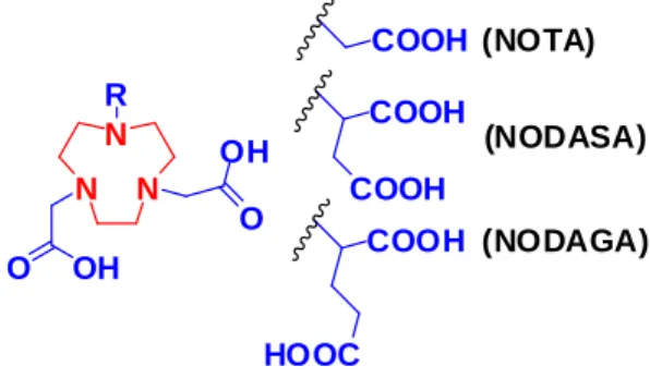
Bifunctional chelators (BFCs) are one of the key elements of a successful radiotracer, which act as a linker between the tracer moiety and the radio isotope. One of the major aims of this study is to develop a method for the synthesis of bifunctional metal chelator NODASA, a potential chelator for radiolabelling. Herein, a facile economic on and off resin method for the synthesis of potential bifunctional chelator “NODASA” functionalized peptide is presented. The seven step synthesis was initiated with a Michael addition reaction between monomethyl fumarate and 1,4,7-triazacyclononane. The final product of NODASA functionalized peptide was obtained with an isolated yield of 84%.
A potential human antimicrobial peptide LL37 possesses impending therapeutic values due to its
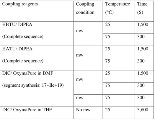
goals of this study. In this regard, a highly efficient and optimized methodology for the synthesis of LL37 on solid phase using microwave energy was developed. During this method development it was concluded that uronium coupling reagents along with standard conditions were inadequate for the synthesis of 20th amino acid residue onwards. Val and Ile amino acid coupling was revealed as the key problematic reaction in the segmentation approach of synthesizing the peptide. It was also found that DIC/OxymaPure in THF is a better combination of reagents for this coupling. The synthesized peptide was further verified for its antimicrobial activity.
In the last part of this study the aim was to explore the radiolabelling potential of LL37 and its usability as a radiotracer. For this purpose LL37 was functionalized with bifunctional chelator NODAGA. NODASA-LL37 was also labelled with cold/hot gallium successfully. This complex was further evaluated in vitro for its bacterial selectivity over mammalian cell line.
With the rapid development of bacterial resistance and the development of “super bugs” there is an urgent need for diagnostic tools which can provide a faster and efficient detection of pathogenic microorganisms. Radiopharmaceutics is an ideal candidate to solve this problem, especially due to its high selectivity, sensitivity and non-invasive nature. In this thesis, an effort was put forward to answer the critical questions regarding the development of synthetic methods, purification and characterization of the antimicrobial peptide LL37. Firstly, a combination of different coupling reagents were used for the optimization of the synthesis, from this study we were able to conclude that the DIC/OxymaPure is better than the HBTU/DIPEA and HATU/DIPEA systems. This method can now be utilized for the large scale production of LL37.
In addition to this, a facile seven step method for the synthesis of the bifunctional chelator NODASA functionalized peptide was developed. In this study a NODASA functionalized peptide was conjugated with cold gallium which, demonstrating it as a potential PET agent for molecular imaging. This route offers a simple and inexpensive alternative to commercially available NODASA and can be coupled with various other peptides. Finally, LL37 was also functionalized with the chelator, NODAGA, and subsequently labelled with natGa. This complex showed significant affinity towards bacterial cells in comparison with mammalian cells and providing evidence that NODAGA-LL37 could be a potential radiotracer for bacterial infection imaging.
Abbreviations: NODAGA: 1,4,7-triazacyclononane,1-glutaric acid-4,7-acetic acid; NODASA:
1,4,7-Triazacyclononane-1-succinic acid-4,7-diacetic acid
Declaration 1- Plagiarism I, Jyotibon Dutta declare that
1. The research report in this thesis, except where otherwise indicated, is my original work.
2. This thesis has not been submitted for any degree or examination at any other university.
3. This thesis does not contain other person’s data, pictures, graphs or other information, unless specifically acknowledged as being sourced from other persons.
4. This thesis does not contain other person’s writing, unless specifically acknowledged as being sourced from other researchers. Where other written sources have been quoted, then:
a. Their words have been re-written but the general information attributed to them has been referenced.
b. Where their exact words have been used, then their writing has been placed in italics and inside quotation marks, and referenced.
5. This thesis does not contain text, graphics or tables copied and pasted from the internet, unless specifically acknowledged, and the source being detailed in the thesis and in the references sections.
Signed
---
Declaration 2- Publication List of publications originated from this Thesis
[1] J. Dutta, T. Naicker, T. Ebenhan, H.G. Kruger, P.I. Arvidsson, T. Govender, Synthetic Approaches to Radiochemical Probes for Imaging of Bacterial Infections, Eur J Med Chem., (2017) Submitted
J. Dutta contributed to the design and wrote the paper.
T. Ebenhan contributed towards adding valuable information into the paper.
The remaining authors are supervisors.
[2] J. Dutta, P.K. Chinthakindi, P.I. Arvidsson, G. Beatriz, H.G. Kruger, T. Govender, T.
Naicker, F. Albericio, A Facile Synthesis of NODASA-Functionalized Peptide, Synlett., 27 (2016) 1685-1688. Published
J. Dutta contributed to the design of the project, synthesized and characterized all compounds, performed the testing of the compounds and wrote the paper.
P.K. Chinthakindi contributed towards writing the paper.
The remaining authors are supervisors.
[3] J. Dutta, S. Ramesh, S.M. Radebe, A.M. Somboro, G. Beatriz, H.G. Kruger, S.Y. Essack, F.
Albericio, T. Govender, Optimized Microwave Assisted Synthesis of LL37, a Cathelicidin Human Antimicrobial Peptide, Int J Pept Res Ther., 21 (2015) 13-20. Published
J. Dutta contributed to the design of the project, synthesized and characterized all compounds, performed the testing of the compounds and wrote the paper.
S. Ramesh and S.M. Radebe contributed towards the synthesis.
A.M. Somboro contributed towards microbiological work.
The remaining authors are supervisors.
[4] J. Dutta, S. Baijnath, A.M. Somboro, S. Nagiah, F. Albericio, G.d.l.T. Beatriz, B.
Marjanovic-Painter, J.R. Zeevaart, M. Sathekge, H.G. Kruger, A. Chuturgoon, T. Naicker, T.
Ebenhan, T. Govender, Synthesis, in vitro evaluation and 68Ga-radiolabeling of CDP1 towards PET/CT imaging of bacterial infection, Chem Biol Drug Des., (2016), Submitted
J. Dutta contributed to the design of the project, synthesized and characterized all compounds, performed the testing of the compounds and wrote the paper.
S. Nagiah contributed towards cell culture work.
The remaining authors are supervisors.
Publications for non-degree purposes
[1] R. Azumah, J. Dutta, A.M. Somboro, M. Ramtahal, L. Chonco, R. Parboosing, L.A. Bester, H.G. Kruger, T. Naicker, S.Y. Essack, In vitro evaluation of metal chelators as potential metallo‐
β‐lactamase inhibitors, J Appl Microbiol., 120 (2016) 860–867.
J. Dutta contributed towards synthesis and characterized few of the compounds utilized in the project; and also wrote the synthesis part.
Acknowledgement I would like to express my most sincere words of gratitude to:
My supervisors, Prof Thavendran Govendor, Dr Tricia Naicker and Prof Gert Kruger, for their remarkable guidance, support and motivation throughout my studies. They have provided me with the ideal platform to hone my research capabilities, while helping me to personally develop.
Prof Fernando Albericio, Prof Glenn Maguire and Prof Beatriz Garcia de la Torre, for the interesting discussions and support during this research.
My role model, Dr Sanil Singh, for constant motivation and personal support during some of the most trying times in my life and career. For always believing in me and pushing me to go further.
My lifelong friend, Sooraj Baijnath, for professional, emotional and personal support.
We met as strangers and will be friends for life.
Dr Byron Peters, Dr Yahya ElSayed Jad and the CPRU group 2015/2016 for their professionalism, eagerness to assist and each having the ability to be a team player.
My father Mr Haresh Dutta, my mother Mrs Kunjalata Hatiboruah, my father in law Late Mr Hiranmay Bhattacharyya, my mother in law Mrs Renu Devi, my wife Dr Neelakshi Bhattacharyya, my younger brother Mr Angshuman Dutta and my little brother Mr Priyam Dutta, for tolerating me and putting up with all the stress that I put them through during the recent years and for continuing to motivating me and for believing in my abilities.
National Research Foundation (NRF, SA), AspenPharmacare and the University of KwaZulu-Natal (College of Health Sciences), for financial support.
Lastly, to my little girl, Adrita Dutta, for her warm presence. A real inspiration and a constant source of motivation for me to achieve anything. This is for you my Baby!
Table of Contents
Abstract ... III Declaration 1- Plagiarism ... V Declaration 2- Publication ... VI Acknowledgement ... VIII Table of Contents ... IX
Chapter 1 Introduction ... 1
1. Preamble ... 1
2. Bifunctional chelators in radiopharmaceutics. ... 1
3. Radio imaging a need for bacterial infection ... 3
4. Peptides as radiotracers ... 4
5. LC-MS as a means for peptide/protein quantification ... 5
6. Research Aims and Objectives ... 6
7. Thesis outline ... 6
Reference ... 6
Chapter 2Synthetic Approaches to Radiochemical Probes for Imaging of Bacterial Infections ... 10
Abstract ... 10
Contents ... 10
1. Introduction... 12
2. Commercially available infection imaging probes: ... 13
2.1. 111In-oxine-Leukocyte ... 13
2.2. 99mTc-HMPAO-Leukocyte: ... 14
2.3. 99mTc-Stannous Colloid ... 14
2.4. 99mTc-Besilesomab ... 15
2.5. 99mTc-Sulesomab ... 15
2.6. 18F-FDG-PET ... 16
2.7. 67/68Ga-Citrate ... 16
3. Novel imaging probes for direct, more specific imaging of bacterial infection ... 19
3.1. Antimicrobial peptides as bacteria-selective imaging probes ... 19
3.1.1. Ubiquicidin (UBI) ... 20
3.1.2. Human neutrophil peptide (HNP) ... 22
3.1.3. Neutrophil elastase inhibitor peptide ... 23
3.1.4. Human β-defensin (HBD) ... 24
3.1.5. Human lactoferrin-derived peptide (hLF) ... 25
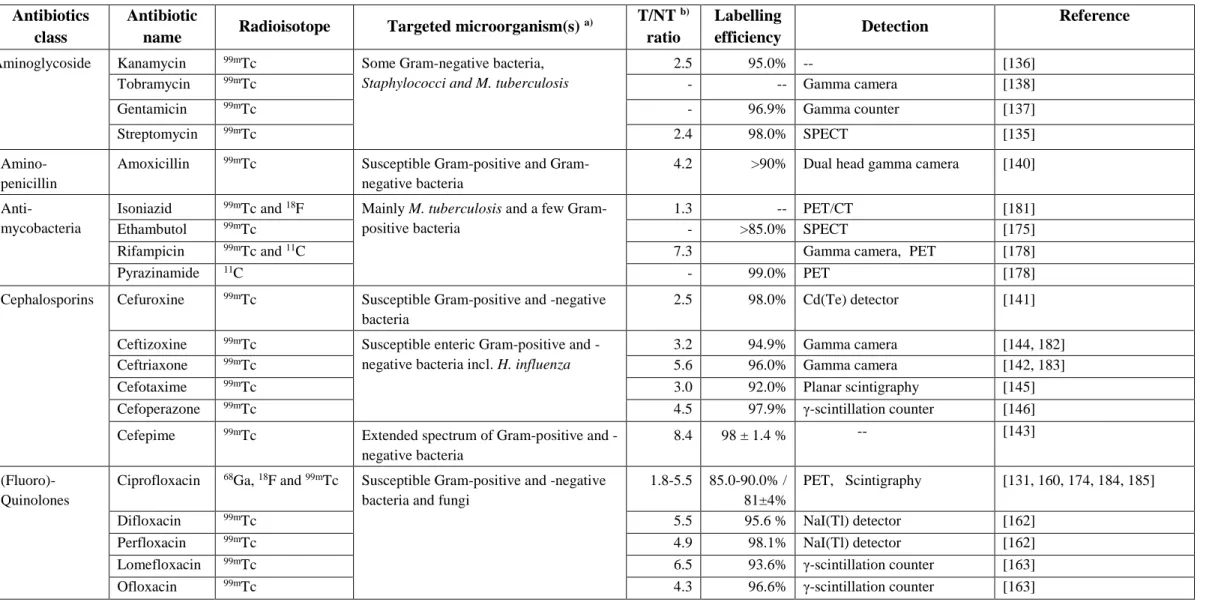
3.2.1. Antibiotics/Antimicrobial drugs ... 27
3.2.1.1. Aminoglycoside ... 27
3.2.1.2. Amino-penicillin 3rd generation ... 28
3.2.1.3. Cephalosporins... 28
3.2.1.4. Glycopeptides ... 29
3.2.1.5. Lincosamide ... 29
3.2.1.6. Macrolides ... 30
3.2.1.7. Nitrofurans ... 30
3.2.1.8. Oxazolidinones ... 30
3.2.1.9. Fluoroquinolones ... 31
3.2.1.10. Anti-mycobacteria ... 33
3.2.1.11. Tetracyclines ... 35
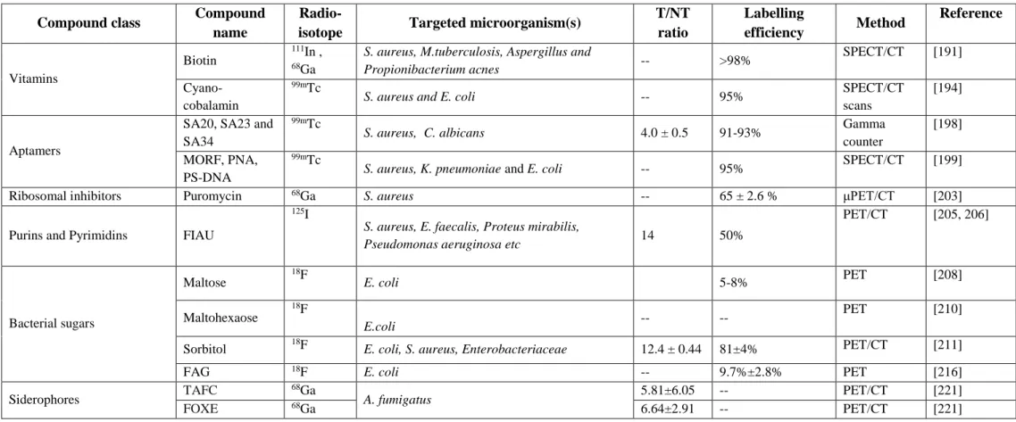
3.2.2. Vitamins ... 39
3.2.2.1. Biotin (Vitamin H) ... 39
3.2.2.2. Cyano-cobalamin (CBL) ... 39
3.2.3. Aptamers (Oligomers) ... 39
3.2.4. Puromycin ... 40
3.2.5. Radiolabeled purines and pyrimidines ... 41
3.2.6. Glycopyranose derivatives ... 41
3.2.7. Siderophores ... 44
4. Challenges and limitations in producing efficient infection imaging probes ... 46
5. Conclusion ... 46
References... 46
Chapter 3A Facile Synthesis of NODASA-Functionalized Peptide ... 62
Abstract ... 63
Acknowledgment ... 67
References and Notes ... 68
Supporting information ... 70
Chapter 4Optimized Microwave Assisted Synthesis of LL37, a Cathelicidin Human Antimicrobial Peptide ... 77
Abstract ... 77
Abbreviations ... 77
1. Introduction... 78
2. Materials and Methods ... 79
2.2.1. Deprotection: ... 79
2.2.2. Coupling ... 80
2.2.3. Cleavage ... 80
2.2.4. Purification of LL37 ... 80
2.3. Peptide analysis ... 81
2.4. Antibacterial test ... 81
3. Results and Discussion ... 81
3.1. LL37 synthetic approaches ... 81
3.2. Antimicrobial activities... 86
4. Conclusion ... 86
Acknowledgements: ... 86
Reference ... 86
Supporting information ... 90
Chapter 5Synthesis, in vitro evaluation and 68Ga-radiolabeling of CDP1 towards PET/CT imaging of bacterial infection ... 92
Abstract ... 92
1. Introduction... 93
2. Methods and Materials ... 94
2.1. Materials ... 94
2.2. Peptide synthesis ... 95
2.1.1. Coupling of NODAGA(tBu)3 to the peptide and cleavage ... 95
2.1.2. CDP1 purification ... 95
2.3. Non-radioactive natGa-labeling of CDP1 ... 95
2.4. LC-MS method for quantification of natGa-CDP1 ... 96
2.5. Uptake of natGa-CDP1 by S.aureus, E.coli and M. smegmatis ... 96
2.6. Uptake of natGa-CDP1 by hepatocellular carcinoma (HepG2) cells ... 96
2.7. 68Ge/68Ga-Generator elution and 68Ga-radiolabeling ... 97
2.8. Identification of the 68Ga-CDP1 using UV/radio-HPLC analysis ... 97
2.9. Biostatistics ... 98
3. Results and Discussion ... 98
3.1. Synthesis of CDP1 and natGa-conjugation ... 98
3.2. Bacterial and Hepatocellular uptake of the natGa-CDP1 ... 98
3.2.1. Uptake of natGa-CDP1 by S.aureus ... 99
3.1.2. Uptake of natGa-CDP1 by E. coli ... 100
3.2.3. Uptake of natGa-CDP1 by M. smegmatis ... 100
3.4 Identification 68Ga-CDP1 ... 101
4. Conclusion ... 102
Acknowledgement ... 102
Funding Sources ... 102
Reference ... 103
Supplementary information: ... 106
Chapter 6Summary ... 108
Chapter 1
Introduction 1. Preamble
Biological processes can be visualized, characterized and measured in vivo with the aid of molecular imaging [1]. This exponentially growing technique is utilized to monitor molecules or cellular processes for the diagnosis and/or management of diseases and disorders. Some of these imaging procedures include positron emission tomography (PET) and single-photon emission computed tomography (SPECT) that acquire images by detecting signals from radiolabelled tracers that have to be injected into the test subjects. However, endogenous molecules or exogenous molecular probes are generally considered in some modalities like optical imaging and magnetic resonance imaging (MRI) to monitor disease progression. Due to its critical role in medical diagnostics, the design and development of radiotracers are becoming a focal area in molecular imaging research [2]. Characteristically, a radiotracer is equipped with a targeting moiety, a signaling radioisotope and a linker connecting these components.
2. Bifunctional chelators in radiopharmaceutics.
Radio-metals carry promise as a tool towards the diagnosis, as well as for monitoring the progression of many diseases. These isotopes have to be impounded by metal chelators in order for use in a biological system. Based on the compatibility, these organic compounds are able to make stable complexation products with a particular radio-metal. An ideal chelator binds to the radio-metal well enough so that it can be delivered to the desired site without trans-chelation thereby improving therapy or in vivo diagnosis [3]. In radiopharmaceutics, typically utilised ligands for metal binding are mostly bifunctional chelators (BFCs). These are distinctive chelators with a functional group to form a covalent bond with the tracer molecules, such as, but not limited to antibodies, nucleotides and peptides. Such functional groups can be carboxylic acids/activated esters, isothiocyanates or maleimides for amide, thiourea and thiol coupling agents, respectively [4, 5]. Moreover, click chemistry based reactions which are either copper- free (Diels-Alder and strain-promoted azide-alkyne cycloadditions click reactions) or copper catalyzed (azide-alkyne Huisgen 1,3-dipolar cycloaddition “click” reactions) are also gaining popularity in bio-conjugate chemistry [6]. Chelators play a major role in the pharmacokinetics of the radiochemical; it has been noted that by only changing the ligand the biodistribution of peptide-conjugates are affected [7]. A simple synthetic strategy which avoids diastereospecific/
non-enantio and stereoisomer reactions are always desirable for BFC synthesis. In addition, it should have a maximum number of modularity possibilities for assigning a variety of
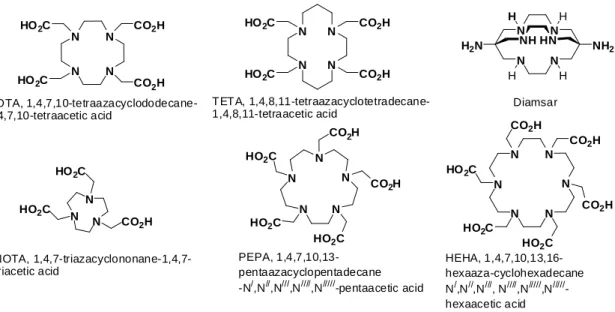
tracers/moieties to impact biodistribution by altering charge and polarity [3]. Broadly, BFCs are categorized into macrocyclic and acyclic chelators. However, macrocyclic chelators have an advantage over its counterpart acyclic ligands as it inherits a pre-organized binding pocket for metal ions which minimizes the entropic loss during radio-isotope coordination. Conversely, acyclic chelators change their physical orientation and geometry drastically for the positioning of the donor atoms to form the conjugate product with the metal ion. This phenomenon leads to a substantial drop in entropy as compared to macrocyclic chelators [8]. A few popular macrocyclic chelators are 1,4,7,10-tetraazacyclododecane-1,4,7,10-tetraacetic acid (DOTA), 1,4,8,11-tetraazacyclotetradecane-1,4,8,11-tetraacetic acid (TETA), diamsar, 1,4,7- triazacyclononane-1,4,7-triacetic acid (NOTA), 1,4,7,10,13,16-hexaaza-cyclohexadecane Nʹ,Nʹʹ,Nʹʹʹ, Nʹʹʹʹ,Nʹʹʹʹʹ,Nʹʹʹʹʹʹ-hexaacetic acid (HEHA), 1,4,7,10,13-pentaazacyclopentadecane- Nʹ,Nʹʹ,Nʹʹʹ, Nʹʹʹʹ,Nʹʹʹʹʹ-pentaacetic acid (PEPA) (Figure 1) and their derivatives; on the other hand diethylenetriaminepentaacetic acid (DTPA), N,N0-bis(2-hydroxybenzyl)-ethylenediamine-N,N- diacetic acid (HBED), N,N-(methylenephosphonate)-N,N0-[6-(methoxycarbonyl)pyridin-2-yl]- methyl-1,2-diaminoethane (H6phospa), bipyridine-chelator (BPCA), desferrioxamine B (DFO) and CP256 (Figure 2) are some examples of widely studied acyclic BFCs [3]. Alongside the chelating moiety, there are a variety of options to choose with regards to the radio-nucleoid which includes 18F, 67/68Ga, 99mTc, 111In, 177Lu, 64Cu, 44Sc, 86Y and 89Zr [3]. These ligands have been widely studied in diagnostic imaging of carcinomas, inflammations and infections.
N N
N
N CO2H
CO2H HO2C
HO2C
DOTA, 1,4,7,10-tetraazacyclododecane- 1,4,7,10-tetraacetic acid
N N
N
N CO2H
CO2H HO2C
HO2C
TETA, 1,4,8,11-tetraazacyclotetradecane- 1,4,8,11-tetraacetic acid
N N
N N
H H
H H
NH2 H2N NHHN
Diamsar
N N
N CO2H HO2C
HO2C
NOTA, 1,4,7-triazacyclononane-1,4,7- triacetic acid
N N N N N
CO2H
CO2H
HO2C HO2C
HO2C
PEPA, 1,4,7,10,13- pentaazacyclopentadecane -N/,N//,N///,N////,N/////-pentaacetic acid
N N
N N N
N
CO2H
HO2C HO2C
HO2C
CO2H
CO2H
HEHA, 1,4,7,10,13,16- hexaaza-cyclohexadecane N/,N//,N///, N////,N/////,N/////- hexaacetic acid
Figure 1 Macrocyclic chelators commonly used for radioisotope conjugation
N N N HO2C
CO2H CO2H CO2H HO2C
DTPA, diethylenetriaminepentaacetic acid
N O HO
N
OH HO
O OH
HBED, N,N0-bis(2-hydroxybenzyl)- ethylenediamine-N,N-diacetic acid
N
COOH
N N
CO2H
HO2C
N HO2C CO2H BPCA
N O
HN O
HN O
OH
HN O N
OH O
HN O
N
HO O
CP256
HN
N O
O
N HO
HN HO
O
O O
N OH
NH2 5
5 5
DFO, desf errioxamine B
N N
N N
P P
HO O OH
O OH OH
O OH
O HO
H6phospa, N,N-(methylenephosphonate)- N,N0-[6-(methoxycarbonyl)pyridin-2-yl]- methyl-1,2-diaminoethane
Figure 2. Acyclic chelators commonly used for radioisotope conjugation 3. Radio imaging a need for bacterial infection
Bacterial infection is a major threat to human health; and is still recognized as one of the most vicious causes of mortality and morbidity worldwide [9]. Because of its heterogeneous nature, infectious diseases are associated with a variety of different clinical signs and symptoms. They may be systemic or localized in one foci and often reoccurs; in many cases these infections require long term treatment [10]. Therefore, in a clinical set up it is difficult to distinguish between infection and aseptic inflammation in addition to its size and distribution throughout the body. This in turn affects the treatment strategies that are employed [11]. In general, the diagnosis of infectious diseases is carried out by using clinical history, physical inspection, pathogen identification in suspected sample and by imaging approaches. Though the application of imaging techniques has advantages due to its non-invasive nature, but there is a significant difference between the uses of non-radionuclide medicine and radionuclide imaging. Imaging techniques such as ultrasound, plain radiography and computed tomography are based on anatomical changes, which arise in the advanced stages of infectious diseases [12]. This highlights the need to develop radio nuclear approaches for the early and precise identification of infectious lesions. Due to the knowledge gained and better understanding, we now have in pathophysiology a number of new promising radiopharmaceuticals have been developed for
imaging bacterial infections [13-15]. This development in radiopharmaceutics is feeding the clinician with invaluable information regarding the infection, thereby giving an opportunity to commence with the best therapeutic strategies for the patients. Up till now, various molecules have been taken into consideration as radio-labelled tracers to detect infection foci; this includes leukocytes, antibiotics, antibodies, peptides, siderophores, bacteriophages, vitamins, carbohydrates and aptamers [16-19]. However, very few from this have been clinically approved for human use; that taken into account includes 111In-oxine-Leukocyte-SPECT, 99mTc-HMPAO- Leukocyte-SPECT, 99mTc-Sn-Colloid “LLK”-SPECT, 99mTc-Besilesomab-SPECT, 99mTc- Sulesomab-SPECT, 18F-FDG-PET and 67Ga-citrate-SPECT [20-24].
4. Peptides as radiotracers
Recently, attention has been drawn to antimicrobial peptides (AMPs) for use as a guiding molecule for the radio-isotope and in PET tracer development [25]. Evolutionarily, AMPs are the ancient ordinance of the immune system. They are widely spread amongst the animal species, as well as in the plant kingdom; this suggests the involvement of AMPs in the evolution of multicellular organisms. These peptides are still an effective and integral part of the primary host defense system regardless of its ancient lineage [26]. They are of greater interest because of the fact that peptides show low toxicity and immunogenicity with high specificity and binding capability towards its desired target [27, 28]. The mechanism of action of AMPs are based on differences in the basic design of the cellular membrane of multicellular organisms and microbes.
The outermost bilayer of bacteria is composed of heavily condensed negatively charged lipids, whereas the outer layer of the animal and plant cells are populated mainly with neutral lipids [29]. This feature gives the AMPs specificity towards bacterial cells and accumulation at the infectious site; making them promising PET and SPECT tracers, for imaging bacterial infections [30]. Even though the use of AMPs in radiopharmaceutics is not a new concept [31, 32]; only a few members of this group have been evaluated so far [33]. Some of the AMPs studied so far include ubiquicidin, human neutrophil peptide, neutrophil elastase inhibitor peptide, human-β- defensin and human lactoferrin-derived peptide. In addition to the aforementioned peptides, human cathelecidin antimicrobial peptide LL37 is also extensively involved with the innate immune system and can be useful in radiopharmaceutics. This antimicrobial peptide is found in various cell lines (human squamous epithelia, granulocytes and neutrophil) and other bodily fluids [34]. LL37 is involved in neutralizing biologically active molecules, which are present in the bacterial cell wall and also acts as growth inhibitor [35, 36]. Moreover, this antimicrobial
5. LC-MS as a means for peptide/protein quantification
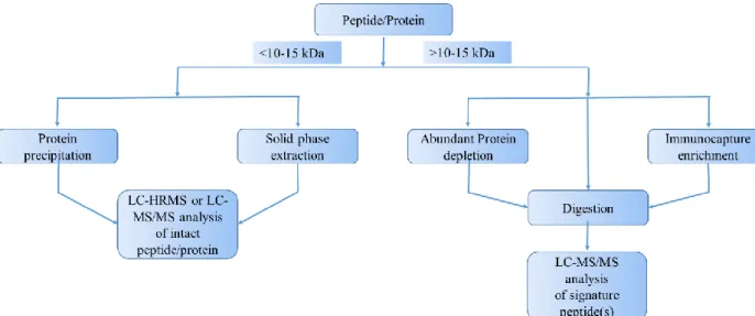
In the current era, peptide and protein quantification is quickly gaining momentum due to the increasing demand for them to be evaluated as diagnostic and therapeutic molecule. Moreover, there is a shift in the trend of drug discovery towards a target oriented approach and therefore a need for the precise and accurate quantification of these molecules [37]. In that context, liquid chromatography–mass spectrometry (LC-MS) which is considered as the gold standard for small molecule quantification is now showing great potential towards larger molecules (such as peptide and protein) determination, due to its inherent advantages [38, 39]. The advantages of this technique over immune based assays include a shorter method development time, lack of antibody requirement, high specificity as well as sensitivity (up to ng/ml or pg/ml concentrations) and its ability to monitor product degradation. Based on the size of the peptide/protein, LC-MS quantification can be broadly categorized into 1) a bottom up approach where the molecule (usually >10-15 kDa), is digested with an enzyme and is followed by the liquid chromatography–
mass spectrometry/mass spectrometry (LC-MS/MS) analysis; and 2) a top down approach where the native molecule (usually <10-15 kDa) is analyzed directly, either by liquid chromatography–
high resolution mass spectrometry (LC-HRMS) or LC-MS/MS (Figure 3). A variety of sample preparation methods such as solid phase extraction, protein precipitation, immuno-capture enrichment and depletion of high-abundant proteins can be integrated into the LC-MS workflow based on the nature of the analyte and to increase sensitivity of the method [37].
Figure 3. Generalized workflow of peptide/protein quantification by LC-MS
6. Research Aims and Objectives
1. What are the current approaches used for the synthesis of radiolabelled probes for bacterial infection identification?
Objective: To assess the currently available radiotracers and evaluate them for imaging of bacterial infections and their synthetic approaches.
2. Till date, there is no method developed for on and off resin synthesis of bifunctional metal chelator 1,4,7-Triazacyclononane-1-succinic acid-4,7-diacetic acid (NODASA); is it possible to synthesize NODASA economically using solid phase synthesis?
Objective: To develop a facile method for the synthesis of NODASA.
3. LL37 is a human antimicrobial peptide, can it be produced efficiently by solid phase peptide synthesis?
Objective: To develop an efficient synthetic route for the production of LL37 by solid phase peptide synthesis.
4. Literature reveals that antimicrobial peptides bears the potential to be used as radiotracer for bacterial infection imaging, can LL37 be radiolabelled and utilized for this purpose?
Objective: To synthesize 1,4,7-triazacyclononane,1-glutaric acid-4,7-acetic acid-LL37 (NODAGA-LL37), investigate its complexation with nat/68Ga and evaluate cellular uptake of
natGa-NODAGA-LL37.
7. Thesis outline
A brief review on radiotracers having potential for bacterial infection imaging will be presented in Chapter 2. A facile approach for the synthesis of bifunctional chelator NODASA will be presented in Chapter 3. The synthesis of antimicrobial peptide LL37 will be discussed in Chapter 4. The synthesis and the evaluation of nat/68Ga complexed NODAGA-LL37 will be discussed in Chapter 5. Finally, in chapter 6 the overview of the results is put into perspective.
Reference
[1] R. Weissleder, U. Mahmood, Molecular Imaging, Radiology, 219 (2001) 316-333.
[2] T.F. Massoud, S.S. Gambhir, Molecular imaging in living subjects: seeing fundamental biological processes in a new light, Genes Dev., 17 (2003) 545-580.
[3] E.W. Price, C. Orvig, Matching chelators to radiometals for radiopharmaceuticals, Chem.
[5] B.M. Zeglis, J.S. Lewis, A practical guide to the construction of radiometallated bioconjugates for positron emission tomography, Dalton Trans., 40 (2011) 6168-6195.
[6] D. Zeng, B.M. Zeglis, J.S. Lewis, C.J. Anderson, The growing impact of bioorthogonal click chemistry on the development of radiopharmaceuticals, J. Nucl. Med., 54 (2013) 829-832.
[7] M. Lin, M.J. Welch, S.E. Lapi, Effects of Chelator Modifications on 68Ga-Labeled [Tyr3]
Octreotide Conjugates, Mol. Imaging Biol., 15 (2013) 606-613.
[8] R.D. Hancock, Chelate ring size and metal ion selection. The basis of selectivity for metal ions in open-chain ligands and macrocycles, J. Chem. Educ, 69 (1992) 615-621.
[9] S. Auletta, F. Galli, C. Lauri, D. Martinelli, I. Santino, A. Signore, Imaging bacteria with radiolabelled quinolones, cephalosporins and siderophores for imaging infection: a systematic review, Clin Transl Imaging, 4 (2016) 229-252.
[10] A. Signore, A.W. Glaudemans, The molecular imaging approach to image infections and inflammation by nuclear medicine techniques, Ann. Nucl. Med., 25 (2011) 681-700.
[11] A. Signore, A.W. Glaudemans, F. Galli, F. Rouzet, Imaging infection and inflammation, BioMed Res. Int., 2015 (2015) 615150.
[12] A. Signore, C. D’Alessandria, E. Lazzeri, R. Dierckx, Can we produce an image of bacteria with radiopharmaceuticals?, Eur. J. Nucl. Med. Mol. Imaging, 35 (2008) 1051-1055.
[13] V. Kumar, D.K. Boddeti, 68Ga-radiopharmaceuticals for PET imaging of infection and inflammation, in: Theranostics, Gallium-68, and Other Radionuclides, Springer, 2013, pp. 189- 219.
[14] A. Glaudemans, F. Galli, M. Pacilio, A. Signore, Leukocyte and bacteria imaging in prosthetic joint infection, Eur. Cell. Mater., 25 (2013) 61-77.
[15] A.W. Glaudemans, E.F. de Vries, F. Galli, R.A. Dierckx, R.H. Slart, A. Signore, The Use of F-FDG-PET/CT for Diagnosis and Treatment Monitoring of Inflammatory and Infectious Diseases, Clin. Dev. Immunol., 2013 (2013) 623036.
[16] C. Tsopelas, Radiotracers used for the scintigraphic detection of infection and inflammation, Scientific World J., 2015 (2015) 676719.
[17] E. Lazzeri, P. Erba, M. Perri, R. Doria, C. Tascini, G. Mariani, Clinical impact of SPECT/CT with In-111 biotin on the management of patients with suspected spine infection, Clin. Nucl.
Med., 35 (2010) 12-17.
[18] C. A Dougherty, W. Cai, H. Hong, Applications of aptamers in targeted imaging: state of the art, Curr. Top. Med. Chem., 15 (2015) 1138-1152.
[19] M. Rusckowski, S. Gupta, G. Liu, S. Dou, D.J. Hnatowich, Investigations of a 99mTc- labeled bacteriophage as a potential infection-specific imaging agent, J. Nucl. Med., 45 (2004) 1201-1208.
[20] D.K. Hughes, Nuclear medicine and infection detection: the relative effectiveness of imaging with 111In-oxine-, 99mTc-HMPAO-, and 99mTc-stannous fluoride colloid-labeled leukocytes and with 67Ga-citrate, J. Nucl. Med. Technol., 31 (2003) 196-201.
[21] W.S. Richter, V. Ivancevic, J. Meller, O. Lang, D. Le Guludec, I. Szilvazi, H. Amthauer, F.
Chossat, A. Dahmane, C. Schwenke, 99mTc-besilesomab (Scintimun®) in peripheral osteomyelitis: comparison with 99mTc-labelled white blood cells, Eur. J. Nucl. Med. Mol.
Imaging, 38 (2011) 899-910.
[22] R. Sousa, M. Massada, A. Pereira, F. Fontes, I. Amorim, A. Oliveira, Diagnostic accuracy of combined 99mTc-sulesomab and 99mTc-nanocolloid bone marrow imaging in detecting prosthetic joint infection, Nucl. Med. Commun., 32 (2011) 834-839.
[23] H. Zhuang, Q.Y. Jian, A. Alavi, Applications of fluorodeoxyglucose-PET imaging in the detection of infection and inflammation and other benign disorders, Radiol. Clin. North Am., 43 (2005) 121-134.
[24] M. Bester, P. Van Heerden, J. Klopper, H. Wasserman, S. Rubow, F. De Klerk, Imaging infection and inflammation in an African environment: Comparison of 99Tcm-HMPAO-labelled leukocytes and 67Ga-citrate, Nucl. Med. Commun., 16 (1995) 599-607.
[25] M. Sathekge, The potential role of 68Ga-labeled peptides in PET imaging of infection, Nucl.
Med. Commun., 29 (2008) 663-665.
[26] M. Zasloff, Antimicrobial peptides of multicellular organisms, Nature, 415 (2002) 389-395.
[27] K. Chen, X. Chen, Design and development of molecular imaging probes, Curr. Top. Med.
Chem., 10 (2010) 1227-1236.
[28] T. Ebenhan, N. Chadwick, M.M. Sathekge, P. Govender, T. Govender, H.G. Kruger, B.
Marjanovic-Painter, J.R. Zeevaart, Peptide synthesis, characterization and 68 Ga-radiolabeling of NOTA-conjugated ubiquicidin fragments for prospective infection imaging with PET/CT, Nucl. Med. Biol., 41 (2014) 390-400.
[29] K. Matsuzaki, Why and how are peptide–lipid interactions utilized for self-defense?
Magainins and tachyplesins as archetypes, Biochim. Biophys. Acta, Biomembr., 1462 (1999) 1- 10.
[30] S.M. Okarvi, Peptide-based radiopharmaceuticals: future tools for diagnostic imaging of cancers and other diseases, Med. Res. Rev., 24 (2004) 357-397.
[31] A. Lupetti, P.H. Nibbering, M.M. Welling, E.K. Pauwels, Radiopharmaceuticals: new antimicrobial agents, Trends Biotechnol., 21 (2003) 70-73.
[32] C.J. Palestro, Radionuclide imaging of infection: in search of the grail, J. Nucl. Med., 50 (2009) 671-673.
[33] M.S. Akhtar, M.B. Imran, M.A. Nadeem, A. Shahid, Antimicrobial peptides as infection imaging agents: better than radiolabeled antibiotics, Int J Pept, 2012 (2012) 965238.
[34] J. Dutta, S. Ramesh, S.M. Radebe, A.M. Somboro, G. Beatriz, H.G. Kruger, S.Y. Essack, F.
Albericio, T. Govender, Optimized Microwave Assisted Synthesis of LL37, a Cathelicidin Human Antimicrobial Peptide, Int. J. Peptide Res. Therapeut., 21 (2015) 13-20.
[35] D.A. Devine, Antimicrobial peptides in defence of the oral and respiratory tracts, Mol.
Immunol., 40 (2003) 431-443.
[36] M.J. Nell, G.S. Tjabringa, A.R. Wafelman, R. Verrijk, P.S. Hiemstra, J.W. Drijfhout, J.J.
Grote, Development of novel LL-37 derived antimicrobial peptides with LPS and LTA neutralizing and antimicrobial activities for therapeutic application, Peptides, 27 (2006) 649-660.
[37] S.W. Zhang, W. Jian, Recent advances in absolute quantification of peptides and proteins using LC-MS, Rev. Anal. Chem., 33 (2014) 31-47.
[39] F. Li, D. Fast, S. Michael, Absolute quantitation of protein therapeutics in biological matrices by enzymatic digestion and LC-MS, Bioanalysis, 3 (2011) 2459-2480.
Chapter 2
Synthetic Approaches to Radiochemical Probes for Imaging of Bacterial Infections
Jyotibon Dutta1, Tricia Naicker1, Thomas Ebenhan2, Hendrik G. Kruger1, Per I.
Arvidsson1,3 andThavendran Govender1*
1Catalysis and Peptide Research Unit, School of Health Sciences and School of Chemistry and Physics, University of KwaZulu-Natal, Durban 4001, South Africa
2University of Pretoria & Steve Biko Academic Hospital, Crn Malherbe and Steve Biko Rd, Pretoria, 0001, South Africa.
3Science for Life Laboratory, Drug Discovery and Development Platform and Division of Translational Medicine and Chemical Biology, Department of Medical Biochemistry and Biophysics, Karolinska Institutet SE-171 77 Stockholm, Sweden
* Corresponding author. E-mail address: [email protected] (Thavendran Govender)
Abstract
This present review provides an account on the available synthetic strategies employed to radiolabel commercial and potential bacteria-selective probes for tomographic imaging. These molecular probes encompass leukocytes, antibodies, small molecules, peptides, antibiotics, macrolides, vitamins, oligomers and siderophores. Although this technique has shown to be a valuable tool for non-invasive infection imaging, more development is required to create easy- to-radiolabel kit solutions/procedures for the preparation of the probes.
Contents
1. Introduction... 12 2. Commercially available infection imaging probes: ... 13 2.1 111In-oxine-Leukocyte ... 13
2.4 99mTc-Besilesomab ... 15 2.5 99mTc-Sulesomab ... 15 2.6 18F-FDG-PET ... 16 2.7. 67/68Ga-Citrate ... 16 3. Novel imaging probes for direct, more specific imaging of bacterial infection ... 19 3.1 Antimicrobial peptides as bacteria-selective imaging probes ... 19 3.1.1 Ubiquicidin (UBI) ... 20 3.1.2 Human neutrophil peptide (HNP) ... 22 3.1.3 Neutrophil elastase inhibitor peptide ... 23 3.1.4 Human β-defensin (HBD) ... 24 3.1.5 Human lactoferrin-derived peptide (hLF) ... 25 3.2 Biomimetics ... 27 3.2.1 Antibiotics/Antimicrobial drugs ... 27 3.2.1.1 Aminoglycoside ... 27 3.2.1.2 Amino-penicillin 3rd generation ... 28 3.2.1.3 Cephalosporins... 28 3.2.1.4 Glycopeptides ... 29 3.2.1.5 Lincosamide ... 29 3.2.1.6 Macrolides ... 30 3.2.1.7 Nitrofurans ... 30 3.2.1.8 Oxazolidinones ... 30 3.2.1.9 Fluoroquinolones ... 31 3.2.1.10 Anti-mycobacteria ... 33 3.2.1.11 Tetracyclines ... 35 3.2.2 Vitamins ... 39 3.2.2.1 Biotin (Vitamin H) ... 39 3.2.2.2 Cyano-cobalamin (CBL) ... 39 3.2.3 Aptamers (Oligomers) ... 39 3.2.4 Puromycin ... 40 3.2.5 Radiolabeled purines and pyrimidines ... 41 3.2.6 Glycopyranose derivatives ... 41 3.2.7 Siderophores ... 44 4. Challenges and limitations in producing efficient infection imaging probes ... 46 5. Conclusion ... 46 References... 46
1. Introduction
Throughout the world, bacterial infections remain a major cause for human morbidity and mortality [1]. Infectious diseases are one of the oldest challengers for humans; and can be ranked with war and famine [2]. In this current era, the growing incident of infection and the development of drug resistance have a huge impact on the global economy and human health [2].
Early detection of bacterial infection is crucial for the prognosis of the disease but insignificant progress has been done in this field. Conventional ways for bacterial diagnosis are based on either or combination of biochemical, physical and bacterial cultures where the bacterial culturing can be considered as the gold standard method [3]. However, bacterial culture methods do not provide in vivo localization of the infection or reservoirs. With the advancement of modern day technology, various diagnostic tools bear the potential for locating the site of infection. Radiological techniques such as computed tomography (CT) can be advantageous for imaging bacterial infections but it is only useful when there is a significant anatomical change in potentially infected tissues. Likewise, magnetic resonance imaging (MRI) can be applied for bacterial imaging aided by specific probes but it is limited to patients without claustrophobia or implanted medical devices. Nuclear medicine techniques such as single photon emission computed tomography (SPECT) and positron emission tomography (PET) stand out as potential candidates because these techniques are able to locate the site of infection with the help of a radiolabeled biomolecule/probe [4]. Available SPECT- or PET-based probes consist of in vitro or in vivo (for example) radiolabeled leukocytes. There are recurring comprehensive reports on the clinical use of commercially available radiotracers targeting infection and inflammation [5, 6]. The radiolabeled probes under current studies showed the potential for targeting molecular components of bacteria or its metabolites resulting in specific identification of the infection site in contrast to the sterile inflammation [7-10]. This field of study is also positively influenced and expanding due to the hybridization of imaging techniques such as PET(SPECT)/CT or PET(SPECT)/MRI [11].
Herein, we aim to provide a concise guide to the synthetic approaches of molecular radiotracers with focus on bacterial imaging aimed at medicinal chemists. This review is divided into commercially available and novel probes as followed: i) clinical/preclinical availability, ii) probe functionalizing via suggested radiolabeling procedures and iii) probe distribution and suggested mechanisms of the probe uptake, internalization and signal amplification. At the end of each class of radiolabeled probe, is a table summary of the transformations that provides information
2. Commercially available infection imaging probes:
Most of the clinically used radiotracers for infection detection are based on leukocytes due to the fact that there is an influx of leukocytes during inflammation. A few of these radiopharmaceuticals are Food and Drug Administration (FDA) approved. These commercially available radiotracers can be divided broadly into i) radiolabeled leukocytes (111In-oxine- leukocyte, 99mTc-HMPAO-leukocute and 99mTc-Stannous Colloid), ii) radiolabeled anti- granulocyte antibodies (99mTc-Besilesomab and 99mTc-Sulesomab), iii) unspecific biomolecules such as 2-deoxy-2-(18F)fluoro-D-glucose(18F-FDG) and 67/68Ga-citrate.
2.1. 111In-oxine-Leukocyte
The current “gold standard” tomographic imaging technique targeting infection is considered to be direct leukocyte labeling with radio metals [12]. Efficient labeling of leukocytes and its imaging can be obtained with 111In-oxine because of its long half-life [13]. Detection and localization of the infectious site is the prime area of interest for 111In-oxine-leukocyte which included various clinical presentations such as pulmonary infection including tuberculosis, endocarditis, neurological infections, fever of unknown origin, diabetic foot, inflammatory bowel disease, infected central venous catheters or other devices; infected joint and vascular prosthesis, postoperative abscesses and osteomyelitis of the appendicle skeleton [14-16]. Roca et al.
described a method of radiolabeling necessitating at least 2 x 10⁸ leukocytes for good labeling efficiencies [14]. A whole-blood sample was collected from the patient in acid-citrate-dextrose (ACD) anticoagulant solution containing vial, at a blood /ACD ratio of 1:5.6. Out of that, 15 ml of the mixture was centrifuged at 2000 g for 10 min at room temperature to separate the blood cells from the cell free plasma (CFP) and the latter was used as re-suspending medium after labeling in the subsequent steps. Leukocyte isolation was initiated by separating the erythrocytes with 10% 2-hydroxyethyl starch (HES) (molecular weight 200/0.5 or 200/0.6) by sedimentation.
Recommended blood-ACD solution to HES ratio was 10:1 and the leukocyte-rich plasma (LRP) was collected with a 20G lumbar needle without disturbing the erythrocyte layer which was followed by centrifugation at 150 g for 5 min. Platelet-rich plasma (PRP) was removed and the mixed leukocyte pellet was gently re-suspended. An optional PBD/saline wash was advised to reduce the number of platelets. For radiolabeling, 20 MBq of 111In-oxine was incubated with the isolated mixed leukocyte cell suspension for 10 min at room temperature with gentle periodic swirling. The authors recommended working with HEPES buffer (about 6 mg/ml final concentration) if 111In-oxine has to be used without the buffer provided by the manufacturer.
After incubation, at least 3 ml (up to a maximum of 10 ml) of PBS/saline was added to the solution and centrifuged at 150 g for 5 min. The supernatant containing the unbound 111In-oxine
was removed and used to calculate the labeling efficiency. Radioactivity was measured in the leukocyte pellet and subsequently dissolved in 3-5 ml of previously extracted CFP [14].
During the process of leukocyte labeling with 111In-oxine, it diffused through the lipid bilayer of the cell because of its neutral and lipophilic nature. Upon internalization, the 111In-complexes (i.e. the radioisotope) irreversibly associate with intercellular and nucleus components and free oxine may experience cellular efflux.
2.2. 99mTc-HMPAO-Leukocyte:
Technetium-99m (99mTc) can be chelated with hexamethylpropyleneamine oxine (HMPAO) and has been used as a non-invasive diagnosis tool for numerous physiological abnormalities [17].
The SPECT radioisotope 99mTc- can be chelated with HMPAO, which facilitates the uptake into cells [18]. Leukocyte labeling with 99mTc-HMPAO follows a similar technique to using 111In- oxine; i.e. it needs the separation of WBC from the whole blood. Once inside the cell, 99mTc- HMPAO changes its lipophilic character to non-diffusible hydrophilic complexes and becomes trapped [19]. This compound has a half-life of 6 h and emits 140-keV γ-rays which can be detected by a SPECT camera [20]. The normal probe bio-distribution can be seen mainly in the kidney, liver and spleen with notable activity in the gastrointestinal tract and bone marrow.
Hence, imaging of hepatic and splenic infections using 99mTc-HMPAO-Leukocyte SPECT may lead to suboptimal image interpretation [20].
2.3. 99mTc-Stannous Colloid
99mTc-labled stannous colloids (99mTc-SnC) is a radiolabeling technique to label neutrophils and monocytes for imaging of inflammation and spatial localization of infection for gram-positive as well as gram-negative bacteria [21-26]. The radiolabeling of leukocytes does not require its separation from the whole blood as 99mTc-SnC can selectively bind to the target immune cells in the infection site which make it simpler and less expensive compared to other labeling agents [21, 27, 28]. The 99mTc-SnC has a shelf-life of up to 6 h [29]. Colloid incubation with a heparinized patient whole blood sample (up to 20 mL) comprises of a 1 h syringe rotation followed by a brief centrifugation step to part cells and plasma supernatant prior to cell re- suspension and re-administration into the patient. It should be noted that 99mTc-SnC labeled blood cells cannot distinguish between sterile inflammation and infection [30]. It has unanimously been reported that its labeling efficiency is more than 95% using whole blood samples [27, 28, 31-33]. The detailed mechanism of action concerning 99mTc-SnC-labeling leukocytes may not be entirely understood [21, 22, 27, 34-36]; it is hypothesized that
2.4. 99mTc-Besilesomab
Besilesomab, a murine derived monoclonal antibody of the IgG1 κ isotype with a molecular weight of 150 kDa, targets the NCA-95 epitope of granulocyte or granulocyte precursors. After injection of 99mTc-Besilesomab to the recipient, 10% of the compound binds to neutrophils whereas 20% of them freely circulate and localize in infected sites through a non-specific mechanism [37, 38]. 99mTc-Besilesomab can be used for the diagnosis of pyrexia of unknown origin, appendicitis, acute myocarditis and diabetic pedal osteomyelitis but it showed variable results in the case of joint infection and inflammatory bowel disease [39-43].
Radiopharmaceutical preparation of 99mTc-Besilesomab is marketed under the trade name Scintimun® and the kit contains Besilesomab with pre-reduced disulfide bonds. Generator-eluted
99mTc-sodium pertechnetate is reduced by the stannous chloride (provided as a kit vial component) yielding 99mTc(IV), which in turn binds to the free thiol groups of the reduced Besilesomab [44]. The usage of Scintimun® might be challenged by: the risk of the pattern of human anti-mouse antibodies (HAMA) response, i.e. eliminates repeated probe administrations, a delayed probe accumulation at the infected site due to the high molecular weight of the antibody, and significant unspecific probe uptake in the liver and bone marrow [45]. 99mTc- Besilesomab-SPECT imaging is unspecific for infection.
2.5. 99mTc-Sulesomab
99mTc-Sulesimab is a sensitive probe primarily used to detect musculoskeletal infections. It also showed to be useful in the case of diagnosing pyrexia of unknown origin as well as infection in soft tissues [46]. Sulesomab is a murine originated fragment antigen binding (Fab) portion of an IgG1 and can couple to 99mTc through its thiol groups. This fragment is 50 kDa and can bind to leukocytes through the normal cross-reactive antigen-90 (NCA-90) [38]. After infusion, 34% of
99mTc-sulesomab can be detected in the blood after 1 h and at 4 h the radioactivity starts to wash out with only 7% of the injected dose remaining in the body after 24 h. This tracer shows higher affinity towards the activated rather than latent granulocytes, thus in vivo radiolabeling is likely to occur at the inflammation site rather than to circulating granulocytes [46]. The commercially available sulesimab (LeukoScan®) kit comprises of sulesomab, stannous chloride dihydrate, potassium sodium tartrate tetrahydrate, sodium acetate trihydrate, sodium chloride, glacial acetic acid (trace), hydrochloric acid (trace) and sucrose under the nitrogen. These lyophilized components are reconstituted in a solution of sodium chloride before adding the radioactivity with a simple 10 min incubation at room temperature is needed to achieve 99mTc-Sulesimab [47].
The final formulation has pH values of 4.5-5.5 with the shelf-life of the probe being 4 h.
2.6. 18F-FDG-PET
Aside from mainly imaging oncologic abnormalities, 18F-FDG-PET is also a technique to image infection and inflammation [48]. The mechanism of 18F-FDG-PET for infection imaging is due to an elevated glucose demand by mononuclear and granulocyte cells during their metabolic eruption when activated [49-53]. Moreover, glucose is also utilized by proliferating fibroblasts [54] but again, this technique cannot distinguish between infection and sterile inflammation.
Hamacher et al. described a nucleophilic substitution of 18F-fluoride for the synthesis of 18F-FDG where they used 1,3,4,6-tetra-O-acetyl-2-O-trifluoromethanesulfonyl-d-manno-pyranose as a precursor (Scheme 1) [55]. The major drawback of this method is the activation of the 18F- fluoride for the nucleophilic substitution reaction which needs an azeotropic drying step [56].
CH3CN, reflux, 5 min
HCl
Tf = -SO2CF3 AcO O
AcO AcO
OTf OAc
AcO O AcO AcO
18F OAc
HO O HO HO
18F OH [K222]+18F-
130oC, 15 min
Scheme 1. Synthesis of 2-deoxy-2-(18F)fluoro-D-glucose
2.7. 67/68Ga-Citrate
The former gold standard of infection imaging, gallium-67 (67Ga) or gallium-68 (68Ga)-labeled citrate has been in use as a probe for the last five decades [57]. However, the mechanism of uptake of 67/68Ga-citrate is still not entirely clear. There are several factors which can affect the accumulation of this compound around the infection site; this includes direct leukocyte binding, complexation to siderophores, or binding to lactoferrin and transferrin [58]. With the advent of germanium-68 (68Ge)/Ga/68Ga-generators, 68Gallium-citrate gained momentum as a PET radiotracer as it is a more advanced technique than 67Ga-citrate-SPECT. Rizzello et. al. described a method for the preparation of 68Ga-citrate for regular clinical use with a commercial semi- automatic labeling module (Eckert & Ziegler F-CON Pharmaitalia) [59]. Ayuob et. al. described the synthesis where 68GaCl3 (3-5 mCi in 150µl) in 0.6 M HCl was heated to dryness. A sodium citrate solution was added to the dried 68GaCl3 and incubated at 50 °C for 10-15 min followed by filtration through a 0.22 micron membrane (Scheme 2). The final pH of the solution was adjusted to 5.5-7 [60]. There is also a kit-based 68Ga-radiolabeling procedure available using ACD as a precursor [61].
ONa ONa O
HO O NaO
O 68GaCl3
O- O- O
O HO
-O O
68Ga3+ 50oC, 15 min
Scheme 2. Synthesis of 68Ga-Citrate
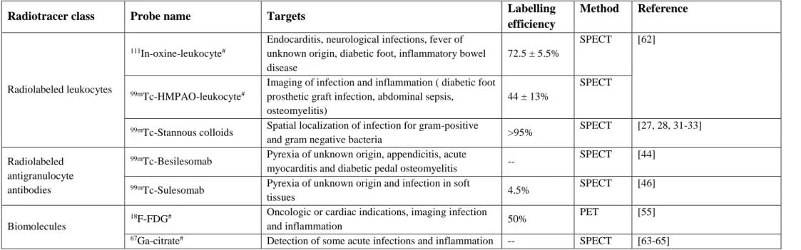
Table 1 Commercially available probes for infection imaging
#) FDA approved
Radiotracer class Probe name Targets Labelling
efficiency
Method Reference
Radiolabeled leukocytes
111In-oxine-leukocyte#
Endocarditis, neurological infections, fever of unknown origin, diabetic foot, inflammatory bowel disease
72.5 ± 5.5%
SPECT [62]
99mTc-HMPAO-leukocyte#
Imaging of infection and inflammation ( diabetic foot prosthetic graft infection, abdominal sepsis,
osteomyelitis)
44 ± 13%
SPECT
99mTc-Stannous colloids Spatial localization of infection for gram-positive
and gram negative bacteria >95% SPECT [27, 28, 31-33]
Radiolabeled antigranulocyte antibodies
99mTc-Besilesomab Pyrexia of unknown origin, appendicitis, acute
myocarditis and diabetic pedal osteomyelitis -- SPECT [44]
99mTc-Sulesomab Pyrexia of unknown origin and infection in soft
tissues 4.5% SPECT [46]
Biomolecules
18F-FDG# Oncologic or cardiac indications, imaging infection
and inflammation 50% PET [55]
67Ga-citrate# Detection of some acute infections and inflammation -- SPECT [63-65]
3. Novel imaging probes for direct, more specific imaging of bacterial infection Compounds having affinity towards a specific pathogen can be utilized for its direct in vivo imaging after being radiolabeled with a compatible radioisotope. In 2016, Auletta et al.
systematically discussed the potential of quinolones, cephalosporins and siderophores as promising imaging agents for infection [66]. In a 2015 review, Tsopelos elaborated on compounds bearing potential towards scintigraphic detection of infection and inflammation [5].
Moreover, a few promising tracers show affinity towards bacteria such as 11In-DPC11870, 11In- DTPA-IgG(14C), 111In/99mTc -DTPA-hpc-IgG, 18F-DPA-714, 99mTc-PEG-liposomes, 99mTc-HAS, labelled fMLFKs and labelled interleukins was explained by Tsopelos, however, these molecules fall out of the scope of this review. Furthermore, 99mTc labelled bacteriophages were also omitted from this discussion. The following text highlights selected compound clusters that bear the largest potential to accomplish bacteria-selective tomographic imaging: i) antimicrobial peptides and ii) biomimetics.
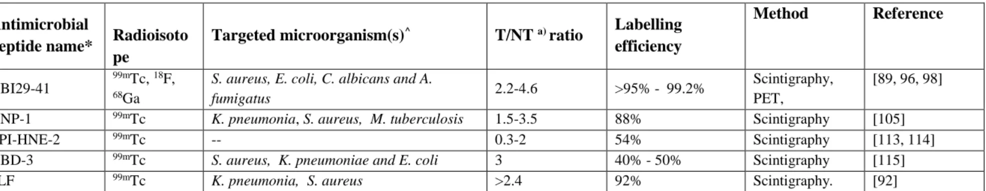
3.1. Antimicrobial peptides as bacteria-selective imaging probes
Peptides are short chains of amino acids linked by amide bonds. These naturally occurring biomolecules are logical options for development as possible targeting vectors for PET tracers as they have unique characteristics such as; high target specificity and binding ability, low toxicity and immunogenicity and easy scale up production in the laboratory [67, 68]. Generally, peptides containing less than 50 amino acids are considered for imaging tracers due to their molecular weight. These peptides are relatively small in size, facilitates rapid accumulation at the target site as well as lead to faster clearance from the recipient which would make them desirable candidates for PET molecular imaging probes [67].
In general, naturally occurring peptides are most suited for designing peptide-based PET tracers;
as they play a vital role in certain physiological conditions by mediating via their high-affinity, specific and massively overexpressed receptors [69]. However, various plasma-containing proteases and peptidases lead to rapid degradation of some of these compounds resulting in a shorter pharmacological half-life and decreased target availability. Thus, to increase the in vivo stability of naturally occurring peptides, these compounds require molecular engineering at the amino acid residues that are involved in degradation without compromising the most desired biological activity. Till date, a number of researchers have described various approaches to modify amino acid residues to increase the efficiency and stability of peptides. These approaches include peptide bond substitution, N- and C-terminus acetylation, side-chain and unnatural amino acid introduction, suitable D-amino acid incorporation and amino alcohol utilization. In addition,