Synthesis and Characterisation of Gold-Based Compounds as Potential DNA Intercalators
Submitted in fulfilment of the requirement for the degree of
Master of Science
By
Desigan Moodley
BSc, (Hons.) (UKZN)
November 2016
School of Chemistry and Physics, University of KwaZulu-Natal
Pietermaritzburg
2
Declaration
I, Desigan Moodley, declare that:
(i) The research reported in this thesis/dissertation, except where otherwise indicated, is my original research.
(ii) This thesis/dissertation has not been submitted for any degree or examination at any other university.
(iii) This thesis/dissertation does not contain other persons’ data, pictures, graphs or other information, unless specifically acknowledged as being sourced from other persons.
(iv) This thesis/dissertation does not contain other persons’ writing, unless specifically acknowledged as being sourced from other researchers. Where other written sources have been quoted, then:
a) Their words have been re-written but the general information attributed to them has been referenced;
b) Where their exact words have been used, their writing has been placed inside quotation marks, and referenced.
(v) Where I have reproduced a publication of which I am author, co-author or editor, I have indicated in detail which part of the publication was actually written by myself alone and have fully referenced such publications.
(vi) This dissertation/thesis does not contain text, graphics or tables copied and pasted from the internet, unless specifically acknowledged, and the source being detailed in the dissertation/thesis and in the References sections.
Signed: ____________ Date: ___________
Desigan Moodley
I hereby certify that this statement is correct to the best of my knowledge:
Signed: ____________ Date: ___________
Dr. Matthew P. Akerman (Supervisor)
3
Acknowledgments
Firstly, I would like to thank Dr. M. Akerman and Prof. O.Q. Munro for all the support, patience and motivation they have provided during the course of this project. Their advice has been essential towards the success of this project. I would especially like to thank Dr. M. Akerman for the time taken from his busy days to mentor me.
I would like to thank my girlfriend for her constant support, motivation and inspiration not just for this project but in everything I do. I will always be grateful to you.
I wish to thank my family and friends for their support and encouragement.
Lastly, I am thankful to N.R.F for the financial support.
4
Research Outputs
The results from this project have been presented as an oral present ation and poster presentation at the following conferences or colloquia.
Title:Gold(III) DNA Intercalators: Targeted Chemotherapeutic Agents.
1. SACI Honours chemistry colloquium 2013.
2. UKZN Research Day 2015.
3. Indaba 8 chemistry conference 2015.
5
List of abbreviations
%Diff. percentage difference
Å angstrom
° degrees
A absorbance
a lattice constant
aq aqueous
BC before Christ
br broad
Calc. calculated
COSY correlation spectroscopy
d doublet
dd doublet of doublets
DNA deoxyribonucleic acid
FTIR fourier transform infra-red
gDNA genomic deoxyribonucleic acid
g gram/ gas
GIAO Gauge-Including Atomic Orbitals
HOMO highest occupied molecular orbital
HSQC heteronuclear single-quantum correlation
spectroscopy
IC50 inhibitory concentration 50%
IR infra-red
J coupling constant
LUMO lowest unoccupied molecular orbital
m multiplet/ medium intensity
MHz megahertz
m/z mass to charge ratio
NMR nuclear magnetic resonance
ppm parts per million
s singlet/ strong
t triplet
TD-DFT time-dependent density functional theory
UV ultraviolet
U.S.A United States of America
vis visible
6
List of figures and schemes
Figure/scheme name
Figure/scheme title Page
number Chapter 1
Figure 1.3.1 Capecitabine (Xeloda), an antimetabolite chemotherapeutic agent.
26
Figure 1.3.2 Cyclophosphamide, an example of an alkylating agent. 27 Figure 1.3.3 Schematic structure of 9-aminoacridine, an example of a
DNA binder.
27
Figure 1.3.4 Structure of topotecan an example of a cytostatic drug. 28 Figure 1.4.1 The three modes of binding for inorganic compounds with
DNA
28
Figure 1.4.2 Structure of Proflavine, a classic intercalator. The fused aromatic ring structure is ubiquitous in DNA intercalators.
29
Figure 1.4.3 Structure of tetraaza macrocyclic complexes, an example of a metallo-intercalator
30
Figure 1.5.1 Structure of cisplatin. 31
Figure 1.5.2 Structure of cisplatin derivatives 32
Figure 1.6.1 Structure of E. coli topoisomerase 33
Figure 1.7.1 Comparison of the structures 34
Figure 1.8.1 Early gold(I)-based drugs for treatment of rheumatoid arthritis
35
Figure 1.8.2 Structures of (AuCl3(Hpm)), AuCl2(pm) and (Au-azpy bidentate
36
Figure1.8.3 Structure of gold(III) porphyrins. 37
Figure 1.8.4 Structure of gold(III) Schiff base complex. 37 Figure 1.8.5 Structure of gold(III) bis(pyridyl)carboxamide. 37 Figure 1.9.1 Scheme for a general Schiff base synthesis. 38 Figure 1.9.2 Structure of Imidazole derived Schiff bases with a zinc(II)
metal centre
39
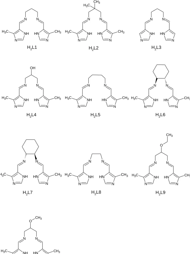
Figure 1.10.1 Structures and abbreviated names of the bis(imidazole- imine) Schiff base ligands proposed for chelation to gold(III).
41
7
Chapter 2Figure 2.3.1 Structure and atom numbering scheme of 2,2,12,12- tetramethyl-3,11-dioxo-4,10-dioxa-5,9-diazatridecan-7-ol.
46
Figure 2.3.2 Structure and atom numbering scheme of 2- ethoxypropane-1,3-diaminium dichloride.
47
Figure 2.3.3 Structure and atom numbering scheme of 2- methoxypropane-1,3-diaminium dichloride.
48
Figure 2.4.1 Structure and atom numbering scheme used for H2L1. 49 Figure 2.4.2 Structure and atom numbering scheme used for H2L2. 50 Figure 2.4.3 Structure and atom numbering scheme used for H2L3. 51 Figure 2.4.4 Structure and atom numbering scheme used for H2L4. 52 Figure 2.4.5 Structure and atom numbering scheme used for H2L5. 53 Figure 2.4.6 Structure and atom numbering scheme used for H2L6. 54 Figure 2.4.7 Structure and atom numbering scheme used for H2L7. 55 Figure 2.4.8 Structure and atom numbering scheme used for H2L8. 56 Figure 2.4.9 Structure and atom numbering scheme used for H2L9. 57 Figure 2.4.10 Structure and atom numbering scheme used for H2L10. 58 Figure 2.5.1 Structure and atom numbering scheme used for
[Au(L1)][PF6]
59
Figure 2.5.2 Structure and atom numbering scheme used for [Au(L2)][PF6]
60
Figure 2.5.3 Structure and atom numbering scheme used for [Au(L3)][PF6]
61
Figure 2.5.4 Structure and atom numbering scheme used for [Au(L4)][PF6]
62
Figure 2.5.5 Structure and atom numbering scheme used for [Au(L5)][PF6]
63
Figure 2.5.6 Structure and atom numbering scheme used for [Au(L6)][PF6]
64
Figure 2.5.7 Structure and atom numbering scheme used for [Au(L7)][PF6]
65
Figure 2.5.8 Structure and atom numbering scheme used for [Au(L8)][PF6]
66
Figure 2.5.9 Structure and atom numbering scheme used for [Au(L9)][PF6]
67
8
Figure 2.5.10 Structure and atom numbering scheme used for[Au(L10)][PF6]
68
Chapter 3
Scheme 3.1 General mechanism for a Schiff base reaction 69 Scheme 3.2.1 Condensation reaction for the synthesis of H2L1-H2L8. 70 Scheme 3.2.2 The protection of primary amines using di-tert-butyl
dicarbonate.
71
Scheme 3.2.3 The different resonance forms of the protected diamine. 71 Scheme 3.2.4 SN2 type reaction of the hydroxyl group to form an ether. 72 Scheme 3.2.5 Deprotection reaction to form the hydrochloride salt. 72 Scheme: 3.3.1 General reaction outline for metalation of ligands H2L1,
H2L2 and H2L10.
74
Scheme 3.3.2 General reaction outline for metalation of ligands H2L4 and H2L9.
75
Chapter 4
Figure 4.1.1 IR spectrum of ligand H2L2. 80
Figure 4.1.2 IR spectrum of complex [Au(L2)](PF6). 80 Figure 4.2.1 Diagram showing possible electronic transitions 81 Figure 4.2.2 Electronic spectrum of H2L2 in methanol showing a
π-π* transition. An n-π * transition is evident as a shoulder at approximately 306 nm.
82
Figure 4.2.3 Electronic spectrum of [Au(L2)](PF6) in DMSO showing an LMCT and π-π* transition.
83
Figure 4.3.1 1H NMR spectrum of the free ligand H2L9. 86 Figure 4.3.2 1H NMR spectrum of the complex [Au(L9)](PF6). 86 Figure 4.3.3 13C NMR spectrum of free ligand H2L9. 87 Figure 4.3.4 13C NMR spectrum of complex [Au(L9)](PF6). 87 Figure 4.3.5 19F NMR spectrum of complex [Au(L10)](PF6). 89 Figure 4.3.6 31P NMR spectrum of complex [Au(L10)](PF6). 90 Figure 4.4.1 Absorption spectrum of [Au(L1)](PF6) with increasing
concentrations of ctDNA.
94
Figure 4.4.3 Proposed structure for gold(III) chelates that would likely improve DNA intercalation.
95
9
Chapter 5Figure 5.1.1 Partially labelled X-ray structures of (a) DAZXOS (b) XUVQAG (c) EFAROU (d)WOBNAD.
98
Figure 5.1.2 One-dimensional network of DAZXOS stabilised by hydrogen bonds between the imine N atoms and the imidazole NH atoms of adjacent molecules in the solid state
99
Figure 5.1.3 Structure of XUVQAG stabilised by hydrogen bonds between the water molecule and imine N atoms and the pyrrole NH groups of adjacent molecules in the solid state.
100
Figure 5.1.4 Heterotetrameric structure of EFAROU stabilised by hydrogen bonds between the water molecules and imine N atoms and pyrrole NH groups.
101
Figure 5.1.5 Two-dimensional hydrogen-bonded network structure of WOBNAD stabilised by hydrogen bonds between the imidazole N atoms and the imidazole NH atoms of adjacent molecules in the solid state.
102
Figure 5.1.6. Partially labeled structure of RIZHAL, RIZHEP, RIZHIT RAVHIG, BAQHEG, MAYQIM, MAYQOS, MAYQUY, BAGHAT, KURQAR, GASQEW.
105
Figure 5.1.7 One dimensional network stabilised by C-H···F interactions in RIZHAL.
107
Figure 5.1.8 One dimensional network stabilised by C-H···O interactions in RIZHEP.
108
Figure 5.1.9 Head-to-tail π-stacked dimers of RIZHIT. 108 Figure 5.1.10 Out-of-plane distortion of the Zn(II) ion from the four-atom
mean plane defined by the imidazole and imine nitrogen atoms for BAQHEG.
109
Figure 5.1.11 A two dimensional network is stabilised by four unique hydrogen bonds for BAQHEG.
110
Figure 5.1.12 Deviation of Mn(II) from planarity as defined by the coordinating nitrogen atoms for MAYQIM.
110
Figure 5.1.13 Crystal structure of MAYQOS showing the nominally octahedral coordination geometry.
111
10
Figure 5.1.14 Deviation of Cu(II) from planarity as defined bycoordinating nitrogen atoms for MAYQUY.
112
Figure 5.1.15 Deviation of Cu(II) from planarity as defined by the N4
donor atoms for BAGHAT.
113
Figure 5.1.16 Deviation of Cu(II) from planarity as defined by the imidazole rings for KURQAR.
114
Figure 5.1.17 Two-dimensional supramolecular structure of KURQAR viewed down the c-axis.
115
Figure 5.1.18 Crystal structure of GASQEW illustrating a distorted square-pyramidal coordination.
116
Figure 5.1.19 Metal chelates of GASQEW are bridged by hydrogen bonding through the nitrate counter ions and imidazole NH groups. This leads to a hydrogen-bonded heterotetramer.
116
Figure 5.3.1 Thermal ellipsoid plot of H2L6, shown at 50% probability level. All hydrogen atoms are represented as spheres arbitrary radius. A single molecule from the asymmetric unit is shown.
120
Figure 5.3.2 Solid state structure of H2L7 illustrating the hydrogen- bonds ed dimer bridged by a water molecule. The dimers are linked by additional hydrogen bonding to form a three- dimensional supramolecular structure
122
Figure 5.3.3 Cross-linking of adjacent dimers through hydrogen bonding between the imidazole NH and imidazole N atoms.
The dimers (as illustrated in Figure 5.3.2) are shown in yellow.
123
Figure 5.3.4 Cross-linked columnar structure of H2L6 the result of a combination of hydrogen bonding and four-fold lattice symmetry. The structure is viewed down the c-axis.
123
Figure 5.3.5: A thermal displacement plot of H2L7 shown at 50%
probability level. All hydrogen atoms are represented as spheres of arbitrary radius.
125
Figure 5.3.6 Solid state structure of H2L7 displaying dimeric structure stabilised by hydrogen bonds.
127
11
Figure 5.3.7 Cross-linking of adjacent dimers through hydrogenbetween the imidazole NH and imidazole N atoms. The dimers (as illustrated in Figure 5.3.6) are shown in yellow.
This hydrogn bonding motif leads to one dimensional columns.
128
Figure 5.3.8 Columnar structure of H2L7 viewed down the c-axis. The structure is stabilised by extensive hydrogen bonding.
129
Figure 5.4.1 A labelled thermal displacement plot of [Au(L1)][PF6], shown at 50% probability level. All hydrogen atoms are represented as spheres of arbitrary radius.
130
Figure 5.4.2 Au···N interactions of [Au(L1)](PF6) measuring 2.811(3) Å link adjacent molecules to form a one-dimensional chain co-linear with the c-axis.
132
Figure 5.4.3 A thermal ellipsoid plot of [Au(L2)][PF6]. It has been rendered at the 50% probability level. All hydrogen atoms are represented as spheres with indiscriminate radii.
133
Figure 5.4.4 Au···N interactions of [Au(L2)](PF6) with an interaction distance measuring 3.21(1) Å.
134
Figure 5.4.5 A thermal ellipsoid plot of [Au(L4)]Cl shown at 50%
probability level. All hydrogen atoms are represented as spheres with indiscriminate radius.
135
Chapter 6
Figure 6.1.1 The general structure of the pyrrolide-imine Schiff base ligands previously studied by DFT methods.
139
Figure 6.1.2 The structure of the bis(pyrrolide-Imine) gold(III) chelate previously studied by DFT studies.
140
Figure 6.3.1 RMSD fit for the DFT calculated and X-ray structures for [Au(L1)]+, [Au(L2)]+ and [Au(L4)]+.
141
Figure 6.3.2 DFT calculated structures for [Au(L9)]+, [Au(L10)]+. 142 Figure 6.3.3 Overlay of the experimental and DFT calculated frequency
data of [Au(L2)][PF6] and [Au(L2)]+.
144
Figure 6.3.4 Plot of vibrational frequencies of [Au(L1)][PF6] versus [Au(L1)]+.
145
Figure 6.3.5 Structure and labelling scheme of [Au(L2)]+. 146
12
Figure 6.3.6 Superposition of calculated and experimental UV/visiblespectra for [Au(L2)]+. The calculated spectra have been scaled.
148
Figure 6.3.7 The electronic orbitals of [Au(L2)]+ involved in most transitions.
149
Figure 6.3.8 Least-squares fit for the DFT calculated and X-ray structures for (a) H2L6 and (b) H2L7. The blue structure represents the X-ray structure and the yellow represents the DFT calculated structure.
151
Figure 6.3.9 Overlay of the experimental and calculated IR spectrum of H2L2.
153
Figure 6.3.10 Plot of calculated versus experimental frequencies for H2L2.
154
Figure 6.3.11 Superposition of calculated and experimental UV/visible spectra for H2L2. The calculated spectra have been scaled.
155
Figure 6.3.12 Superposition of DFT-calculated and experimental UV/visible spectra for H2L6. The calculated spectra have been scaled.
157
Figure 6.3.13 Shows that all the molecular orbitals involved in the transitions of H2L6 were of π-symmetry. The assignment of the primary absorption band as π-π* is therefore appropriate.
158
Chapter 7
Figure 7.1.1 Structures and abbreviated names of the gold(III) chelates synthesized in this work.
161
Figure 7.2.2 Proposed structure of the chelate with biotin attached. 165
13
List of tables
Table name Table title Page
number Chapter 4
Table 4.1.1 Imine stretching frequencies of the free ligands and gold(III) chelates.
78
Table 4.1.2 Imdazole NH stretching frequencies for the free ligands. 79 Table 4.2.1 Extinction Coefficients for the free ligands and gold(III)
chelates.
80
Table 4.3.1 A summary of the chemical shifts of the 1H NMR for H2L9 and [Au(L9)](PF6).
84
Table 4.3.2 A summary of the chemical shifts of the 13C NMR spectra for H2L9 and [Au(L9)](PF6).
88
Table 5.4.3 Bond lengths and angles for [Au(L4)][PF6]. 88
Chapter 5
Table 5.1.1 Reported X-ray structures of similar bis(imidazole- imine)ligands
97
Table 5.1.2 Summarised bond lengths for XUVQAG. 99
Table 5.1.3 Hydrogen bond lengths (Å) and bond angles (˚) of XUVQAG. 100 Table 5.1.4 Hydrogen bond parameters (Å,˚) for EFAROU. 101 Table 5.1.5 Hydrogen bond parameters (Å,˚) for WOBNAD. 102 Table 5.1.6 Reported X-ray structures of related gold(III) chelates. 103 Table 5.1.7 Summary of bond lengths and bond angles describing the
coordination sphere of RIZHAL.
106
Table 5.1.8 Bond parameters for the C-H···F interactions (Å,˚) for RIZHAL. 106 Table 5.1.9 Bond parameters for the C-H···O interactions (Å,˚) for
RIZHEP.
107
Table 5.1.10 Hydrogen bond parameters (Å,˚) for BAQHEG. 109 Table 5.1.11 Hydrogen bond parameters stabilising the two-dimensional
network of KURQAR.
115
Table 5.1.12 Hydrogen bond parameters (Å,˚) for GASQEW. 116
14
Table 5.2.1 Crystal refinement data for ligands. 118
Table 5.2.2 Crystal refinement data for chelates. 119
Table 5.3.1 Key bond lengths and bond angles for H2L6. 121 Table 5.3.2 Hydrogen bond lengths (Å) and bond angles (˚) of H2L6. 128 Table 5.3.3 Bond lengths and bond angles describing the imidazole ring
and imine moieties of H2L7.
126
Table 5.3.4 Hydrogen bond lengths (Å) and bond angles (˚) of H2L7. 127 Table 5.4.1 Bond lengths and bond angles describing the coordination
sphere of [Au(L1)][PF6].
131
Table 5.4.2 Bond lengths and bond angles describing the coordination sphere of [Au(L2)][PF6].
134
Table 5.4.3 Bond lengths and angles for [Au(L4)][PF6]. 136
Chapter 6
Table 6.3.1 Summary of DFT and experimental bond lengths, angles and torsion angles of [Au(L2)][PF6].
143
Table 6.3.2 Summary of calculated and experimental frequencies for [Au(L2)]+.
145
Table 6.3.3 Summary of the calculated and experimental chemical shifts of 1H NMR for [Au(L2)]+.
147
Table 6.3.4 Summary of the calculated ad experimental chemical shifts of
13C NMR for [Au(L2)]+.
147
Table 6.3.5 Summary of the DFT calculated electronic transitions for [Au(L2)]+.
150
Table 6.3.6 Summary of DFT and experimental bond lengths, angles and torsion angles of H2L6 and H2L7.
152
Table 6.3.7 Summary of the calculated ad experimental chemical shifts of
1H NMR for H2L2.
155
Table 6.3.8 Summary of the calculated ad experimental chemical shifts of
13C NMR for H2L2.
156
Table 6.3.8 Summary of the DFT calculated electronic transitions for H2L2.
159
15
Abstract
Five novel bis(imidazole-imine) gold(III) chelates were synthesized and characterized by NMR (
1H,
13C, COSY, HSQC,
19F and
31P), FTIR and UV/visible spectroscopy as well as high resolution mass spectrometry and in some cases X-ray crystallography. The ligands comprised methyl imidazole moieties bridged by a synthetically variable di(azomethine) linkage. Five of the ten synthesised ligands were successfully chelated to gold(III), these ligands contained propyl, 2,2-dimethyl, 2-hydroxy, 2-methoxy-propyl and 2-ethoxy-propyl linkages. The ligands were synthesised by the condensation of two equivalents of 4-methyl-5-imidazolecarbaldehyde and the respective diamines. The chelate synthesis was achieved with direct metalation using [Bu
4N][AuCl
4].
Solid state structures were determined by single crystal X-Ray crystallography for two ligands and three gold(III) chelates. The ligands were found to form stable hydrogen-bonded networks with the water molecule. The water molecules are found in the centre of hydrogen-bonded columns forming hydrogen bonds with the surrounding ligand molecules. The anti-configuration of the ligands is required for formation of the polymers. The hydrogen-bonded columns are co-linear with the c-axis. The three- dimensional structure is a result of a cross-linking the columns through additional hydrogen bonds. The gold(III) chelates all had a square planar coordination geometry.
This is the regular geometry of d
8metal ion chelates. Metal ion chelation occurs with concomitant deprotonation of the imidazole NH groups; this is in contrast to previously reported metal chelates of the related ligands. The deprotonation of the imidazole NH yields a dianionic tetradentate N-donor ligand. The average Au-N
iminebond measures 2.007 Å and the average Au-N
imidazolebond measures 1.993 Å. The coordination geometry differed slightly from the ideal square planar geometry with an acute bond angle of 80.73˚ (N
imine-Au-N
imidazole). This bond angle is constrained by the bite angle of the ligands and resulting 5-membered chelation ring. Au···π and π···π interactions were present in the chelates, stabilising various supramolecular structures.
The chelates were designed to exploit the square planar nature of the gold(III) ion to
produce metal aromatic metal chelates suitable for DNA intercalation to control the
proliferation of tumour cells. Both direct DNA binding titrations and competitive DNA
binding studies did not show any significant affinity towards DNA. The likely reason for
16 this is that the methyl groups on the imidazole rings, which are required to stabilise the metal ion through their inductive effect, prevent the intercalation process. This class of compounds therefore needs further development to achieve the final goal of chemotherapeutic agent.
Density Functional Theory (DFT) was used to simulate various
propertiesof the ligands
and gold(III) chelates. The DFT calculated data was compared to the experimental data
to determine the efficiency of the chosen basis set and hybrid functional used. DFT was
also used to deconvolute the experimental data obtained, with particular emphasis on
the electronic spectra. The level of theory used for ligands was B3LYP/6-311G and for
the gold(III) chelates PBE1PBE/LanL2DZ. DFT was used to calculate the optimized
geometry, NMR and electronic spectra. The vibrational frequencies for both ligands and
chelates showed no negative Eigen values which implies that the optimized geometry is
indeed the true minimum on the global potential energy surface.
17
Table of Contents
Declaration ... 2
Acknowledgments ... 3
Research Outputs ... 4
List of abbreviations... 5
List of figures and schemes ... 6
List of tables ... 13
Abstract ... 15
Chapter 1| Introduction ... 23
1.1 Preface ... 23
1.2 Causes of cancer ... 24
1.3 Treatment of cancer ... 25
1.3.1 Cytotoxic chemotherapeutic agents ... 26
1.3.2 Cytostatic chemotherapeutic agents ... 27
1.4 DNA Intercalating agents ... 28
1.4.1 Organic DNA Intercalators ... 29
1.4.2 Inorganic DNA Intercalators ... 29
1.5 Cisplatin ... 30
1.5.1 Mechanism of action of cisplatin ... 31
1.6 DNA topoisomerase I ... 32
1.7 DNA topoisomerase I inhibitors ... 33
1.8 Gold in medicine ... 34
1.9 Schiff bases ... 38
1.10 Proposed Research ... 39
1.11 Objectives ... 42
1.12 References ... 43
18
Chapter 2| Experimental ... 45
2.1 General Methods ... 45
2.2 Instrumentation ... 45
2.3 Synthesis of Ligand Precursors ... 46
2.3.1 Synthesis of 2,2,12,12-tetramethyl-3,11-dioxo-4,10-dioxa-5,9-diazatridecan-7-ol ... 46
2.4 Synthesis of Ligands ... 49
2.4.1 Synthesis of N,N'-bis[(E)-(4-methyl-1H-imidazol-5-yl) methylidene]propane- 1,3-diamine (H
2L1)... 49
2.4.2 Synthesis of 2,2-dimethyl-N,N'-bis[(E)-(4-methyl-1H-imidazol-5-yl) methylidene] propane- 1,3-diamine (H
2L2) ... 50
2.4.3 Synthesis of N,N'-bis[(E)-1H-imidazol-5-ylmethylidene]propane- 1,3- diamine (H
2L3) ... 51
2.4.4 Synthesis of N,N'-bis[(E)-1H-imidazol-5-ylmethylidene]propane-1,3- diamine (H
2L4) ... 52
2.4.5 Synthesis of N,N'-bis[(1E)-(4-methyl-1H-imidazol-5-yl)methylene]butane- 1,4-diamine (H
2L5)... 53
2.4.6 Synthesis of (1S,2S)-N,N'-bis[(E)-(4-methyl-1H-imidazol-5-yl) methylidene]cyclohexane-1,2-diamine (H
2L6) ... 54
2.4.7 Synthesis of (1R,2R)-N,N'- bis[(E)- (4-methyl-1H-imidazol-5-yl) methylidene]cyclohexane-1,2-diamine (H
2L7) ... 55
2.4.8 Synthesis of N,N'-bis[(1E)-(4-methyl-1H-imidazol-5-yl)methylene]ethane- 1,2-diamine (H
2L8)... 56
2.4.9 Synthesis of 2-ethoxy-N,N'-bis[(1E)-(4-methyl-1H-imidazol-5- yl)methylene]propane- 1,3-diamine (H
2L9) ... 57
2.4.10 Synthesis of 2-methoxy-N,N'-bis[(1E)-(4-methyl-1H-imidazol-5-
yl)methylene]propane-1,3-diamine (H
2L10) ... 58
19 2.5 Synthesis of Complexes ... 59
2.5.1 Synthesis of 5,5'-{propane-1,3-diylbis [nitrilo(E) methylylidene]} bis(4-
methylimidazol-1-ide) gold(III) hexafluorophosphate(V) [Au(L1)](PF
6) ... 59 2.5.2 Synthesis of 5,5'-{(2,2-dimethylpropane-1,3-diyl)bis[nitrilo(E)
methylylidene]}bis(4-methylimidazol-1-ide)gold(III) hexafluorophosphate(V)
[Au(L2)](PF6) ... 60 2.5.3 Attempted synthesis of N,N'-bis[(E)-1H-imidazol-5-yl methylidene]propane-1,3- diamine gold(III) hexafluorophosphate(V) [Au(L3)](PF
6) ... 61 2.5.4 Synthesis of 5,5'-{(2-hydroxypropane-1,3-
diyl)bis[nitrilo(E)methylylidene]}bis(4-methylimidazol-1-ide) gold(III)
hexafluorophosphate(V) [Au(L4)](PF
6) ... 62 2.5.5 Attempted synthesis of 5,5'-{butane-1,4-diylbis[nitrilo(E)methylylidene]}bis(4- methylimidazol-1-ide) gold(III) hexafluorophosphate(V) [Au(L5)](PF
6) ... 63 2.5.6 Attempted Synthesis of 5,5'-{(1S,2S)-cyclohexane-1,2-
diylbis[nitrilo(E)methylylidene]}bis(4-methylimidazol-1-ide) gold(III)
hexafluorophosphate(V)[Au(L6)](PF
6) ... 64 2.5.7 Attempted synthesis of 5,5'-{(1R,2R)-cyclohexane-1,2-
diylbis[nitrilo(E)methylylidene]}bis(4-methylimidazol-1-ide) gold(III)
hexafluorophosphate(V) [Au(L4)](PF
6) ... 65 2.5.8 Attempted synthesis of 5,5'-{(2,2-dimethylethane-1,3-diyl)bis[nitrilo(E)
methylylidene]}bis(4-methylimidazol-1-ide)gold(III) hexafluorophosphate(V)
[Au(L8)](PF
6) ... 6666 2.5.9 Synthesis of 5,5'-{(2-ethoxypropane-1,3-
diyl)bis[nitrilo(E)methylylidene]}bis(4-methylimidazol-1-ide) gold(III)
hexafluorophosphate(V) [Au(L9)]Cl ... 67 2.5.10 Synthesis 5,5'-{(2-methoxypropane-1,3-
diyl)bis[nitrilo(E)methylylidene]}bis(4-methylimidazol-1-ide) gold(III)
hexafluorophosphate(V) [Au(L10)](PF
6)] ... 68
20
Chapter 3| Synthesis ... 69
3.1 Introduction ... 69
3.2 Synthesis of the Schiff Base Ligand ... 70
3.2.1 Synthesis of H2L1- H2L8 ... 70
3.2.2 Synthesis of H
2L9 and H
2L10 ... 71
3.3 Metallation of Schiff Base Ligands ... 73
3.3.1 Gold(III) salts ... 73
3.3.2 Metallation of H
2L1, H
2L2 and H
2L10 ... 73
3.3.3 Metallation of H
2L4 and H
2L9 ... 74
3.3.4 Metallation of H
2L3, H
2L5, H
2L6, H
2L7 and H
2L8 ... 75
3.4 References... 76
Chapter 4| Spectroscopy ... 77
4.1 Infrared spectroscopy ... 77
4.1.1 Introduction ... 77
4.1.2 Results and discussion ... 77
4.2 UV/visible spectroscopy ... 81
4.2.1 Introduction ... 81
4.2.2 Results and discussion ... 82
4.3 NMR spectroscopy ... 85
4.3.1 Introduction ... 85
4.3.2 Results and discussion ... 86
4.4 DNA binding studies ... 90
4.4.1 Introduction ... 90
4.4.2 Experimental ... 91
4.4.3 Results and discussion ... 94
4.5 References... 96
21
Chapter 5| X-Ray Crystallography ... 97
5.1 Introduction ... 97
5.1.1 Previously reported ligands ... 97
5.1.2 Previously reported metal chelates ... 102
5.1.3 Objectives ... 117
5.2 Experimental ... 117
5.3 X-Ray Crystallography of Ligands ... 120
5.3.1 X-Ray structural analysis of H
2L6 ... 120
5.3.2 X-Ray structural analysis of H
2L7 ... 125
5.4 X-Ray Crystallography of Complexes... 130
5.4.1 X-Ray structural analysis of [Au(L1)][PF
6] ... 130
5.3.2 X-Ray structural analysis of [Au(L2)][PF
6] ... 133
5.3.3 X-Ray structural analysis of [Au(L4)][PF
6] ... 135
5.4 Conclusions ... 136
5.5 References... 137
Chapter 6| Computational Studies ... 138
6.1.1 Introduction ... 138
6.1.2 Previous computational studies on related compounds ... 139
6.2 Computational method ... 141
6.3 Results and discussion ... 141
6.3.1 Gold(III) chelates ... 141
6.3.2 Ligands ... 151
6.4 Conclusion... 159
6.5 References... 160
Chapter 7| Conclusions and Future Work ... 161
7.1 Conclusion... 161
22
7.2 Future Work ... 163
7.3 References... 166
23
Chapter 1| Introduction 1.1 Preface
Cancer has been well documented in animals and humans alike throughout history. The earliest evidence of cancer comes from ancient Egypt where bone cancer was discovered in mummified human remains [1]. Cancer has now been dated to 3000 B.C., yet this disease from biblical times still greatly affects the modern population. In fact, it is the second leading cause of death in the United States of America.
Cancer is a group of diseases defined as the uncontrollable and rapid growth of cells in the body.
There are many forms of cancer such as bladder, breast and prostate cancers. Statistics obtained from the National Cancer Institute (NCI, U.S.A.) clearly show a decrease in the number of deaths from 1975 until 2010. It can safely be assumed that the decrease in mortality is due to the advancement in anti-cancer drugs and chemotherapy. Statistics for the African continent still show a high mortality rate. This is due to lack of early detection and limited access to treatment [2]. Although statistics based on the U.S.A. population show a decrease in mortality rates, there is still a need to continue research into new anti-cancer agents as there are problems associated with current treatments. These include unpleasant side-effects and drug resistance in secondary tumours [3].
It is hypothesised that by including metal ions in the anti-cancer agents it will potentially alter their mechanism of action and helps overcome the current issues associated with chemotherapy. Cisplatin was the first anti-cancer drug to be used that had a metal centre.
Although it shows good efficacy, it has several harmful side-effects and tumours can become resistant to the drug. These drawbacks have prompted the search for alternative metal-based anti-cancer drugs [4].
The medicinal properties of gold were first discovered in the 1890s when it was found that K[Au(CN)2] could kill the bacteria responsible for tuberculosis. It was later found that gold(I) thiolate complexes could be used in the treatment of rheumatoid arthritis. Gold(III) complexes have until recently not been as intensely studied as those of gold(I) because gold(III) complexes are highly reactive, have a high redox potential and poor stability. These properties meant that gold(III) would be reduced to gold(I) or metallic gold under physiological conditions. Recently, more stable gold(III) complexes have been synthesised using strong sigma-donor ligands to stabilise the gold(III) ion. Gold(III) is isoelectronic with platinum(II) i.e. they have a d8
24
configuration which leads to a square-planar geometry and is therefore an ideal scaffold around which ligands with DNA recognition elements can be assembled. These factors have allowed the applications of gold(III) to expand into medicinal chemistry [4][5].The aim of this project is to synthesise novel bis(imidazole-imine) gold(III) anti-cancer agents that will intercalate DNA thus preventing DNA replication and tumour cell growth. The complexes have been designed to include a planar aromatic region, a hydrogen bonding region and carry a positive charge. They are therefore anticipated to be effective DNA intercalators and chemotherapeutic agents.
1.2 Causes of cancer
Cancer does not have one main cause, but is rather caused by a combination of several factors.
These can include diet, pollution, alcohol, viral infections, geophysical factors, occupational factors, tobacco and genetic factors. These external stimuli lead to a change in the arrangement of genes, therefore causing a mutation to occur. Cancer cells arise when critical genes are mutated; these mutations lead to the unregulated proliferation of cells forming tumours [6][7].
There are many forms of cancer and these grow and divide at different rates. The most common form of cancer among men is prostate cancer, the most common form of cancer among women is breast cancer and the most common form of preventable cancer in both sexes is lung cancer [3].
There are many mutations that could lead to cancer, but the most common are defective START checkpoint (vida infra), apoptosis malfunction and oncogenes. The cell cycle has three periods:
cell growth, DNA synthesis and cell division. The length of each period is controlled by chemical signals. If the signals malfunction the cell can become cancerous. There are four phases in the cell cycle: G1, synthesis, G2 and mitosis. Translation into each phase is regulated by checkpoints.
Each checkpoint halts the progression through the cycle until important processes such as DNA replication are complete or damaged cells are repaired. Two proteins are important to the functioning of these checkpoints i.e. cyclin and cyclindependantkinases (CDK). CDK regulates the activity of other proteins and cyclin enables CDK to function. One of the most important checkpoints is the START checkpoint which is found in mid G1. The START checkpoint determines if it is appropriate to move from the G1 phase to the synthesis phase. If the cell is driven past the START checkpoint it is committed to another round of DNA replication. In tumour cells, checkpoints are deregulated due to defects in genes that control the abundance of cyclin and CDK. In normal cells if DNA is damaged the cell pauses before DNA replication to
25
repair the DNA, but if the START checkpoint is damaged the cell doesn’t pause for damaged DNA and the damaged DNA is replicated. These cells are highly prone to becoming cancerous [9].Damaged cells can be terminated through apoptosis; controlled cell death. If the apoptosis pathway is inhibited abnormal cells can replicate, potentially leading to tumours. Proteolytic enzymes called caspases play an import role in apoptosis. These enzymes attack proteins and cleave them which results in cells losing their integrity, their chromatin becomes fragmented and blebs of cytoplasm form at their surfaces and the cell shrinks. The cell is then engulfed by phagocytes. If the apoptosis mechanism is damaged or inactive, a cell that would be terminated because of a compromise in genetic integrity will continue to live and replicate and this can lead to a tumour [9].
Researchers have identified two classes of genes which when mutated can contribute to the development of cancer. The first class is called oncogenes; these genes have been altered such that they are constantly active promoting uncontrolled cell division. The second class is tumour suppressor genes, when these falter cell division can no longer be suppressed leading to a tumour [6].
1.3 Treatment of cancer
There are a range of treatment options available for cancer patients, but the three major forms of treatment are radiation, surgery and chemotherapy.
Radiation therapy is a treatment that uses high energy particles such as X-rays to destroy cancer cells. There are three methods for radiation therapy: external, internal and systematic radiation.
External radiation uses equipment that focuses high energy particles from outside of the patient’s body into the tumour. Internal radiation uses a radiation source that is placed near the tumour. Since the radiation source is close to the tumour fewer cells are damaged when compared to external radiation methods. The radiation source can be placed in a body cavity, near the tumour. Systematic radiation uses radioactive drugs to treat the tumour. These drugs bind to antibodies that attach to the cancer cells and then radiate the tumour cells [10].
Surgery as a cancer treatment is referred to as curative surgery and refers to the surgical removal of a tumour. There are several curative surgery types, for example preventive surgery which is the removal of benign tissue that is likely to become cancerous and staging surgery to determine how much cancer is present and how much has spread [11].
Chemotherapy involves using a drug to treat a disease; recently it has been associated with the drug treatment of cancer. In chemotherapy, drugs are given to a patient in an effort to slow or
26
stop tumour cell growth. The first anti-cancer drug was discovered surreptitiously. Mustard gas was first used as a chemical weapon in World War I and was further studied in World War II. In World War II when a group of people were accidentally exposed to this chemical weapon, it was discovered that their white blood cell count was very low. It was theorized that if it could affect the production of white blood cells it could have a similar effect on cancer cells. In the early 1940’s several patients diagnosed with advanced lymphomas, which is cancer of the white blood cells, were treated by intravenous administration of a solution of 1,1-thiobis(2- chloroethane) (commonly referred to as mustard gas). The treatment proved effective in terms of reducing tumour cell growth, but the success was only partial due to the high toxicity of the compound [8].Other treatments such as surgery and radiation act only on localised parts of the body targeted, for example the lung, whereas chemotherapy can be used to as a local (treat a targeted area) or systematic treatment (travels through the body treating cancer cells wherever they are found).
One of the major advantages of chemotherapy is that it can be used as a treatment for remote areas such as the brain where other treatment methods may not be possible. Chemotherapy can also be administrated with other treatments, for example: neoadjuvant chemotherapy, where chemotherapy is administrated before surgery [13].
Chemotherapeutic drugs administered fall into two main categories: cytotoxic and cytostatic [14].
1.3.1 Cytotoxic chemotherapeutic agents
Cytotoxic drugs work by interfering with DNA replication [14]. Under the cytotoxic category there are three main groups:
Antimetabolites: Molecules that appear to be nucleotides and are therefore integrated into the DNA molecule. Once part of the molecule it prevents DNA replication resulting in non-functional DNA [12]. Shown below in Figure 1.3.1 is Xeloda, an example of a cytotoxic chemotherapeutic agent.
C O
H3 N
O
H OH
N
O NH
F
O O
CH3
Figure 1.3.1: Capecitabine (Xeloda), an antimetabolite chemotherapeutic agent.
27
Alkylating agents: Compounds that attach to DNA molecules and distort the shape of the DNA molecule preventing replication. The downside to this group is that it also binds to other molecules in the cell [12]. Shown below in Figure 1.3.2 is Cyclophosphamide, an example of an alkylating agent.NH P O
N
Cl Cl
O
Figure 1.3.2: Cyclophosphamide, an example of an alkylating agent.
DNA binders: This group of drugs attach to DNA, break the DNA, detach and move to the next DNA molecule. Once the drug has bonded to the DNA molecule it blocks transcription or prevents enzymes such as topoisomerase from working. They usually work by inhibiting an enzyme [13]. An example of this class of chemotherapeutics is 9-aminoacridine which is shown in Figure 1.3.3.
N NH
2Figure 1.3.3: Schematic structure of 9-aminoacridine, an example of a DNA binder.
These drugs are not cell specific, but target any rapidly dividing cell such as bone marrow and hair follicles. This is the reason for the side-effects of this treatment.
1.3.2 Cytostatic chemotherapeutic agents
Cytostatic drugs target altered biochemical pathways which enable the cancer cells to reproduce at a fast rate. Cytostatic drugs are designed to deactivate the altered enzymes and not to kill the cell, but instead just to prevent cell reproduction [14]. Shown below in Figure 1.3.4 is topotecan which is an example of commercially available cytostatic drug.
28
Figure 1.3.4: Structure of topotecan an example of a cytostatic drug.1.4 DNA Intercalating agents
The novel gold(III) chelates proposed in this work are planar with an extended aromatic region.
They are therefore anticipated to act as DNA intercalators [12][13].
DNA is a tightly wound molecule which has a negatively charged phosphate backbone. It requires a cationic species for stability. This is provided by histone proteins, but it can also be stabilised by metal salts and other small organic molecules [15].
Lerman first proposed this DNA binding mode to explain the strong affinity of certain heterocyclic dyes for DNA. When DNA is intercalated by a small molecule, it is stiffened, stabilised, lengthened and partially unwound. Different intercalators cause the DNA to unwind to different degrees. Since the structure of the intercalated DNA has been altered, it leads to functional change such as inhibition of replication [16].
Drug/DNA interactions can be classified into two major groups: intercalators and groove binders. Intercalation involves the insertion of a planar molecule between DNA base pairs, which results in a decrease in DNA helical twist and increasing the length of the DNA strand.
Figure 1.4.1 illustrates the three modes of binding for inorganic compounds with DNA [17].
(a) (b) (c)
29
Figure 1.4.1: The three modes of binding for inorganic compounds with DNA. (a) Groove binding. (b) Intercalation. (c) Insertion [17].1.4.1 Organic DNA Intercalators
Organic DNA intercalators are typically polyaromatic compounds. These compounds are able to intercalate DNA and inhibit nucleic acid synthesis in vivo. Organic anti-cancer drugs can interact with DNA in a number of ways such as minor groove binding, major groove binding, intercalation between base pairs and threading intercalation. Molecules without any bulky functional groups can intercalate without binding to the minor or major groove. Intercalators that bind almost entirely with their aromatic region inserted between the guanine-phosphate- guanine bases are refered to as classic intercalators [18]. An example of a classic intercalator is proflavine as shown in Figure 1.4.2.
N N
H2 NH2
Figure 1.4.2: Structure of Proflavine, a classic intercalator. The fused aromatic ring structure is ubiquitous in DNA intercalators.
1.4.2 Inorganic DNA Intercalators
DNA intercalators are characterized by the presence of an extended electron-deficient planar aromatic ring system. When DNA intercalators bind they extend and unwind the negatively charged phosphate backbone. The DNA/drug conjugate is stabilised by π-π stacking interactions with the planar aromatic region [19].
A metallo-intercalator is a metal complex with one or more intercalating ligands. These intercalators orientate themselves parallel to base pairs and unwind the DNA strands. The aromatic region of the complex intercalates the DNA strand while the metal centre protrudes out of the DNA helix. The intercalated ligand anchors the metal centre with respect to the DNA strand. These complexes are rigid and have well-defined symmetry. Selective recognition of a DNA sequence by a metallo-intercalator can be achieved by matching the functionality of the ligands positioned in the major groove with that of the target base pairs [17].
Intercalation by the ligands of metal chelates was first illustrated using photophysical methods, but by using more extensive techniques like NMR and high resolution crystal structure, the
30
complex binding modes are now well understood. It is found that the intercalating molecule enters the DNA double strand via the major groove and then acts as a new base pair. During this process no existing base pair is ejected. This intercalation results in the widening of the major groove and binding site but overall distortion is minimal [15].N+ N+
R N+ N+
NH
CH3
N H
C H3
R = Ni,Cu
Figure 1.4.3: Structure of tetraaza[14]macrocyclic complexes, an example of a metallo-intercalator [34].
1.5 Cisplatin
Cisplatin (structure shown in Figure 1.5.1) is one of the most widely used chemotherapeutic agents. Cisplatin was synthesised in 1945, but its anti-cancer properties were only discovered in the mid 1960’s. Since then cisplatin has been extensively used in the fight against cancer. It is most effective in the treatment of testicular and ovarian cancer, though it is widely used for treating other forms of cancer such as bladder, cervical, head and neck cancers [22]. Although effective, cisplatin does have disadvantages. The first disadvantage is severe side-effects, for example nausea, vomiting, ototoxicity, neurotoxicity, and nephrotoxicity. These major side effects limit the dosage that can be prescribed to patients. A second disadvantage is cisplatin has limited aqueous solubility therefore the drug has to be given intravenously as a suspension.
Finally, the drug is only effective against a few types of cancer. Cancer types such as non-small cell lung are resistant to cisplatin while cancers such as ovarian can develop resistance after the first treatment [21].
Cisplatin is a highly relevant molecule for comparison with gold(III) chemotherapeutics as it is one of the metal-based drugs commercially available. The Pt(II) metal centre is also
31
isoelectronic and shares the same coordination geometry with the gold(III) chelates proposed in this work.Cl Cl
Pt
NH3+ N
H3 + Figure 1.5.1: Structure of cisplatin.
1.5.1 Mechanism of action of cisplatin
Cisplatin works in a two-step process: firstly it enters the cell and binds to non-DNA targets and secondly it binds to the DNA of the cell.
In the non-DNA binding the Pt(II) coordinates to the constituents in the lipid bilayer, which contains nitrogen and sulphur atoms, as it passes through the cell wall. In the cytoplasm many cellular components have soft nucleocytoplasm sites such as proteins. The most important target in the non-DNA aspect of the mechanism is the tripeptide glutathione (GSH). Cisplatin may also alter enzymes, receptors, and proteins via sulphur co-ordination [21].
In the DNA binding mechanism the main target for cisplatin is gDNA. The cisplatin attaches to the N7 nitrogen atoms of guanine and adenine in the major groove of the double helix because this is the most accessible and most reactive site for cisplatin. When bound to the DNA it prevents DNA replication from occurring by blocking transcription [21].
Due to the severe side-effects and acquired resistance to cisplatin, derivatives i.e. carboplatin and oxaliplatin (structures shown in Figure 1.5.2) were developed and are available for commercial use. Carboplatin was developed in the 1980’s to reduce the side-effects of cisplatin while retaining the anticancer properties [20]. In this analogue, the two chlorides are replaced with a cyclobutanedicarboxylate ligand. Replacing the chlorides does not eliminate the drugs ability to be an anti-cancer agent. Oxaliplatin is a third generation platinum compound and was developed to overcome cisplatin resistance. Oxaliplatin shows good antitumour effects in vivo and in vitro [22]. The chlorides are replaced by an oxalate ligand and the ammines are replaced with a diaminocyclohexane ring [21].
32
O O
O O
Pt
NH3+ N
H3 +
O O
O O
Pt
NH2+ N
H2 +
(a) (b)
Figure 1.5.2: Structure of cisplatin derivatives. (a) Carboplatin and (b) Oxaliplatin.
Although cisplatin is not an example of a DNA intercalator, as are the gold(III) chelates in this work, it is an example of how effective the inclusion of metal ions can be in the synthesis of anti- cancer agents.
1.6 DNA topoisomerase I
DNA topoisomerase enzymes manage the topological state of the DNA strand during DNA replication and are a common target for chemotherapeutic agents. There are two classes of the topoisomerase enzyme i.e. topoisomerase I and topoisomerase II. The first class regulates the supercoiled DNA structure and relieves the tension in the DNA strand caused by unwinding and winding during DNA replication. It works by causing a single strand break in the DNA which allows the torque to spin the broken strand before the relaxed strand is religated. The second class passes a region of duplex DNA from the same or a different molecule through a double- stranded break generated in the DNA helix [23]. The structure of E. coli topoisomerase is illustrated below in Figure 1.6.1.
33
Figure 1.6.1: Structure of E. coli topoisomerase [32].1.7 DNA topoisomerase I inhibitors
DNA topoisomerase is the target for many anticancer drugs. Topoisomerase inhibitors have two categories: catalytic inhibitors and topoisomerase poisons. The topoisomerase poisons are cytotoxic and work by stabilising the covalent complexes between the enzyme and DNA. They interfere with the religation step and leave the DNA strands unligated. Catalytic inhibitors stop the enzyme activity by preventing the enzyme inducing DNA strand breaks. A well known topoisomerase inhibitor is camptothecin [24].
Camptothecin is found in the bark of the Chinese camptotheca tree. It was used in China as a treatment for many illnesses like psoriasis, leukemia, diseases of the liver and stomach and infection of the spleen. In modern medicine, it is used as an anti-cancer agent. The mode of action of camptothecin is that it prevents the topoisomerase I enzyme from producing reversible single-strand breaks in DNA during DNA replication. These strand breaks relieve torsional strain and allow DNA replication to continue. When camptothecin is present it binds to the topoisomerase I enzyme and prevents the topoisomerase I enzyme from functioning and thus the enzyme is released from the DNA strand and DNA replication is stopped [25].
The gold(III) chelates that will be synthesised in this work share many structural features with topotecan and other topoisomerase inhibitors such as an aromatic planar region, a hydrogen bonding region and have a positive charge. Shown in Figure 1.7.1 is a comparison of the
34
structure of camptothecin and [Au(L4)]+. The red regions are aromatic planar regions and the green atoms indicate potential hydrogen bonding.N
N O
O
OH O C
H3
Au3-
N N
N+ N+
C H3
N N
CH3 OH
(a) (b)
Figure 1.7.1: Comparison of the structures of (a) Camptothecin, an example of a topoisomerase inhibitor and (b) [Au(L4)]+.
1.8 Gold in medicine
Gold has always been viewed as an extraordinary metal which denotes status, wealth and royalty, so naturally ancient civilizations that had access to gold began to explore the medicinal properties of this precious metal. Ancient civilizations such as China in 2500 B.C. believed that gold could cure or heal people suffering from small pox, ulcers and measles. Japanese traditional healers said gold should be placed in food items and in tea to ensure good health. Gold(I) was used clinically for the first time in the 1920’s for its in vitro bacteriostatic effect [35].
In recent years gold(I) complexes have been used to treat rheumatoid arthritis, which is an autoimmune inflammatory disease. Rheumatoid arthritis is characterised by the erosion of joints. The first gold compounds used in treatment were aurothioglucose (ATG) and aurothiomalate (ATM). The structures of these two drugs are shown below in Figure 1.8.1.
These drugs were given intramuscularly and were cleared quickly from the plasma, but sufficient amounts of the drug were spread throughout the body to allow the compounds to be therapeutic. The highest concentration of the drug is found in the kidney which results in unwanted nephrotoxicity. ATM and ATG have serious side effects which prompted the synthesis of new drugs [26].
35
(a) (b)
Figure 1.8.1: Early gold(I)-based drugs for treatment of rheumatoid arthritis. (a) aurothiomalate and (b) aurothioglucose [26].
Gold(III) was not seen as a viable option in the fight against cancer due to its high reactivity, high redox potential and low stability. It has subsequently been determined that the gold(III) ion can be sufficiently stabilised by the use of ligands which donate electron density to the electron deficient gold(III) ion. Some of the ligands used for this purpose are polyamines.
Nitrogen donor ligands, particularly anionic N-donor ligands, are very effective for gold(III) ion stabilisation. This breakthrough opened the door for new research. One area the gold(III) ion could be used in is anticancer research. Cisplatin has dominated this field and is the benchmark for future drugs. Both Pt(II) and gold(III) are d8 metals and both have a square planar geometry.
So the circumstantial evidence suggests that gold(III) complexes should mimic the anti-cancer properties of the popular cisplatin [33].
Gold(III) complexes that have been synthesized as anti-cancer drugs contain ligands with nitrogen, oxygen and sulphur donor atoms. There are two species of pyridine complexes [AuCl3(Hpm)] and [AuCl2(pm)](structures shown in Figure 1.8.2). Both these complexes show good cytotoxicity against Tlymphoblastoid and human ovarian cell lines. [Au(phen)Cl2]Cl, [Au(terpy)Cl]Cl2, [AuCl(dien)Cl2] and [Au(en)2]Cl3 all showed reasonable stability under physiological conditions. The IC50 value for [Au(phen)Cl2]Cl is 7.4 µM and for [AuCl(dien)Cl2] is 6.0 µM. The Au-azpy bidentate (structure shown in Figure 1.8.2) complex also shows good cytotoxicity against cisplatin-resistant ovarian tumour cells [26].
36
Au3-Cl Cl
Cl N+
OH
Au
3-Cl Cl
N
+O
(a) (b)
Au3- Cl
N+ Cl N+ N
Cl
(c)
Figure 1.8.2: Structures of (a) (AuCl3(Hpm)), (b) AuCl2(pm) and (c) Au-azpy bidentate.
Gold(III) tetraarylporphyrins, Schiff bases and bis(pyridyl)carboxamidechelates all exhibit anti-cancer properties. The IC50 values of the gold(III) porphyrin (general structure shown in Figure 1.9.2.) complexes were found to be in the range of 0.11-0.73 µM. It was also noted that the gold(III) porphyrins complexes were effective against cisplatin resistant cells , having IC50
values of 0.17, 0.14 and 0.11 µM against promyelocytic leukemia, nasopharyngeal carcinoma, cervical epithelioid carcinoma, hepatocellular carcinoma and oral epidermoid carcinoma. The gold(III) Schiff base complexes (structure shown in Figure 1.8.4.) and bis(pyridyl)carboxamide (general structure shown in Figure 1.8.5.) compounds displayed IC50 values in the range of 10- 30 µM. These are comparable to cisplatin which has a mean IC50 value of approximately 15 µM [26].
The gold(III) complexes proposed in this study will share three structural similarities with the gold complexes mentioned above. The complexes have a planar aromatic region, a hydrogen bonding region and will have a positive charge. They are therefore anticipated to be effective DNA intercalators and chemotherapeutic agents.
37
Au3- N N+
R
N+ N
R R R
+
R = H = Me = OMe = Br = Cl
Figure 1.8.3: Structure of gold(III) porphyrins.
Au3- O
N+ N+
O
+
Figure 1.8.4: Structure of gold(III) Schiff base complex.
Au3- N N
N+ N+
O O
NO2
Cl
Figure1.8.5: Structure of gold(III) bis(pyridyl)carboxamide.
Both the gold(III) porphyrins and bis(pyridyl)carboxamide complexes have dianionic, tetradentate N-donor ligands. These ligands have been shown to stabilise the gold (III) ion
38
under physiological conditions. This suggests that the ligands proposed in this work which are also examples of dianionic tetradentate N-donor ligands will similarly stabilise the gold(III) ion.1.9 Schiff bases
The ligands that will be coordinated to gold(III) in this work are examples of Schiff bases. Schiff bases are derived from primary amines and carbonyl compounds. These ligands coordinate through the azomethine nitrogen atoms. Schiff bases have many applications and are found in the food industry, analytical chemistry, catalysis and biological applications [30].
A scheme for the general synthesis of a Schiff base is shown in Figure 1.9.1. Schiff base reactions are mostly reversible and the use of an acid/base catalyst or heating is employed during the reaction. This reaction does not follow a nucleophilic addition, but forms an unstable intermediate compound, carbinolamine. Due to the unstable nature of the carbinolamine, it eliminates a water molecule. This step leads to the formation of the Schiff base and is the rate determining step [29].
R R
O
+ R
1NH
2R R
N R
1+ H
2O
N H
R R
O
R
1H
Figure 1.9.1: Scheme for a general Schiff base synthesis.
Schiff bases derived from pyrrole are mainly used in the pharmaceutical field and in electroconducting polymers. Imidazole derived Schiff bases are used in a wide range of applications in the pharmaceutical field such as anti-fungals, anti-inflammatories, anti-tubular activities, anti-depressants, anti-cancer agents and anti-virals. 4-methyl imidazole, one of the key compounds in this work, is similarly used in pharmaceuticals as a raw material, but is also found in chemical dyes and rubber. Imidazole and 4-methyl imidazole have also been commonly
39
used as starting material for ligands [26]. These ligands coordinate through the nitrogen atoms with a wide variety of transition metals, for example copper(II), manganese(II), nickel(II) and zinc(II) [31].Figure 1.9.2: Structure of Imidazole derived Schiff bases with a zinc(II) metal centre [31].
The methyl imidazole analogues of the ligands proposed for chelation to gold(III) have been successfully coordinated to a range of metal ions including zinc(II) and copper(II) [31]. A notable feature of the chelates is that the coordination sphere is square planar. This coupled with the planarity of the imidazole-imine moiety suggests that these compounds will effectively intercalate DNA. The addition of a methyl group at the 4-position of the imidazole would have an inductive effect making the nitrogen atoms stronger sigma donors and will likely further stabilise the gold(III) ion.
1.10 Proposed Research
The aim of this project is to synthesise a range of bis(imidazole-imine) ligands. The ligands and complexes are varied by the structures of the di(azomethine) linkage. Seven of the ten ligands are novel, ligands H2L4, H2L7 and H2L7 have previously been synthesised. Metal chelation will require deprotonation of the imidazole NH leading to a dianionic ligand. Coordination of these dianionic ligands to the gold(III) ion will yield a monocationic complex. The compounds have been designed to intercalate DNA and inhibit the topoisomerase enzyme thus preventing tumour cell replication. Ten ligands are proposed for chelation to gold(III) (Figure 1.10.1).
Zn
N1 N3
N1(a) N3(a)
40
The gold(III) complexes have a planar aromatic region, a hydrogen bonding region (in some cases) and an overall positive charge. They are therefore anticipated to be effective DNA intercalators and chemotherapeutic agents. Both gold(III) porphyrins and gold(III) bis(pyridyl)carboxamide complexes have dianionic, tetradentate N-donor ligands. These ligands have been shown to stabilize the gold(III) ion under physiological conditions. This suggests that the ligands proposed in this work will sufficiently stabilise the gold(III) ion.41
N NH
N N
C H3
N H
N CH3
N NH
N N
C H3
N H
N CH3 CH3
C H3
N NH
N N
N H
N
N NH
N
![Figure 1.7.1: Comparison of the structures of (a) Camptothecin, an example of a topoisomerase inhibitor and (b) [Au(L4)] +](https://thumb-ap.123doks.com/thumbv2/pubpdfnet/10721561.0/34.892.121.767.183.424/figure-comparison-structures-camptothecin-example-topoisomerase-inhibitor-au.webp)
![Figure 1.8.1: Early gold(I)-based drugs for treatment of rheumatoid arthritis. (a) aurothiomalate and (b) aurothioglucose [26]](https://thumb-ap.123doks.com/thumbv2/pubpdfnet/10721561.0/35.892.132.744.119.396/figure-early-based-treatment-rheumatoid-arthritis-aurothiomalate-aurothioglucose.webp)


.](https://thumb-ap.123doks.com/thumbv2/pubpdfnet/10721561.0/59.892.255.634.322.612/figure-structure-atom-numbering-scheme-used-au-l1.webp)
.](https://thumb-ap.123doks.com/thumbv2/pubpdfnet/10721561.0/60.892.241.615.250.544/figure-structure-atom-numbering-scheme-used-au-l2.webp)
.](https://thumb-ap.123doks.com/thumbv2/pubpdfnet/10721561.0/62.892.235.638.220.540/figure-structure-atom-numbering-scheme-used-au-l4.webp)
.](https://thumb-ap.123doks.com/thumbv2/pubpdfnet/10721561.0/63.892.224.667.254.544/figure-structure-atom-numbering-scheme-used-au-l5.webp)
.](https://thumb-ap.123doks.com/thumbv2/pubpdfnet/10721561.0/64.892.259.625.214.526/figure-structure-atom-numbering-scheme-used-au-l6.webp)
