© 2015 Muchtaridi Muchtaridi et al. This is an open access article distributed under the terms of the Creative Commons Attribution License -NonCommercial-ShareAlikeUnported License (http://creativecommons.org/licenses/by-nc-sa/3.0/).
Journal of Applied Pharmaceutical Science Vol. 5 (Suppl 1), pp. 065-073, May, 2015 Available online at http://www.japsonline.com
DOI: 10.7324/JAPS.2015.54.S11 ISSN 2231-3354
Potential activity of some natural products compounds as
Neuraminidase inhibitors based on molecular docking simulation and
in vitro
test
Muchtaridi Muchtaridi
*, Ady Aliyudin, Holis Abdul Holik
Department of Pharmaceutical Analysis and Medicinal Chemistry, Faculty of Pharmacy, UniversitasPadjadjaran, Jl. Bandung-Sumedang KM 21, Jatinangor, 45363, Indonesia.
ARTICLE INFO ABSTRACT
Article history:
Received on: 19/02/2015 Revised on: 11/03/2015 Accepted on: 30/03/2015 Available online: 15/05/2015
Neuraminidase (NA) plays an important role in replication and the release of a new avian influenza virion. In consequence, NA has been considered as a valid target in drug design against influenza virus. The aim of this study was to identify the new neuraminidase inhibitors using molecular docking simulation based on virtual screening from natural products compounds. The X-ray crystal structure of neuraminidase type N1 (PDB id: 3B7E) and N1 mutant (PDB id: 3NNS) using Autodock 4.2 program. Zanamivir was used as the control ligand and docked against neuraminidase type N1, further plotted between log IC50 value experiments of sialic acid
derivatives compound versus log of Ki value of molecular docking. Molecular docking simulation was performed
on 113 herb compounds along with zanamivir and oseltamivir as the control ligands. The result showed that the best interaction against of neuraminidase N1 and N1 mutant from herbs compound is katsumadain-A withfree energy value -7,54 kcal/mol and -7,46 kkal/mol, respectively. Katsumadain-Aformed hydrogen bond with amino acid residue Arg118 and Arg371 on neuraminidase and neuraminidase N1 mutant Katsumadain-A was also connected with Arg118 through hydrogen binding interaction. This in silico results also was proved by in vitro
MUNANA assay.
Key words:
Nueramindase, Molecular Docking Simulation, Autodock, Katsumadain.
INTRODUCTION
Various types of the influenza virus have infected nearly 20% of the world’s population (De Filette et al., 2005). Based on the antigenic variation on nucleoprotein (NP) and protein matrix, the influenza virus is divided into three types: A, B, and C(Scholtissek, 1994). Type A is the most harmful influenza virus and has become an epidemic and pandemic in animals and humans. Type A influenza virus has 15 HA subtypes and 9 NA subtypes. Some subtypes that have caused a worldwide pandemic were H2N2 in 1957, H1N1 in 1918, also known as Spanish flu and H3N2 in 1968(Fanning et al., 2002; Ma et al., 2008). Prevention and cure of influenza virus infection can be carried out by inhibiting or terminating the functions of
.
* Corresponding Author
Muchtaridi Muchtaridi, Department of Pharmaceutical Analysis and Medicinal Chemistry, Faculty of Pharmacy, Universitas Padjadjaran, Indonesia. Email: [email protected]
glicoproteinhemaglutinin (HA) and neuraminidase (NA)(Stoll et al., 2003). The amino acid composition in the active side of HA is easily changed or prone to mutation, while the amino acid composition in the active side of NA is relatively unchanged or not prone to mutation. This stability of the active side of NA is the determinant factor in the activity of those viruses in infecting other cells, because it is able to break the sialic acid bond with virus cell wall upon completion of virus synthesis. This piqued attracts the researchers to formulate an anti-influenza drug that can function as a NA inhibitor(von Itzstein et al., 1996). The failure of enzyme protein NA function causes new viruses to remain bonded with sialic acid, thus other viruses are constrained from infecting other cells.
However, the rise of NA resistance cases causes a decline in NA-inhibiting drugs, for example in NA type N1 that mutates thereby resulting in a resistance to oseltamivir (Collins et al., 2008). Therefore, it is necessary to find a new NA-inhibiting compound that responds well to both unmutated and mutated NAs. Natural products plants contain many biosynthetic compounds with a high variety of chemical compounds to help find a new NA-inhibitor. In general, the compounds from these natural products plants are highly flexible to protein targets, hence the resistance is kept to a minimum(Grienke et al., 2012).
The potential for antivirus activities from natural products compounds is very promising, but their high variety will prolong the search for drug candidates and increase the cost. To avoid the matters, the search for NA-inhibiting drugs from natural products compounds based on the research conducted by Grienke (2012) is carried out by using virtual screening method assisted by computerized modeling. Molecular docking simulation is one of the computerized modelling methods that is able to efficiently predict non-covalent macromolecules: a large molecule (receptor) and a small molecule (ligand) (Yanuar, 2012). The molecular docking principle, which is the placement of ligand in the active side of receptors, is supplemented by evaluation of molecules based on matching form and traits(Kroemer, 2003).
METHODS
The 3D-chemical structures of the compounds were built using Hyperchem 7.0 (Ref) and energy minimization using MM+. Subsequently, the compound conformations were generated using the Discovery Studio 2.5 with CATALYST best conformation module. CHARMM forced field was adopted for energy optimization. The generated compounds which had higher than 20 kcal/mol as compared to the global minimum for conformation l minimum were rejected. The maximum number of conformations was set to 255 (Li et al., 2000). The neuraminidase protein of subtype N1 binding with zanamivir and oseltamivir complex (PDB code : 3B7E (Xu et al., 2008) and 3NNS (Li et al., 2010)) were used as the target. Molecular docking simulations were performed with AutoDock 4.2 (Morris et al., 2009). The AutoDockTools (ADT) script was used to convert the ligand PDB to the pdbq format by adding Gasteiger charges, checking polar hydrogens and assigning ligand flexibility. In addition, the ADT was also performed to prepare the protein targets for the simulations. Using ADT interface, the Kollman charges were added for the macromolecule and a grid box of 60 x 60 x 60 points, with a spacing of 0.375 Å, centered on the binding site for the co-crystallized ligand (x=-29,793; y=12,515; and z=-21,927 for 3B7E and x=32,563; y=14,201; and z=19,027 for 3NNS) was setup for AutoGrid and AutoDock calculations.
MUNANA Assay
Catechin, epicatechin, gallocatechin, and gallic acid were utilized in assay as neuraminidase inhibitors that purchased from Sigma-Aldrich St. Louis, USA. The assays were carried out on the
bacterial neuraminidase. Neuraminidase from bacteria Clostridium
perfingens (Sigma®) were diluted in 2-(N-morpholino)
ethanesulfonic acid (MES) buffer (Sigma®) followed by dilution of the substrate MUNANA (Sigma®), and the inhibitors. Due to solubility problems, all the compounds were diluted in 2.5% DMSO (Merck®). NA activity towards inhibitors was measured via fluorogenic substrate, MUNANA (2’2-(4-Methylumbelliferyl)-a-D-N-acetylneuraminic acid sodium salt hydrate) excitation at 365 nm and fluorescence emission at 450(Hurt, 2007). The data results was analysed by GraphPad Prism 5.0.
RESULTS AND DISCUSSION
Receptor structure of neuraminidase enzyme used in the research was obtained from Protein Data Bank (PDB), with the code PDB 3B7E(Xu et al., 2008) for type N1 neuraminidase enzyme and 3NSS for its mutant counterpart. The completeness of amino acid conserved was further examined of amino acid in neuraminidase enzyme structure by using Discovery Studio 2.5 Client software. The choosing of neuraminidase structure is preferred towards neuraminidase of A-H1N1 influenza virus and contains less missing amino acids, especially the amino acid in its active side. Neuraminidase has an active side in amino acids Arg118, Arg118, Arg292, Arg 371, Glu227, Glu119, Trp178, Ile222, Arg152, Ala246, Arg224, and Glu276 (von Itzstein, 2007). Amino acid residues in this active side tend to have positive and polar charge, thereby preferring negative-charged ligand residues(Xu et al., 2008). Wild-type N1 neuraminidase is isolated from H1N1 virus that caused the 1918 influenza(Xu et al., 2008). The chosen N1 neuraminidase mutant (PDB 3NSS) is a neuraminidase isolated from the H1N1 virus that caused the 2009 influenza virus pandemic (H1N1/09). This influenza virus mutates in amino acid number 149. It means that there is a change from amino acid Val149 to Ile149(Li et al., 2010).
The Autodock 4.2 software was capable of pointing out the amino acid coordinate in the active side of neuraminidase, as well as in the molecular docking simulation phase Therefore, the central coordinate of grid box could be determined. Grid box had to be able to cover all amino acid of the active side of neuraminidase. In neuraminidase code PDB 3B7E, zanamivir ligand was crystallized in the active side of neuraminidase in coordinate x=-29,793; y=12,515; and z=-21,927, while in neuraminidase code PDB 3NSS, acetate ion ligand is crystallized in the active side of neuraminidase in coordinate x=32,563; y=14,201; dan z=19,027.
(Morris et al., 1998). At this point, to carry out the molecular docking simulation, it was necessary to add polar hydrogen atom and Kollman load using Autodock 4.2 software(Morris and Lim-Wilby, 2008). This hydrogen atom addition was essential in the interaction between ligand and a receptor. The considered hydrogen atom had a polar trait, because this atom was involved in a hydrogen bond (Morris et al., 2008). The .pdb file format of neuraminidase enzyme was further converted into pdbqt using the Autodock 4.2 software(Morris et al., 2009).
Ligand Preparation
The natural products compound used in the research consisted of kumarin, coumestan, diarylheptanoid, flavonoid, stilbenes, sesquiterpen, and phenylpropanoid derivatives(Grienke et al., 2009). The structure of that natural product compounds molecule were drawn using ChemDraw Ultra 8.0 which is then saved as a ‘mol’ file thus it could be opened in other software without converting it into other formats. In the molecule modeling process, geometry optimization was carried out. This geometry optimization caused molecules to have a lower energy than a system and the geometry structure of compounds to have a low model structure total energy. In addition, it was expected to mirror the real-life counterpart, such as the condition of human body by producing a more stable conformation.
Validation of Molecular Docking Simulation Methods
Validation software used was carried out by calculating the value of Root Mean Square Deviation (RMSD) from the result of zanamivirmodel ligand docked against zanamivir ligand co-crystallized with neuraminidase type N1 receptor and comparing the value of log IC50 of sialic acid compound derivation that
functions as neuraminidase inhibitor type N1 with the value of Ki
from the result of molecular docking simulation. The crystallized ligand compound was re-docked with the active side of neuraminidase type N1. Afterwards, the free energy (∆G) was
scored and the interaction with amino acid residue is examined.
Re-docking of Co-crystallized Ligand with Neuraminidase N1 and N1 Mutant Bond Pocket
In the re-docking process of zanamivir ligand and acetic ion against neuraminidase N1 and N1 mutant receptor bond pocket, the parameter used was the default grid size, which is 40 x 40 x 40, with the spacing of 0.375 Å. The amount of runs was 100. The central position of grid box was in the coordinate x=-29,793; y=12,515; z=-21,927 inneuraminidase N1 dan x=32,563; y=14,201; z=19,027 in neuraminidase N1 mutant. Following the re-docking process, the zanamivir ligand and acetic ion with the best conformation that produce the smallest free energy value were isolated. The co-crystallized zanamivir and redockedzanamivirwere overlaid using DS 2.5 Client software, and an RMSD value of 0.528 Å was obtained. Another overlaying of crystallized acetic ion from the redockingwas carried outas an RMSD value of 0.137 Å was obtained. The RMSD value from the crystallized ligand overlay and ligand value from the re-docking
were less than 2 Å. In this regards,it could be concluded that the use of molecular docking simulation method and Autodock 4.2 software might be satisfactorily valid (Kontoyianni et al., 2004). Based on the acetic ion re-docking result, the ligand was able to bond with amino acid residues Glu276, Glu277, Arg292, Tyr406 and Asn294, and had one hydrogen bond with Glu276(Stoll et al., 2003).
In crystallized acetic ion, the interaction occurred only with amino acid residues Glu276, Glu277, Arg292, and Tyr406 and had the same hydrogen bond with Glu276. Zanamivir ligand re-dockedagainst the active side of neuraminidase had a free energy of -9.04 kcal/mol and Ki of 234.75 nM. It also had seven
hydrogen bonds with amino acid residues Arg118, Arg152, Arg292, Arg371, Trp178, Asp151, and Glu276. Moreover, re-docked zanamivir did not bond with only three amino acid residues, Arg224, Glu227, and Ser246. The interaction between co-crystallized zanamivir ligand interaction and redockedzanamiviragainst the active side of neuraminidase was shown in Figure 1.
Fig. 1: Zanamivir of Crystal (blue carbon) was imposed with zanamivir of model (grey carbon). Amino acid residues are depicted in cyan wherebyshowing hydrophobic interaction.
Re-docking result showed that some essential amino acid residues were able to interact with neuraminidase receptor in the predetermined grid dimension. Zanamivirwas able to bond with important amino acids were Arg118, Arg292, Arg371, Arg156, Arg152, Glu119, Glu276, Glu277, Asp151. In particular, Tyr406 became the benchmark in determining the appropriate grid dimension and the central coordinate of the grid box for further docking simulations on some natural products compounds.
Comparing the Values of IC50 and Ki based on Molecular
Docking Simulation
This method was carried out to test the validity of a program and used for virtual screening. The parameter for comparing the values of log IC50 and log Ki was correlation
coefficient value. Correlation coefficient value (r2) was a value
variables. The closer correlation coefficient value wasthe better regression model will appear.Therefore, the program and method used for this molecular docking simulation was satisfactorily valid. Regarding to a literary review, the activity data (IC50) as
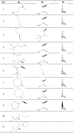
a result of in vitro experiment of eleven sialic acid derivative compounds on neuraminidase type N1 as was retrieved from www.bindingdb.org(Liu et al., 2007; Chen et al., 2002). This sialic acid derivative compoundswas chosen, since sialic acid is the natural ligand of neuraminidase and functions as neuraminidase inhibitor as shown in Table1.
Table 1: Alkyls substitution in Sialic Acid Derivative Compounds.
No R1 R2 R3
1
2
3
4
5
6
7
8
9
10 - -
11 - -
This sialic acid derivativeswere used for validation method process and Autodock 4.2 software by plotting the values of IC50 and Ki obtained from molecular docking simulation result.
The sialic acid derivative compound log IC50 data was plotted with
the value of log Ki obtained from the molecular docking
simulation result, thus linearity curve is obtained as shown in Table 2 and Fig. 2.
Fig. 2: Plot graph of IC50 experiment (literature) of sialic acid derivative compound versus log Ki.based on molecular docking simulation result.
Table 2: Correlation of IC50 experiment versus Ki of molecular docking results.
No Ki (Docking)
(µM)
IC50(experiment)
(µM) Log Ki Log IC50
1 63.77 200 1.804 2.301
2 29.32 130 1.467 2.113
3 203.37 100 2.308 2
4 94.29 225 1.974 2.352
5 262.11 450 2.418 2.653
6 37.49 100 1.573 2
7 5.25 25 0.720 1.397
8 5.34 26 0.727 1.414
9 4.74 22 0.675 1.342
10 3.4 15 0.531 1.176
11 8.64 40 0.936 1.602
Based on the linear equation result obtained, the correlation coefficient value (r2) was 0.861 It means that 86.1% of the value of
Ki from the docking result had a linear correlation and parallel with the value of IC50 from the in vitro experiment. From this
result, it can be summarized that the Autodock 4.2 software and the molecular docking simulation method was satisfactorily valid.
Result of Molecular Docking Simulation of Natural products Compounds on Neuraminidase N1 and N1 Mutant
In this study, molecular docking simulation was examined insome natural product compounds on neuraminidase N1 and N1 mutant. The natural products compound used in this molecular docking simulation was a natural product compounds that has been tested in vitro and has a good activity as influenza virus neuraminidase inhibitor (Grienke et al., 2012). Details on each natural products compound was shown in Supplementary Table 1. Besides natural products compounds, molecular docking simulation wasalso examined inoseltamivir and zanamivir— used as the control, since both compounds have been used as anti-influenza drugs. In the natural products compound molecular docking process against neuraminidase N1 and N1 mutant, the parameter used was the default grid size, which is 40 x 40 x 40 point, with the spacing of 0,375 Å. This size was able to include every essential amino acid residues. The amount of runs was 100. The coordinate for the central position in the grid box is
y = 0.6545x + 0.9496 R² = 0.8618
0 0.5 1 1.5 2 2.5 3
0 0.5 1 1.5 2 2.5 3
Lo
g
IC
5
0
x=29,793; y=12,515; dan z=-21,927 for N1 and x=32,563; y=14,201; dan z=19,027 for N1 mutant. This docking simulation used the Lamarckian Genetic Algorithm (LGA) search method with evaluation energy up to 250.000(Morris et al., 1998). In the docking process, the receptor was set as a rigid while the ligand was set in a flexible state, thus it can move and rotate freely. The molecular docking simulation was carried out in order to find a precise bond conformation between a ligand and a receptor. This process was aimed at screening some natural product compound candidates; hence a compound with the best affinity that could bond with its receptor’s active side was obtained. The PyRx software was employed for virtual screening.
Interpretation Result and Data Analysis
The parameters that could be analyzed from the
ligand-protein interaction include free energy bond (ΔG), inhibition
constant (Ki) and hydrogen bond. The chosen protein-ligand complex was the one with the smallest bond energy value and inhibition constant. It was further compared with neuraminidase inhibitor standard, which werezanamivir and oseltamivir.
The value of bond free energy showed the bond strength of ligand and receptor. There was a connection between the values of bond free energy with inhibitor constant (Ki) which value corresponds with thermodynamic equation(Morris and Lim-Wilby, 2008; Kroemer, 2003).
∆G0 = -RT ln K A
KA = Ki-1 = [ ] [ ][ ]
Some natural product compounds used in the research were 117 compounds isolated from various plants. Data analysis was carried out in the first-rank molecular docking simulation result in its population with the grid dimension 40 x 40 x 40 point and the spacing of 0,375 Å. Based on this molecular docking simulation result, the natural products compound that has a lower free energy value was katsumadain A (9), both in neuraminidase type N1 and N1 mutant, consecutively –7.54 kcal/mol and –7.46 kcal/mol.
The negative sign in the free energy value shows the possibility of a spontaneous interaction between a ligand and a receptor (Morris et al., 2008). This result showed the strongest bond on neuraminidase as compared to other natural products compounds. Katsumadain A had an inhibition constant of 2.98 µM on neuraminidase N1 and 3.38 µM on neuraminidase N1 mutant. Inhibition constant (Ki) shows the concentration that is required by the ligand in inhibiting macromolecules (protein) (Morris et al., 1998). The smaller value of Ki is the better result due to the smaller ligand concentration was required to inhibit the spread of newly-formed influenza viruses.
Katsumadain A is a compound of diarylheptanoid class which is isolated from the plant Alpinia kasumadai Hayata (Grienke et al., 2012). Generally, diarylheptanoid-class compound has two phenyl groups that are connected with seven carbons. Other Alpinia katsumadai Hayata-isolated diarylheptanoid compounds include (3S)-1,7-diphenyl-(6E)-6-hepten-3-ol (5),
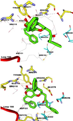
(E,E)-5-hydroxy-1,7-diphenyl-4,6 heptadien3-one (7), (3S,5S)-trans-3,5-dihydroxy-1,7-diphenyl1-heptene (6), and alnustone (8). They have free energy values of over –7 kcal/mol in both neuraminidase N1 and N1 mutant. This means that a poor affinity is in contrast tokatsumadain A. As shown in Figure 3, katsumadain A carbonyl group accepts hydrogen atom from Arg118 amino group with a distance of 2.306 Å. The hydrogen bond in between katsumadain A and neuraminidase N1 also occurs in amino acid residue Arg371 with a distance of 1.680 Å.
Fig. 3: Molecule interaction between katsumadain A and neuraminidase types N1 (a) and N1 mutant (b). Ligand is depicted in green. Hydrogen bond is depicted in red dashes. Amino acid residue is depicted in cyan in which showing hydrophobic interaction. Pi-cation interaction is depicted in orange.
ligand are. This change of properties affects the biological activities of a compound. However, interaction katsumadain A-N1 was stabilized by pi-pi cation interaction between ring aromatic of katsumadain A and the both amino acid residue of Arg 118 and Arg 152. The interaction between katsumadain A and neuraminidase types N1 and N1 mutant could be seen in Figure 3.
In comparison with oseltamivir (116) and zanamivir (117) standards, katsumadain A had a larger free energy value on neuraminidase type N1. This result showed a better affinity on oseltamivir and zanamivir standards to bond with neuraminidase type N1 as compared with katsumadain A.
In neuraminidase type N1 mutant, katsumadain A has a higher free energy value in comparison to zanamivir with only – 7.43 kcal/mol and oseltamivir with–7.37 kcal/mol. This showed that katsumadain A has a better affinity to bond with neuraminidase type N1 mutant than zanamivir and oseltamivir. Katsumadain A was able to form ligand-enzyme complex in a more stable conformation than standard drugs (zanamivir and oseltamivir) and to better inhibit neuraminidase N1 mutant enzyme.
Zanamivir and oseltamivir have a more larger free energy value on neuraminidase N1 mutant than N1 mutant; hence it is less capable of inhibiting neuraminidase as compared to neuraminidase N1. The interaction between hydrogen bonded with katsumadain A, zanamivir, and oseltamivir, and other best natural products compounds with neuraminidase types N1 and N1 mutant could be seen in Table S1 (Supplementary File).
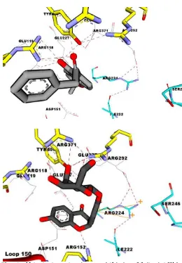
In addition to katsumadain A, two other natural products compounds that had the lowest free energy value are 1-(5-hydroxy-2,2-dimethyl-2H-1-benzopyran-6-yl)-2-phenyl-ethanone (103) andliquiritin (38), with free energy values of –7.36 kcal/mol and –7.23 kcal/mol, consecutively, on neuraminidase type N1. (103) is a flavonoid-derived compound that has the best affinity to bond with neuraminidase N1 while (38) is a flavonon-group compound isolated from the plant Glycyrrhiza uralensis Fisch(Grienke et al., 2012).
Based on Figure 4, compound no. 103 had two hydrogen bonded with amino acids Arg152 and Arg292, while in (38), it had six hydrogen bonded with amino acid residues Arg152, Arg156, Arg292, Asn347, Arg371, and Tyr406.
In neuraminidase type N1 mutant, two natural products compounds with the lowest free energy value following katsumadain A are chlorogenicacid (117) and ferulic acid (115) that obtained free energy values of –7.37 kcal/mol and –7.31 kcal/mol, consecutively.
Chlorogenic acid ispresent in many plants. This compound is found in fruits and seeds of plants and is also widely distributed as conjugates including many foods and beverages (Clifford, 2003). Currently, this compound significantly enhances the inhibitory effect on NA but less than oseltamivir(Yang et al., 2011), even it is one of the neuraminidase inhibitory components in sweet potato against influenza virus which was patented by Koji et al.(Koji et al., 2011). Ferulic acid had five hydrogens bonded with amino acids: Arg118, Arg152, Arg292, Arg371, Tyr406 and
Arg 371 as shown in Table 3. To be specific, the amino acids were on the active side of neuraminidase N1 mutant.
Fig. 4: Molecule interaction between 1-(5-hydroxy-2.2-dimethyl-2H-1-benzopyran-6-yl)-2-phenyl-ethanone (103) (a) andliquiritin (38) (b) with neuraminidase type N1. Ligand is depicted in green. Hydrogen bond is depicted in red dashes. Amino acid residue is depicted in cyan in whichshowing hydrophobic interaction. Pi-cation interaction is depicted in orange.
Table 3: Neuraminidase amino acid residues that form hydrogen bonded with potential natural products compounds and neuraminidase inhibitor standard.
Ligand Amino acid residue
NA N1 NA N1 mutant
(9) Arg118. Arg371 Arg118
(17) Glu119, Arg 152, Tyr 406 Glu119, Arg 152, Tyr 406
(38) Arg152. Arg156. Arg292. Asn347. Arg371. Tyr406
Arg152. Arg156. Ser179. Arg292. Arg371. Tyr406
(103) Arg118. Arg371. Glu119 Arg118. Arg371. Glu119
(79) Arg292. Asn347. Arg371 Asn221
(115) Arg118. Arg152. Arg292.
Arg371. Tyr406 Arg118. Arg152. Arg292. Arg371. Tyr406 (116) Arg118. Arg152. Arg292.
Arg371. Trp178. Asp151. Glu276
Arg118. Arg152. Arg292. Arg371. Tyr406
(117) Arg118. Arg152. Arg292. Arg371. Asp151. Glu119
Arg118. Arg152. Arg292. Arg371. Asp151. Glu119
liquiritin (38), 1-(5-hydroxy-2,2-dimethyl-2H-1-benzopyran-6-yl)-2-phenyl-ethanone (103), catechin(17), chlorogenic acid (115)and ferulic acid (117) are, consecutively, 3.18; 2.00; 1.03; 0.,56; -0.60 and -0.63.
This means that katsumadain A is the most lipophilic ligand as compared to other ligands with the better affinities. There was a significant difference between the values of log P katsumadain A with log P liquiritin. In the case of liquiritin, there were five hydroxyl groups that caused the liquiritin to have a higher polarity than katsumadain A that has no hydroxyl group. In addition, four natural products compounds had log P value and molecule mass that correspond with Lipinski’s Rules of Five, which have the log P value of between -2to5 and molecule mass of less than 500 Da as shown in Table S2(Lipinski et al., 2001). There was a natural product compound which had a small enough IC50 based on in vitro tests, but satisfactorily has a lower affinity
based on in silico tests by using molecular docking simulation method.
An example of such natural product compound was artocarpin (46). Based on in vitro tests, artocarpin has an IC50
value of 0.18 µM (Grienke et al., 2012).In contrast to the docking result, it only had a free energy value of –5.83 kcal/mol; Ki of 53.22 µM on neuraminidase type N1, and –5.49 kcal/mol; 94.19 µM on neuraminidase N1 mutant.
Catechin natural product compound (17) has free energy value of –5.69 on neuraminidase N1 and –5.64 on neuraminidase N1 mutant. This free energy value is larger than the best natural products compound—katsumadain A. Catechin has been reported to be present in some Garcinia sp such as G. kola (Ejele et al., 2012) and G. penangiana (Lim, 2005) but have not been reported in Garcinia celebica. Catechin has been evaluated as anti-influenza by inhibiting anti-influenza virus replication in cell culture (Song et al., 2005; Song and Seong, 2013).
In addition, herbal tea consisted catechin have the capability to halt influenza virus infection in elderly nursing home residents (Yamada et al., 2006). Kuzuhara et al. (2009) explained that catechin inhibited the endonuclease activity of influenza A virus RNA polymerase, thus potential to be an anti-influenza A drug (Kuzuhara et al., 2009). The mechanism of catechin as an anti-influenza also corresponded to the antioxidant activity. The study of anti-influenza virus activity of catechin against neuraminidase has been discussed by Liu et al (2008); nevertheless they did not conduct molecular docking studies of catechin against NA.
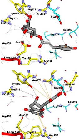
Catechin favorably docked against neuraminidase. 2-catechol (ring C) of catechin interacted well with the arginine triad through hydrogen bond and pi-cation interactions. As shown in Figure 5c, it appeared that the compound lost interaction with hydrophobic pocket (Ileu222, Arg224, and Ser246), but formed hydrogen bond with Glu276. 3-OH of catechin formed hydrogen bond with Trp178 (2.3 Å). 3-OH in catechin linked to 3-gallocyl to form epicatechingallate (ECG). Uchide and Toyoda (2011) discussed that the activity of ECG as anti-influenza virus is contributed mainly by 3-gallloyl moiety of this compound,
whereas the 5'-OH at the trihydroxybenzyl moiety at the 2-position plays a minor role.
This molecular mechanism is related to the capability as antioxidant that scavenge for superoxide anion and hydroxyl radicals (Uchide and Toyoda, 2011). Catechin has an IC50 value of more than 100 µM on neuraminidase N1, based on in vitro test (Grienke et al., 2012).
Fig. 5: Molecule interaction between chlorogenic acid (117) (a) and catechin(17) with neuraminidase N1 mutant. Ligand is depicted in green. Hydrogen bond is depicted in red dashes. Amino acid residue is depicted in cyanin which showing hydrophobic interaction. Pi-cation interaction is depicted in orange.
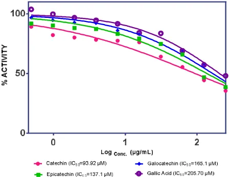
In vitro Assay for Some Natural Compounds
value of catechin was 93.92μM. Epicatechin (18), galocatechin (19), and gallic acid (20) had137.1µM, 165.1 µM, and 205.7 µM, respectively. It was higher than catechin which is in line with the results of in silico methods that Ki catechin (67.16 µM) has smaller
than epicatehcin (71.52 µM), galocatechin (80.87 µM) and gallic acid (84.02µM) as shown in Fig. 6.
Fig. 6: Neuraminidase Activity of Catechin (pink). Epicatechin (green). Galocatechin (blue).and Gallic Acid (purple).
CONCLUSION
In conclusion, the research on molecular docking simulation was able to examine amino acid residue’s interaction on the active side of neuraminidase enzyme types N1 and N1 mutant with a number of natural product compounds. The result of the research showed that katsumadain A had the best affinity to bond with neuraminidase types N1 and N1 mutant. This is based on the lowest free energy value of –7.46 kcal/mol on neuraminidase N1 mutant. Furthermore, katsumadain A has two hydrogen bonds to the active side of neuraminidase N1, namely amino acids Arg118 and Arg371. While on neuraminidase N1 mutant, it only has one hydrogen bond to Arg118.
ACKNOWLEDGMENTS
We would gratefully acknowledge Dean of Faculty of Pharmacy Universitas Padjadjaran, for funding this project through Seeds Grant Faculty 2014.
REFERENCES
Chen, X., Lin, Y., Liu, M., and Gilson, M. K.. The Binding Database: data management and interface design. Bioinformatics, 2002; 18
(1):130-139.
Clifford, M. The analysis and characterization of chlorogenic acids and other cinnamates. In Methods in Polyphenol Analysis, edited by
G. Williamson. Cambridge: Royal Society of Chemistry, 2003; 314–337. Collins, P. J., Haire, L. F., Lin, Y. P., Liu, J., Russell, R. J., Walker, P. A., Skehel, J. J., Martin, S. R., Hay, A. J., and Gamblin, S. J. Crystal structures of oseltamivir-resistant influenza virus neuraminidase mutants. Nature, 2008; 453 (7199):1258-1261.
De Filette, M., Min Jou, W., Birkett, A., Lyons, K., Schultz, B., Tonkyro, A., Resch, S., and Fiers, W. Universal influenza A vaccine: optimization of M2-based constructs. Virology, 2005; 337 (1):149-161.
Ejele, A. E., Iwu, I. C., Enenebeaku, C. K., Ukiwe, L. N., and Okolue, B. N. Bioassay-Guided Isolation, Purification and Partial Characterization of Antimicrobial Compound from Basic Metabolite of
Garcinia Kola. JETEAS, 2012; 3 (4):668-672.
Fanning, T. G., Slemons, R. D., Reid, A. H., Janczewski, T. A., Dean, J., and Taubenberger, J. K. 1917 avian influenza virus sequences suggest that the 1918 pandemic virus did not acquire its hemagglutinin directly from birds. J Virol, 2002; 76 (15):7860-7862.
Grienke, U., Schmidtke, M., Kirchmair, J., Pfarr, K., Wutzler, P., Dürrwald, R., Wolber, G., Liedl, K. R., Stuppner, H., and Rollinger, J. M. Antiviral Potential and Molecular Insight into Neuraminidase Inhibiting Diarylheptanoids from Alpinia katsumadai. Journal of
Medicinal Chemistry, 2009; 53 (2):778-786.
Grienke, U., Schmidtke, M., von Grafenstein, S., Kirchmair, J., Liedl, K. R., and Rollinger, J. M. Influenza neuraminidase: A druggable target for natural products. Natural Product Reports, 2012; 29 (1):11-36.
Hurt, A. 2007. Fluorometric Neuraminidase Inhibition Assay. Australia. : WHO Collaborating Centre for Reference and Research on Influenza, 1-10.
Koji, I., Makoto, Y., and Takehiko, N. 2011. Neuraminidase Inhibitory Component, edited by N. A. F. R. ORGANIZATION. Japan.
Kontoyianni, M., McClellan, L. M., and Sokol, G. S. Evaluation of docking performance: comparative data on docking algorithms. J Med
Chem, 2004; 47 (3):558-565.
Kroemer, R. T. Molecular modelling probes: docking and scoring. Biochem Soc Trans, 2003; 31 (Pt 5):980-984.
Kuzuhara, T., Iwai, Y., Takahashi, H., Hatakeyama, D., and Echigo, N. Green tea catechins inhibit the endonuclease activity of influenza A virus RNA polymerase. PLoS Curr, 2009; 1:RRN1052.
Li, H., Sutter, J., and Hoffmann, R. 2000. HypoGen: An Automated System fro Generating 3D predictive Pharmacophore Models.
In Pharmacophore Perception, Development, and Use in Drug Design,
edited by O. F. Guner. La Jolla: IUL Biotechnology Series, 171-189. Li, Q., Qi, J., Zhang, W., Vavricka, C. J., Shi, Y., Wei, J., Feng, E., Shen, J., Chen, J., Liu, D., He, J., Yan, J., Liu, H., Jiang, H., Teng, M., Li, X., and Gao, G. F. The 2009 pandemic H1N1 neuraminidase N1 lacks the 150-cavity in its active site. Nat Struct Mol Biol, 2010; 17
(10):1266-1268.
Lim, C. K. 2005. Phytochemicals From Garcinia, Mesua and Jatropha Species and Their Biological Activities, [Thesis], Faculty Science, Univerisi Putra Malaysia, Kuala Lumpur.
Lipinski, C. A., Lombardo, F., Dominy, B. W., and Feeney, P. J. Experimental and computational approaches to estimate solubility and permeability in drug discovery and development settings. Adv Drug Deliv Rev, 2001; 46 (1-3):3-26.
Liu, T., Lin, Y., Wen, X., Jorissen, R. N., and Gilson, M. K. BindingDB: a web-accessible database of experimentally determined protein-ligand binding affinities. Nucleic Acids Res, 2007; 35 (Database
issue):D198-201. Lamarckian genetic algorithm and an empirical binding free energy function. Journal of Computational Chemistry, 1998; 19 (14):1639-1662.
Morris, G. M., Huey, R., Lindstrom, W., Sanner, M. F., Belew, R. K., Goodsell, D. S., and Olson, A. J. AutoDock4 and AutoDockTools4: Automated docking with selective receptor flexibility. J Comput Chem,
2009; 30 (16):2785-2791.
Morris, G. M., Huey, R., and Olson, A. J. 2008. Using AutoDock for ligand-receptor docking. Curr Protoc Bioinformatics,
Chapter 8:Unit 8 14.
Morris, G. M., and Lim-Wilby, M. Molecular docking. Methods
Mol Biol, 2008; 443:365-382.
Russell, R. J., Haire, L. F., Stevens, D. J., Collins, P. J., Lin, Y.
P., Blackburn, G. M., Hay, A. J., Gamblin, S. J., and Skehel, J. J. The structure of H5N1 avian influenza neuraminidase suggests new
Scholtissek, C. Source for influenza pandemics. Eur J
Epidemiol, 1994; 10 (4):455-458.
Song, J.-M., and Seong, B.-L. 2013. Chapter 99-Anti-Influenza Viral Activity of Catechins and Derivatives. In Tea in Health and Disease
Prevention: Academic Press, 1185-1193.
Song, J. M., Lee, K. H., and Seong, B. L. Antiviral effect of catechins in green tea on influenza virus. Antiviral Res, 2005; 68
(2):66-74.
Stoll, V., Stewart, K. D., Maring, C. J., Muchmore, S., Giranda, V., Gu, Y. G., Wang, G., Chen, Y., Sun, M., Zhao, C., Kennedy, A. L., Madigan, D. L., Xu, Y., Saldivar, A., Kati, W., Laver, G., Sowin, T., Sham, H. L., Greer, J., and Kempf, D. Influenza neuraminidase inhibitors: structure-based design of a novel inhibitor series. Biochemistry, 2003; 42
(3):718-727.
Uchide, N., and Toyoda, H. Antioxidant Therapy as a Potential Approach to Severe Influenza-Associated Complications. Molecules,
2011; 16 (3):2032-2052.
Von Itzstein, M. The war against influenza: discovery and development of sialidase inhibitors. Nat Rev Drug Discov, 2007; 6
(12):967-974.
Von Itzstein, M., Dyason, J. C., Oliver, S. W., White, H. F., Wu, W. Y., Kok, G. B., and Pegg, M. S. A study of the active site of influenza virus sialidase: an approach to the rational design of novel anti-influenza drugs. J Med Chem, 1996; 39 (2):388-391.
Xu, X., Zhu, X., Dwek, R. A., Stevens, J., and Wilson, I. A. Structural characterization of the 1918 influenza virus H1N1 neuraminidase. J Virol, 2008; 82 (21):10493-10501.
Yamada, H., Takuma, N., Daimon, T., and Hara, Y. Gargling with tea catechin extracts for the prevention of influenza infection in elderly nursing home residents: a prospective clinical study. J Altern
Complement Med, 2006; 12 (7):669-672.
Yang, F., Jin, L., Huang, N. Y., Chen, F., Luo, H. J., and Chen, J. F. [Design, synthesis and activity of a new type of influenza virus N1 neuraminidase inhibitors]. Yao Xue Xue Bao, 2011; 46 (11):1344-1348.
Yanuar, A., 2012, Penambatan Molekuler: Praktek dan Aplikasi
pada Virtual Screening. . Vol. 1. Depok: UI Press .
How to cite this article:
Muchtaridi M., Aliyudin A., Holik H.A.Potential Activity of Some Natural products Compounds as Neuraminidase Inhibitors Based on Molecular Docking Simulation and in vitro Test. J App Pharm