Enhancing Dental Pulp Stem Cell Viability for Treatment of Spinal Cord Injury via Immune Cell Preconditioning
A thesis submitted in partial fulfilment of the
HONOURS DEGREE of BACHELOR OF HEALTH AND MEDICAL SCIENCES in
The Discipline of Pathology Adelaide Medical School The University of Adelaide
By Sandra Jenkner November 2020
Abstract
Spinal cord injury (SCI) is a devastating condition that affects more than 15,000 people in
Australia. As there is no treatment or cure available, patients are often burdened with a lifetime of disability and co-morbidity. Growing evidence supports the use of stem cells to repair the spinal cord following injury, with dental pulp stem cells (DPSCs) in particular showing superior regenerative capacity and modulation of immune responses following injury. However, studies demonstrate that only approximately 1% of transplanted DPSCs survive due to harsh inflammatory conditions during SCI, thus limiting their full therapeutic benefits. Therefore, attempts to enhance the survival and viability of DPSCs should be investigated. Utilising fluorescence activated cell sorting, the present study characterised the inflammatory responses of peripheral blood
mononuclear cells (PBMCs) in a clinically relevant rat model of contusion SCI, showing
phenotypic variations between SCI, Sham and non-injured animals. Moreover, a novel approach was used to precondition DPSCs with PBMCs taken 3 days post-injury. Cytotoxicity analysis revealed that cytotoxicity is significantly higher following 7 days of culture than 3, particularly following pro-inflammatory TNF- conditioning (p<0.05), but less cytotoxic than when DPSCs were cultured alone. Interestingly, following pro-inflammatory conditioning, co-cultured DPSCs were found to release significant amounts of IL-6 (p<0.0001), which may be involved in
modulating the pro-inflammatory environment. This data suggests that preconditioning of DPSCs and PBMCs in co-culture for 3 days is a suitable ex vivo strategy to enhance the viability and immunomodulatory behaviour of DPSCs.
Introduction
Spinal cord injury (SCI) is a debilitating condition caused by direct insult to the spinal cord. It results in the long-term loss of functional mobility and/or sensation due to impaired conduction of descending motor and ascending sensory signals, and greatly increases the chance of morbidity and mortality.1, 2 It is estimated that there are 15,000 Australians currently living with SCI3 with 374 new cases in 2016-2017 alone.4 Despite a wealth of knowledge on the pathophysiology of SCI, there is still no treatment to stop or revert the neurological deficits within the spinal cord following injury.
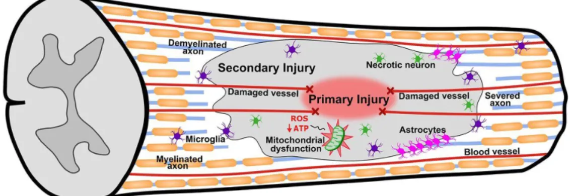
Central nervous system (CNS) injuries elicit secondary injury cascades (Fig. 1) within minutes that exacerbate neurological damage, creating a non-permissive environment for physiological regeneration.5 This includes acute haemorrhagic swelling,6 excitotoxicity,7 oxidative stress, ionic imbalances,8, 9 inflammatory cell recruitment and neuroinflammation,10 all of which lead to neural and glial apoptosis and axon demyelination,7, 11 thus worsening functional outcomes.
Figure 1. Multiple secondary injury cascades (including inflammation and cell death) follow the primary injury and exacerbate damage. Schematic extracted from Scholpa and Schnellmann.12
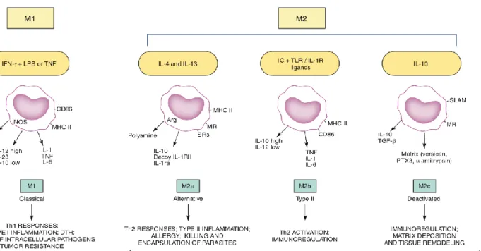
Inflammation is upstream of many secondary injury cascades and is both a driver and consequence of excitotoxicity, oxidative stress and ionic imbalances.13 However, immune cells can change their
phenotype based on environmental cues (Fig. 2) and thus have varying effects on CNS tissue. Pro- inflammatory M1 polarised cells are activated by interferon-gamma (IFN-), lipopolysaccharide (LPS) and tumour necrosis factor (TNF),14, 15 releasing cytokines that contribute to pathogen destruction and phagocytosis, but cause collateral damage to healthy neurons and glia.15, 16 Conversely, anti-inflammatory M2 cells are activated by interleukin-4 (IL-4), IL-13 and IL-1017 and work to promote tissue protection and repair18, 19 through functions such as angiogenesis, axon growth, tissue remodelling20 and suppression of pro-inflammatory effects.15
These inflammatory responses involve immune cells including lymphocytes (T-cells, B-cells), neutrophils and monocytes (microglia, macrophages). In the spinal cord following injury, the first responders are resident microglia which clear debris and fight infection.21, 22 Neutrophils appear in the SCI lesion within 4-6h23 and release proteases6 and oxidative factors that injure neurons and glia and cause demyelination.24 Monocytes then infiltrate the spinal cord, reaching maximal numbers within 7-10 days and persist for weeks to months after injury.23 Microglia/macrophages, which are indistinguishable within the CNS, exhibit mainly pro-inflammatory phenotypes in SCI, leading to excessive oxidative factor release and neurotoxicity.20 Microglia/macrophages also function as antigen presenting cells, recruiting T and B-cells to the injury site which release further pro- inflammatory cytokines.25
Figure 2. Pro-inflammatory (M1) and anti-inflammatory (M2) immune cell polarisation and their respective cell products (cytokines) and responses. Schematic extracted from Martinez and
Gordon.15
Furthermore, immune function becomes dysregulated in chronic SCI due to insufficient dampening of the pro-inflammatory response, leading to increased neural and glial degeneration and death.21, 23,
26 Blocking TNF-27 and the pro-inflammatory IL-1 cytokine28 has been shown to reduce apoptosis and demyelination in the spinal cord, highlighting the detrimental effects of pro-inflammation in SCI.23, 24 Therefore, inflammatory responses within SCI greatly impact functional outcomes,29, 30 supporting the premise that targeting pro-inflammatory responses acutely would aid in SCI recovery and prevent chronic dysregulation.13
Despite inflammation and immune cells having a notorious reputation in injury and disease, studies have demonstrated the efficacy of activated immune cell engraftment as a potential targeted treatment strategy for SCI which circumvents the adverse effects of pharmaceuticals,31, 32 directly promoting recovery whilst encouraging appropriate inflammatory responses. Prewitt et al. first showed that
implantation of microglia/macrophages into transected nerves in rodents significantly promoted axon regeneration.33 Following this, studies engrafted preconditioned anti-inflammatory peripheral macrophages following SCI, which increased wound healing and nerve regeneration in rodent studies13, 34 and showed safety in human trials.35 Contradictory to long-standing knowledge of immune cell populations, studies have shown that unlike microglia, infiltrating peripheral macrophages perform anti-inflammatory roles,36, 37 including wound healing and decreasing lesion size, and improve locomotor function following SCI.38 However, treatment with anti-inflammatory macrophages alone has limitations, supported by findings describing a 20-40% phenotype loss of transplanted cells, favouring pro-inflammatory phenotypes once within the harsh acute SCI microenvironment.20 Additionally, low functional recovery within Phase 1 and 2 human clinical trials limits translation from bench-to-bedside.35, 39 Therefore, further studies are needed to develop novel strategies to increase the viability of anti-inflammatory immune cell populations, particularly blood- derived macrophages, following SCI.
Mesenchymal stem cells (MSCs) have been investigated within the field of neuroregeneration for decades, and, importantly, interact with immune cells to modulate inflammatory responses.40-44 This includes decreasing apoptosis and lesion size, reducing the effects of pro-inflammatory cytokines such as IL-1 and TNF-, inhibiting immune cell proliferation45, 46 and releasing anti-inflammatory cytokines,42 peripherally and within the spinal cord.47, 48 Notably, a bidirectional relationship exists in which anti-inflammatory immune cells support the viability of stem cells18 by providing trophic effects,49 inducing cell renewal50 and promoting growth.51
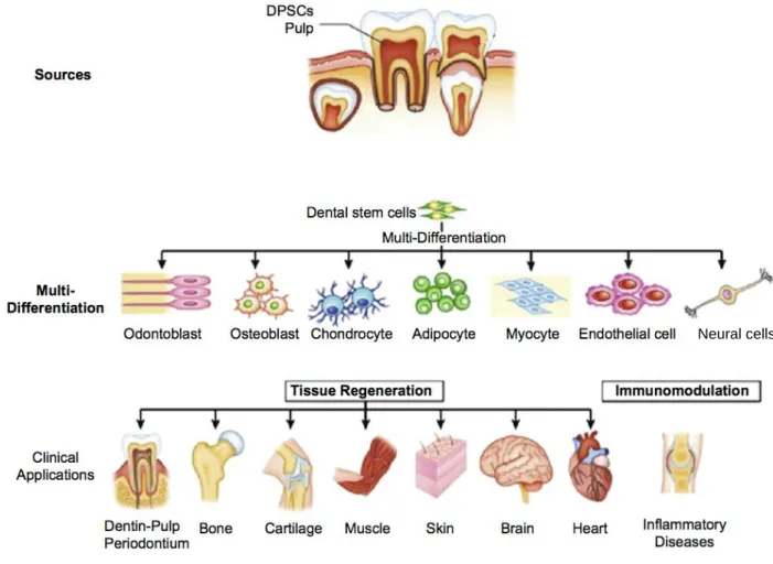
Neural crest derived dental pulp stem cells (DPSCs) are multipotent, self-renewing stem cells with MSC-like characteristics52, 53 (Fig. 3), that exert similar modulatory effects on immune cells as MSCs.54-58 These cells have a high regenerative capacity and higher proliferation rate than other MSC
populations,59 and can be extracted in a non-invasive and autologous manner from dental tissue,60, 61 thus limiting considerations of ethics and cell rejection.
Figure 3. DPSCs can be extracted non-invasively from dental tissue and have various differentiation potentials and clinical applications. Schematic extracted from Li et al.62
As in MSC populations,47, 63 studies utilising DPSCs show survival rates of approximately 1-2%
following CNS engraftment,57, 64 validating further investigation. Despite low survival, DPSCs have shown significant regeneration within the CNS and following SCI. In a rat stroke model, DPSCs promoted neuro-behavioural improvement with a ~2% survival rate.64 Similarly, DPSCs engrafted into SCI rat models significantly increased regeneration of nerve tracts and improved locomotor function.65, 66
Neural cells
The independent administration of the two aforementioned preclinical SCI therapies (immune cell administration and DPSC administration) has great potential; however, issues concerning phenotype loss and low survival create gaps in knowledge. An increasing number of studies have outlined the potential of combinatorial treatment strategies for SCI that overcome the harsh inflammatory microenvironment whilst preserving neural and glial function.9, 67, 68 Therefore, the present study employed a novel approach of merging the two therapies into one, multifaceted treatment mechanism.
This strategy takes advantage of the symbiotic relationship between DPSCs and immune cells in order to increase their viability and immunomodulatory behaviour in vivo, and could enhance their neuroregenerative and neuroprotective potentials following engraftment. However, little is known regarding DPSC and immune cell co-culture and its effects on cell viability and immunomodulation ex vivo. Additionally, a limited understanding of the behaviour of peripheral immune cell populations following SCI limits the knowledge of when these cells can be extracted and administered as combinatorial therapies.
Hypothesis and aims
We hypothesise that preconditioning DPSCs and peripheral blood mononuclear cells (PBMCs), comprised of T-cells, B-cells and monocytes, will reveal immunomodulatory activities and enhance the viability of both DPSCs and PBMCs for future engraftment into a SCI model.
The aims of this project are, therefore, 1) to characterise how SCI changes the proportion of PBMC populations at different time-points following injury; 2) to identify a time-point for PBMC extraction and utilisation for novel preconditioning paradigms; and, 3) to investigate the effects of DPSC and PBMC preconditioning on cytotoxicity and immunomodulation ex vivo.
By showing that DPSCs and PBMCs can be preconditioned through co-culture to induce immunomodulatory activity and increase viability, their regenerative potential can then be further investigated in vivo. Knowledge gained from these studies could be used to develop a novel SCI
treatment mechanism that not only boosts cellular regeneration, but also limits the negative effects of inflammation. On a wider scale, this project could aid in the development of a viability model for DPSC survival, which may be used in a range of chronic pathologies both within the CNS and peripherally, particularly in those linked to an imbalance of inflammatory responses.
Methods
Animal surgeries. Adult female Sprague-Dawley rats (10-12 weeks; Bioresources, SAHMRI) were housed in a 12h light/dark cycle and fed ad libitum. Animals were sedated with Diazepam (1.75 mg/kg) and anaesthetised using isoflurane (2.0-2.5% v/v). Surgical sites were triple swabbed with ethanol and betadine solution and a longitudinal incision was made through the skin to reveal the spinal column. Partial laminectomy at T9-T11 was conducted to expose the spinal cord without disrupting the meninges. Using an Infinite Horizon impactor device (Precision Systems), animals received a moderate-severe T10 contusion (200kDyne) to the dorsal surface of the spinal cord.69 The incision site was closed with nylon sutures and wound clips. Surgical control (Sham) animals received laminectomies only, whilst non-injured animals served as negative controls. Saline and Buprenorphine (0.01mL/100g bodyweight; 0.0324mg/kg) were administered post-operatively and then twice daily until 5 days post-injury (dpi), then once daily until 7 dpi. Benacillin (0.02mL/100g bodyweight; 64mg/kg [150mg/mL procaine penicillin, 150mg/mL benzathine penicillin, 20mg/mL procaine hydrochloride]) was administered immediately after and at 2, 4 and 6 dpi. Bladders of SCI rodents were manually expressed twice daily until normal bladder function returned (7-10 dpi). All procedures performed complied with the Australian Code for the Care and Use of Animals for Scientific Purposes (National Health and Medical Research Council (8th edition, 2013) with approval from the SAHMRI Animal Ethics Committee (SAM247; SAM413.19).
PBMC isolation. Animals were deeply anaesthetised with isoflurane (5% v/v) and humanely killed via cardiac puncture: whole blood samples (4-8mL) from non-injured (n=5), Sham (n=30) and SCI animals (n=30) were harvested using a 25-gauge needle and split into two heparinised vacutainers (Greiner Bio-One) per animal, at 1, 2, 3, 7, 14 and 28 dpi. PBMCs were isolated via density gradient centrifugation using Lymphoprep (STEMCELL Technologies), according to the manufacturer instructions. Briefly, blood samples were diluted 1:1 with Dulbecco’s phosphate-buffered saline (DPBS; Thermo Fisher), centrifuged at 1200g for 10min, washed with DPBS and centrifuged at 500g
for 10min twice and resuspended in 50% (v/v) foetal bovine serum (FBS) (Thermo Fisher; Australian Origin), 40% (v/v) RPMI 1640 (Thermo Fisher) and 10% (v/v) dimethyl sulfoxide (Sigma). Each animal sample was split into four cryotubes, placed in Mr Frosty freezing containers (Thermo Fisher) overnight at -80C, and stored until further processing.70
Fluorescence activated cell sorting (FACS). FACS analysis utilising a modified 9-colour staining panel71 was conducted to sort PBMC populations and obtain proportions of each as percentages of the total PBMC sample, to investigate aims 1 and 2. PBMC samples were thawed at 37C and washed with RPMI and 20% FBS and centrifuged at 500g for 10min twice. After incubating with viability assay and blocking with bovine serum albumin and CD32 (0.5g/L) for 15min at room temperature, 1x10⁶ cells/100L were incubated with conjugated antibodies obtained from BD Biosciences against CD45 (BUV 395 OX-1; 0.1g/L), CD4 (Horizon V450; 0.025g/L), CD3 (BV605 1F4;
0.2g/L), CD45R (BUV 737 HIS24; 0.1g/L), RT1D (BV480 OX-17; 0.2g/L), CD161a (Alexa 647 10/78; 0.1g/L), CD8a (BV786 OX-8; 0.2g/L), and from BioLegend against CD43 (PC7;
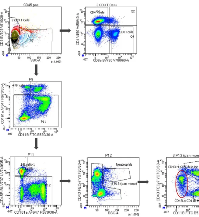
0.2g/L), CD11b (FITC RUO; 0.5g/L), and CD172a (PE OX-41; 0.5g/L), as well as Brilliant Stain Buffer Plus (1:10; BD Biosciences) for 30min at 4C. Cell sorting was conducted using a FACS Aria Fusion cytometer (BD Biosciences). The gating strategy involved first removing debris, non- living cells, cells stuck together and cells not positive for the hemopoietic marker CD45. PBMCs were then sorted sequentially, first using CD3 to sort T-cells, which were also separated by CD4 and CD8 expression. B-cells were sorted by CD45R expression and monocytes by CD43 and CD11b expression.
PBMC and DPSC co-culture. Human DPSCs were donated by the Mesenchymal Stem Cell Laboratory (University of Adelaide) and isolated as previously described.59, 72 DPSCs were cultured in -MEM (Thermo Fisher) supplemented with 10% FBS, 2mM glutamine (Thermo Fisher), 100 units/mL penicillin plus 100g/mL streptomycin (Thermo Fisher) and 100M L-ascorbate (Sigma)
and incubated at 37C in 5% CO2. 24h before co-culture, DPSCs at passage 6 were transferred to 48- well plates at 5x10³ cells/well resuspended in RPMI culture medium supplemented as above and incubated further. After 24h, 3d SCI (n=3) and 3d Sham (n=3) rat PBMC samples were thawed, washed twice with RPMI and seeded at 5x10³ cells/well, on top of DPSC cultures or as monocultures, and supplemented with fresh culture medium. Culture medium only wells served as negative controls.
Culture media was changed 24h following PBMC addition, and then every 48h, thus leaving adherent monocytes73 and DPSCs within wells.
Cell culture preconditioning and cytotoxicity analysis. To investigate aim 3, cytotoxicity analysis was conducted after conditioning with inflammatory cytokines to determine the effects of co-culture, as well as DPSC-only and rat PBMC-only culture, on cell viability, using the CytoTox96® Non- Radioactive assay (Promega) according to manufacturer instructions. Wells were stimulated with pro- (TNF-, IFN-, granulocyte-macrophage colony-stimulating factor; GM-CSF) or anti-inflammatory (macrophage colony-stimulating factor; M-CSF) rat recombinant proteins (25ng/L) (STEMCELL Technologies) after 48h of culture. Non-stimulated wells served as negative controls. Half of the culture media was replaced with fresh recombinant protein media every 48h. Cell culture supernatants (n=3/condition) were collected and used to determine the level of lactate dehydrogenase release in each well as a measure of cytotoxicity74 after 3 and 7 days of culture, measuring absorbance at 490nm on a GloMax® Discover (Promega) plate reader.
Measurement of DPSC cytokine release (Cytometric Bead Array). To determine the effect of preconditioning on DPSC inflammatory cytokine release as part of aim 3, supernatants collected from 3 day co-culture wells were analysed using a Cytometric Bead Array - Human Soluble Protein Master Buffer Kit (BD Biosciences), according to manufacturer instructions. Briefly, 50L of each sample supernatant (n=3/condition) was added to a 96-well plate, followed by 50L of mixed IL-6, IL-1
and TNF- Capture Bead solution and incubated for 1 hour. 50L of the corresponding mixed
Detection Reagent solution was then added and incubated for 2 hours. Fluorescence of each sample was analysed using a BD LSRFortessa™ X20 Flow Cytometer (BD Biosciences).
Data and Statistical analysis. FACS cytometric data were analysed using FlowJo v10.7.1 (Becton Dickinson & Company). PBMC population proportions at different time-points and between Sham and SCI groups were analysed using mixed ANOVA. Cytotoxicity following inflammatory conditioning after 3 and 7 days and DPSC cytokine release were analysed using two-way ANOVAs.
Sidak’s post hoc tests were utilised for multiple comparisons. Statistical analyses were conducted using GraphPad Prism v8.0 and were expressed as group mean ± SEM. Results were considered statistically significant at p<0.05. All data assumes normality unless otherwise stated.
Results
Characterisation of PBMC populations reveals trends within Sham and SCI responses, and identifies a novel monocyte sub-population.
Quantification of the proportion of PBMC samples utilising FACS analysis (Fig. 1) revealed no significant differences between non-injured, Sham or SCI groups in the proportion of B-cells (CD45R+) at any time-point over 28 dpi (p=0.15; Fig. 2A). Similarly, no significant differences were observed within total T-cell (CD3+) proportions (p=0.12) or cytotoxic (CD8+; p=0.41) and helper (CD4+; p=0.070) T-cell sub-populations (Fig. 2B-D). However, trends were noticed whereby proportions of B-cells were consistently reduced when comparing SCI to Sham animals up to 14 dpi.
At 28 dpi, the proportion of B-cells in SCI increased (27.12% 11) compared to Sham (10.78% 0.77). T-cell proportions trended higher at 2 (44.92% 8.0) and 3 dpi (37.28% 4.4) in SCI compared to Sham (24.15% 3.0 and 26.43% 4.7, respectively), with helper T-cell proportions increasing at 3 dpi. Conversely, cytotoxic T-cell proportions were reduced at 2, 3 and 7 dpi following SCI.
Although there were no significant differences between non-injured, Sham or SCI groups in total monocyte proportions (p=0.77), or CD43highCD11bhigh (p=0.10) and CD43lowCD11bhigh (p=0.35) monocyte sub-populations at any time-point, notable trends were observed (Fig. 2E-G). At 1 and 2 dpi, CD43lowCD11bhigh (44.53% 13) and CD43highCD11bhigh (35.77% 6.8) monocyte sub- populations increased following SCI when compared to Sham (28.70% 8.4 and 10.79% 6.8, respectively). Notably, a novel monocyte sub-population (CD43midCD11blow) not previously reported in SCI literature in rats was identified, which made up the greatest proportion of the total monocyte population (54.95% 8.6 in non-injured animals). CD43midCD11blow sub-populations were consistently lower in SCI animals when compared to Sham at 1, 2, 3, 14 and 28 dpi (Fig. 2H).
Figure 1. FACS analysis identified distinct immune cell populations. Representative gating strategy utilised to sort and identify CD3+ and CD4+ or CD8+ T-cells, CD45R+ B-cells, CD43+/SSC-A monocytes and CD43highCD11bhigh and CD43lowCD11bhigh monocyte sub-populations. Red ellipses indicate a novel monocyte sub-population (CD43midCD11blow) not previously identified in rats.
Figure 2. FACS analysis reveals non-significant trends between SCI and Sham PBMC proportions at different time-points. A, B, E Total B-cell, T-cell and monocyte populations, respectively, as proportions of the total PBMC sample. C, D CD8+ and CD4+ T-cell sub-populations as proportions of the total T-cell amount. F-H CD43highCD11bhigh, CD43lowCD11bhigh and CD43midCD11blow sub- populations as proportions of the total monocyte amount. Time-point 0 indicates the mean of non- injured animal PBMC proportions (n=5). Values expressed as mean SEM; p>0.05. n=5.
Conditioning of PBMCs, DPSCs and DPSC+PBMC co-cultures ex vivo with various rat inflammatory cytokines for 7 days increases cytotoxicity.
Following conditioning with TNF-, IFN-, TNF-+IFN-, GM-CSF and M-CSF, PBMCs within both Sham (p<0.0001) and SCI groups (p<0.0001) showed significantly increased cytotoxicity following 7 days of conditioning when compared to 3 days (Fig. 3A-F). However, no significant differences were recorded between conditions at day 3, in both Sham and SCI groups (p=0.86), nor were Sham conditions significantly different from SCI conditions (p=0.29).
Figure 3. Conditioning of PBMCs from SCI and Sham animals with rat inflammatory cytokines has higher cytotoxicity following 7 days of conditioning. A No conditioning and conditioning with B TNF-
, C IFN-, D TNF-+IFN-, E GM-CSF and F M-CSF is significantly more cytotoxic following 7 days of culture than 3 days, within both Sham and SCI PBMCs. Values expressed as mean SEM;
**p<0.01, ***p<0.001, ****p<0.0001. n=3.
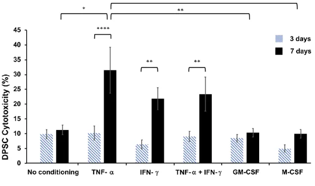
Following conditioning with TNF-, IFN-, TNF-+IFN-, GM-CSF or M-CSF, cytotoxicity of DPSCs was significantly higher after 7 days of TNF- (p<0.0001), IFN- (p=0.0022) and TNF-
+IFN- (p=0.0071) stimulation, compared to 3 days. Following 7 days of conditioning, TNF-
stimulated DPSCs had significantly higher cytotoxicity than those stimulated with GM-CSF (p=0.0068) or M-CSF (p=0.0053), and non-conditioned DPSCs (p=0.014) (Fig. 4).
Figure 4. Conditioning of DPSCs with rat pro-inflammatory cytokines has higher cytotoxicity following 7 days of conditioning. Conditioning with the pro-inflammatory cytokines TNF-, IFN-
and TNF-+IFN- is significantly more cytotoxic at 7 days of culture than 3. Values expressed as mean SEM; *p<0.05, **p<0.01, ****p<0.0001. n=6.
Cytotoxicity analysis was conducted following 3 and 7 days of co-culture of DPSCs and PBMCs.
Following 7 days of conditioning, cytotoxicity was significantly increased in non-conditioned Sham co-cultures (p=0.007), and non-conditioned (p=0.005) and TNF- stimulated (p=0.02) SCI co- cultures, compared to 3 days (Fig. 5A-F). No statistical significance was observed between different stimulation conditions following 3 days of conditioning (p=0.28) in either Sham or SCI groups.
Figure 5. Conditioning of DPSC+PBMC co-cultures with rat inflammatory cytokines reveals increased cytotoxicity following 7 days in TNF- and control conditions only. A No conditioning and conditioning with B TNF- is significantly more cytotoxic at 7 days of culture than 3 in co-cultures containing SCI PBMCs. No conditioning has greater cytotoxicity following 7 days of culture in co- cultures containing Sham PBMCs. Conditioning with C IFN-, D TNF-+IFN-, E GM-CSF and F M-CSF reveals no statistical differences between culture time-points. Values expressed as mean SEM; *p<0.05, **p<0.01. n=3.
Ex-vivo conditioning of DPSC+PBMC co-cultures with rat pro-inflammatory cytokines increases the release of IL-6 protein from DPSCs.
Quantification of cytokines released from DPSCs co-cultured with PBMCs from Sham and SCI animals following 3 days of conditioning was performed utilising Cytometric Bead Array assays.
Stimulation of DPSCs with TNF- and TNF-+IFN- significantly increased the release of IL-6 cytokine by DPSCs, within both Sham (p<0.0001) and SCI co-culture groups (p<0.0001), when compared to no conditioning (Fig. 6A). Additionally, IL-6 release was significantly higher following TNF-+IFN- stimulation in SCI co-cultures compared to Sham (p<0.0001). No significant differences in IL-1 release were observed between Sham and SCI animals (p=0.93; Fig. 6B) or within any stimulation condition (p=0.95). No changes in TNF- were detected due to the abundance of protein being below the lower detectable limits of the array assay (data not shown).
Figure 6. DPSC+PBMC co-culture with rat inflammatory cytokines increases the release of IL-6 protein from DPSCs. A IL-6 release is significantly increased following conditioning with TNF-
and TNF-+IFN-. TNF-+IFN- conditioning in SCI co-culture has significantly higher IL-6 release compared to Sham co-culture. B Changes in IL-1 release were not significant within any condition. Values expressed as mean SEM; ****p<0.0001. n=3.
Discussion
DPSC engraftment has previously been investigated as a strategy to regenerate the injured spinal cord65, 66 and address the lack of effective SCI treatment. However, low cell survival rates and viability within this hostile tissue environment limits translation from bench-to-bedside. Similarly, immune cell engraftment following SCI regulates pathological pro-inflammation following SCI,13, 34 but is limited in its regenerative efficacy. Therefore, this study aimed to characterise how SCI changes PBMC populations in a clinically relevant contusion model of SCI, and to identify critical timepoints for PBMC extraction and utilisation in novel DPSC preconditioning co-culture models that could increase survival and viability of engrafted cells. Additionally, co-culture was conducted to further develop the limited understanding of the immunomodulatory effects of DPSCs and PBMCs ex vivo.
Immune cells infiltrate the spinal cord within the first week following SCI, persisting at upregulated numbers for weeks to months75 and contribute to the exacerbation of secondary damage. FACS analysis showed acute, biphasic trends within each PBMC population following SCI, suggesting that PBMCs are recruited at these time-points, which could correspond to the biphasic opening of the blood-spinal cord-barrier at 1-2 and 6-10 dpi.76, 77 In parallel, the increases in T-cell and monocyte proportions at 1 and 7 dpi within our study coincide with T-cell and monocyte influxes within the spinal cord at the same time-points.26, 75, 78 Conversely, the decreased proportion of other PBMCs following SCI, such as B-cells, may be explained by immunodeficiency following SCI that limits the recruitment of immune cells to the spinal cord.79
Acute trends in Sham responses may be useful to decipher a functional immune response that, if mimicked, could promote healing following SCI, addressing the gap of no viable SCI treatment. In particular, because studies have shown that peripheral monocyte/macrophage activation is beneficial following SCI,13, 33, 38 their responses within Sham peripheral blood could be pivotal. In our study, CD43lowCD11bhigh and CD43midCD11blow monocyte sub-populations varied most between Sham and
SCI groups. CD43low monocytes have previously been described as ‘classically activated,’ and are involved in pathogen clearance and more pronounced pro-inflammatory functions.71, 80, 81
Contrastingly, CD43high ‘non-classically activated’ monocytes are smaller and less readily recruited within pro-inflammatory responses.82, 83 CD43lowCD11bhigh proportions remained higher in SCI compared to Shams, particularly at 3 dpi, thus corroborating CD43low descriptions. CD43mid monocytes have been suggested to be a transitional sub-population between the two subsets in humans and mice, but have not previously been identified in rats.83, 84 Additionally, low CD11b expression indicates an earlier state of monocyte/macrophage maturation.81, 85 Therefore, the novel CD43midCD11blow sub-population may be a developing subset more amenable to modulation.
However, further studies are needed to determine which level of CD43/CD11b expression is most beneficial for SCI recovery. Co-culture of monocytes with DPSCs within specific inflammatory conditions could then be a viable mechanism to achieve this expression. Additionally, further studies are needed to identify whether monocyte sub-populations exhibit anti- or pro-inflammatory phenotypic genes20, 86 following SCI utilising real-time quantitative polymerase chain reaction, in order to support the selection of a time-point for their extraction that is most viable for SCI immune cell administration therapies.
As no statistical significance was recorded, 3 dpi was chosen as the PBMC extraction time-point for culture experiments, due to consistent variations between Sham and SCI trends at this time-point in multiple PBMC populations. Additionally, this was done to provide ample time for co-culture whilst still remaining clinically relevant, in terms of administration of PBMC+DPSC cell therapy during a sub-acute time-point, shown to be most beneficial for stem cell engraftment.87 Within PBMC monoculture, 7 days of culture significantly increases cytotoxicity in all conditions. Interestingly, however, following 7 days of culture, only pro-inflammatory treatment with TNF-, IFN- and TNF-
+IFN- significantly increased cytotoxicity in DPSC monoculture. This is in line with previous studies that have highlighted the detrimental effects of TNF- and trophic effects of anti-
inflammatory cytokines on DPSC viability,88 and suggests that GM-CSF and M-CSF conditioning may limit cytotoxicity of DPSCs beyond 3 days of culture.
DPSC+PBMC co-culture was conducted to address the limited understanding of DPSC and PBMC interactions on cytotoxicity and immunomodulation. Once again, 3 days of culture was less cytotoxic than 7, particularly following TNF- conditioning. Compared to DPSC monoculture, cytotoxicity following TNF-, IFN- and TNF-+IFN- conditioning was reduced during the co-culture experiment, highlighting symbiotic immunomodulatory interactions between PBMCs and DPSCs reported previously.51, 54 This provides evidence that preconditioning of DPSCs and SCI PBMCs through co-culture may be beneficial before engraftment into the injured spinal cord. Similarly, 3 day culture is most effective at maintaining cell viability, answering one of the main gaps addressed in this study. However, further studies are necessary to confirm the exact mechanisms by which cytotoxicity is reduced following co-culture.
The cytokine assay showed, for the first time, that within TNF- and TNF-+IFN- pro- inflammatory conditions, DPSCs significantly upregulate their secretion of IL-6, particularly following co-culture with SCI PBMCs. IL-6 protects DPSCs and MSCs from apoptosis, aiding wound healing in vitro following inflammation89 and increasing proliferation.90 Importantly, IL-6 is released during DPSC and MSC immunoregulatory functions,91 and works to inhibit the synthesis of pro- inflammatory factors such as IL-1 and TNF-.92 Additionally, DPSC IL-6 secretion aids in anti- inflammatory macrophage polarisation.93 These findings support the premise that co-culture of SCI PBMCs with DPSCs under pro-inflammatory conditions induces anti-inflammatory functional behaviour of DPSCs, which may modulate PBMCs to acquire anti-inflammatory phenotypes, thus answering a key gap in knowledge. However, conflicting studies suggest that IL-6 release is detrimental to recovery following SCI, due to its involvement in exacerbating pro-inflammation,94 recruiting inflammatory cells,95, 96 and neuropathic pain.97 Therefore, further in vivo studies are
needed to identify whether engraftment of DPSCs secreting high levels of IL-6 into a SCI model worsens recovery, or whether the immunomodulatory properties of DPSCs inhibit the negative effects of IL-6. In parallel, investigation should be conducted into whether particular time-points following SCI, during which TNF- levels are highest, should be avoided for DPSC engraftment.
This study has characterised changes in PBMC populations following SCI, and has shown that PBMCs extracted 3 dpi are suitable to achieve co-culture of PBMCs and DPSCs that has low cytotoxicity when cultured for 3 days, thus supporting our main hypothesis. Furthermore, we have shown that co-culture within pro-inflammatory conditions may be beneficial to induce anti- inflammatory functions of DPSCs. Future in vivo studies are necessary to elucidate the effects of this preconditioning on DPSC and PBMC survival and viability following engraftment into a SCI model, which may lead to the development of a novel neuroregenerative and immunomodulatory SCI treatment.
Acknowledgment
This study was financially supported by the AOSpine Discovery and Innovation Research Grant (AOSDIA2019_081_SCI_OHareDoig), AOSpine Asia Pacific Research Grant (AOSAUNZR2019- 05) and the Neil Sachse Centre for Spinal Cord Research. Thank you to Dr Randall Grose at SAHMRI Flow and Laser Scanning Facility for assistance in flow data capture, as well as Honours candidate Aleesha Searle for assistance in animal handling and initial tissue collection. Thank you also to my supervisors, Dr Ryan O’Hare Doig, A/Prof Jillian Clark and Prof Stan Gronthos.
References
1. McDonald JW & Howard MJ (2002). Repairing the damaged spinal cord: a summary of our early success with embryonic stem cell transplantation and remyelination. Prog Brain Res 137, 299-309.
2. Migliorini C, Tonge B & Taleporos G (2008). Spinal cord injury and mental health. Aust N Z J Psychiatry 42, 309-14.
3. New PW, Baxter D, Farry A & Noonan VK (2015). Estimating the incidence and prevalence of traumatic spinal cord injury in Australia. Arch Phys Med Rehabil 96, 76-83.
4. Tovell A, Spinal cord injury, Australia, 2016–17. 2020, Australian Institute of Health and Welfare: Canberra
5. York EM, Petit A & Roskams AJ (2013). Epigenetics of neural repair following spinal cord injury. Neurotherapeutics 10, 757-70.
6. Schwab ME & Bartholdi D (1996). Degeneration and regeneration of axons in the lesioned spinal cord. Physiol Rev 76, 319-70.
7. Rowland JW, Hawryluk GW, Kwon B & Fehlings MG (2008). Current status of acute spinal cord injury pathophysiology and emerging therapies: promise on the horizon. Neurosurg Focus 25, E2.
8. Sekhon LH & Fehlings MG (2001). Epidemiology, demographics, and pathophysiology of acute spinal cord injury. Spine (Phila Pa 1976) 26, S2-12.
9. O'Hare Doig RL, Santhakumar S, Fehily B, Raja S, Solomon T, Bartlett CA, Fitzgerald M &
Hodgetts SI (2020). Acute Cellular and Functional Changes With a Combinatorial
Treatment of Ion Channel Inhibitors Following Spinal Cord Injury. Front Mol Neurosci 13, 85.
10. Pineau I & Lacroix S (2007). Proinflammatory cytokine synthesis in the injured mouse spinal cord: multiphasic expression pattern and identification of the cell types involved. J Comp Neurol 500, 267-85.
11. Lou J, Lenke LG, Ludwig FJ & O'Brien MF (1998). Apoptosis as a mechanism of neuronal cell death following acute experimental spinal cord injury. Spinal Cord 36, 683-90.
12. Scholpa NE & Schnellmann RG (2017). Mitochondrial-Based Therapeutics for the
Treatment of Spinal Cord Injury: Mitochondrial Biogenesis as a Potential Pharmacological Target. J Pharmacol Exp Ther 363, 303-313.
13. Rapalino O, Lazarov-Spiegler O, Agranov E, Velan GJ, Yoles E, Fraidakis M, Solomon A, Gepstein R, Katz A, Belkin M, Hadani M & Schwartz M (1998). Implantation of stimulated homologous macrophages results in partial recovery of paraplegic rats. Nat Med 4, 814-21.
14. Biswas SK & Mantovani A (2010). Macrophage plasticity and interaction with lymphocyte subsets: cancer as a paradigm. Nat Immunol 11, 889-96.
15. Martinez FO & Gordon S (2014). The M1 and M2 paradigm of macrophage activation: time for reassessment. F1000Prime Rep 6, 13.
16. McTigue DM & Tripathi RB (2008). The life, death, and replacement of oligodendrocytes in the adult CNS. J Neurochem 107, 1-19.
17. Cherry JD, Olschowka JA & O'Banion MK (2014). Neuroinflammation and M2 microglia:
the good, the bad, and the inflamed. J Neuroinflammation 11, 98.
18. Mantovani A, Biswas SK, Galdiero MR, Sica A & Locati M (2013). Macrophage plasticity and polarization in tissue repair and remodelling. J Pathol 229, 176-85.
19. Thompson CD, Zurko JC, Hanna BF, Hellenbrand DJ & Hanna A (2013). The therapeutic role of interleukin-10 after spinal cord injury. J Neurotrauma 30, 1311-24.
20. Kigerl KA, Gensel JC, Ankeny DP, Alexander JK, Donnelly DJ & Popovich PG (2009).
Identification of two distinct macrophage subsets with divergent effects causing either neurotoxicity or regeneration in the injured mouse spinal cord. J Neurosci 29, 13435-44.
21. Schwartz M & Baruch K (2014). The resolution of neuroinflammation in
neurodegeneration: leukocyte recruitment via the choroid plexus. EMBO J 33, 7-22.
22. David S, Greenhalgh AD & Kroner A (2015). Macrophage and microglial plasticity in the injured spinal cord. Neuroscience 307, 311-8.
23. Fleming JC, Norenberg MD, Ramsay DA, Dekaban GA, Marcillo AE, Saenz AD, Pasquale- Styles M, Dietrich WD & Weaver LC (2006). The cellular inflammatory response in human spinal cords after injury. Brain 129, 3249-69.
24. Taoka Y, Okajima K, Uchiba M, Murakami K, Kushimoto S, Johno M, Naruo M, Okabe H
& Takatsuki K (1997). Role of neutrophils in spinal cord injury in the rat. Neuroscience 79, 1177-82.
25. Trivedi A, Olivas AD & Noble-Haeusslein LJ (2006). Inflammation and Spinal Cord Injury:
Infiltrating Leukocytes as Determinants of Injury and Repair Processes. Clin Neurosci Res 6, 283-292.
26. Donnelly DJ & Popovich PG (2008). Inflammation and its role in neuroprotection, axonal regeneration and functional recovery after spinal cord injury. Exp Neurol 209, 378-88.
27. Ferguson AR, Christensen RN, Gensel JC, Miller BA, Sun F, Beattie EC, Bresnahan JC &
Beattie MS (2008). Cell death after spinal cord injury is exacerbated by rapid TNF alpha- induced trafficking of GluR2-lacking AMPARs to the plasma membrane. J Neurosci 28, 11391-400.
28. Nesic O, Xu GY, McAdoo D, High KW, Hulsebosch C & Perez-Pol R (2001). IL-1 receptor antagonist prevents apoptosis and caspase-3 activation after spinal cord injury. J
Neurotrauma 18, 947-56.
29. Batchelor PE, Tan S, Wills TE, Porritt MJ & Howells DW (2008). Comparison of
inflammation in the brain and spinal cord following mechanical injury. J Neurotrauma 25, 1217-25.
30. Zhang B & Gensel JC (2014). Is neuroinflammation in the injured spinal cord different than in the brain? Examining intrinsic differences between the brain and spinal cord. Exp Neurol 258, 112-20.
31. Ito Y, Sugimoto Y, Tomioka M, Kai N & Tanaka M (2009). Does high dose
methylprednisolone sodium succinate really improve neurological status in patient with acute cervical cord injury?: a prospective study about neurological recovery and early complications. Spine (Phila Pa 1976) 34, 2121-4.
32. Casha S, Zygun D, McGowan MD, Bains I, Yong VW & Hurlbert RJ (2012). Results of a phase II placebo-controlled randomized trial of minocycline in acute spinal cord injury.
Brain 135, 1224-36.
33. Prewitt CM, Niesman IR, Kane CJ & Houlé JD (1997). Activated macrophage/microglial cells can promote the regeneration of sensory axons into the injured spinal cord. Exp Neurol 148, 433-43.
34. Bomstein Y, Marder JB, Vitner K, Smirnov I, Lisaey G, Butovsky O, Fulga V & Yoles E (2003). Features of skin-coincubated macrophages that promote recovery from spinal cord injury. J Neuroimmunol 142, 10-6.
35. Knoller N, Auerbach G, Fulga V, Zelig G, Attias J, Bakimer R, Marder JB, Yoles E, Belkin M, Schwartz M & Hadani M (2005). Clinical experience using incubated autologous macrophages as a treatment for complete spinal cord injury: phase I study results. J Neurosurg Spine 3, 173-81.
36. Shechter R & Schwartz M (2013). Harnessing monocyte-derived macrophages to control central nervous system pathologies: no longer 'if' but 'how'. J Pathol 229, 332-46.
37. London A, Cohen M & Schwartz M (2013). Microglia and monocyte-derived macrophages:
functionally distinct populations that act in concert in CNS plasticity and repair. Front Cell Neurosci 7, 34.
38. Shechter R, London A, Varol C, Raposo C, Cusimano M, Yovel G, Rolls A, Mack M, Pluchino S, Martino G, Jung S & Schwartz M (2009). Infiltrating blood-derived
macrophages are vital cells playing an anti-inflammatory role in recovery from spinal cord injury in mice. PLoS Med 6, e1000113.
39. Lammertse DP, Jones LA, Charlifue SB, Kirshblum SC, Apple DF, Ragnarsson KT, Falci SP, Heary RF, Choudhri TF, Jenkins AL, Betz RR, Poonian D, Cuthbert JP, Jha A, Snyder DA & Knoller N (2012). Autologous incubated macrophage therapy in acute, complete spinal cord injury: results of the phase 2 randomized controlled multicenter trial. Spinal Cord 50, 661-71.
40. Yeung TY, Seeberger KL, Kin T, Adesida A, Jomha N, Shapiro AM & Korbutt GS (2012).
Human mesenchymal stem cells protect human islets from pro-inflammatory cytokines.
PLoS One 7, e38189.
41. Kolios G & Moodley Y (2013). Introduction to stem cells and regenerative medicine.
Respiration 85, 3-10.
42. Meirelles Lda S & Nardi NB (2009). Methodology, biology and clinical applications of mesenchymal stem cells. Front Biosci (Landmark Ed) 14, 4281-98.
43. Phinney DG & Prockop DJ (2007). Concise review: mesenchymal stem/multipotent stromal cells: the state of transdifferentiation and modes of tissue repair--current views. Stem Cells 25, 2896-902.
44. Kim J & Hematti P (2009). Mesenchymal stem cell-educated macrophages: a novel type of alternatively activated macrophages. Exp Hematol 37, 1445-53.
45. Krampera M, Glennie S, Dyson J, Scott D, Laylor R, Simpson E & Dazzi F (2003). Bone marrow mesenchymal stem cells inhibit the response of naive and memory antigen-specific T cells to their cognate peptide. Blood 101, 3722-9.
46. Di Nicola M, Carlo-Stella C, Magni M, Milanesi M, Longoni PD, Matteucci P, Grisanti S &
Gianni AM (2002). Human bone marrow stromal cells suppress T-lymphocyte proliferation induced by cellular or nonspecific mitogenic stimuli. Blood 99, 3838-43.
47. White SV, Czisch CE, Han MH, Plant CD, Harvey AR & Plant GW (2016). Intravenous Transplantation of Mesenchymal Progenitors Distribute Solely to the Lungs and Improve Outcomes in Cervical Spinal Cord Injury. Stem Cells 34, 1812-25.
48. Nakajima H, Uchida K, Guerrero AR, Watanabe S, Sugita D, Takeura N, Yoshida A, Long G, Wright KT, Johnson WE & Baba H (2012). Transplantation of mesenchymal stem cells promotes an alternative pathway of macrophage activation and functional recovery after spinal cord injury. J Neurotrauma 29, 1614-25.
49. Gyorki DE, Asselin-Labat ML, van Rooijen N, Lindeman GJ & Visvader JE (2009).
Resident macrophages influence stem cell activity in the mammary gland. Breast Cancer Res 11, R62.
50. London A, Itskovich E, Benhar I, Kalchenko V, Mack M, Jung S & Schwartz M (2011).
Neuroprotection and progenitor cell renewal in the injured adult murine retina requires healing monocyte-derived macrophages. J Exp Med 208, 23-39.
51. Freytes DO, Kang JW, Marcos-Campos I & Vunjak-Novakovic G (2013). Macrophages modulate the viability and growth of human mesenchymal stem cells. J Cell Biochem 114, 220-9.
52. Shi S, Robey PG & Gronthos S (2001). Comparison of human dental pulp and bone marrow stromal stem cells by cDNA microarray analysis. Bone 29, 532-9.
53. Ashri NY, Ajlan SA & Aldahmash AM (2015). Dental pulp stem cells. Biology and use for periodontal tissue engineering. Saudi Med J 36, 1391-9.
54. Wada N, Menicanin D, Shi S, Bartold PM & Gronthos S (2009). Immunomodulatory properties of human periodontal ligament stem cells. J Cell Physiol 219, 667-76.
55. Pierdomenico L, Bonsi L, Calvitti M, Rondelli D, Arpinati M, Chirumbolo G, Becchetti E, Marchionni C, Alviano F, Fossati V, Staffolani N, Franchina M, Grossi A & Bagnara GP (2005). Multipotent mesenchymal stem cells with immunosuppressive activity can be easily isolated from dental pulp. Transplantation 80, 836-42.
56. Demircan PC, Sariboyaci AE, Unal ZS, Gacar G, Subasi C & Karaoz E (2011).
Immunoregulatory effects of human dental pulp-derived stem cells on T cells: comparison of transwell co-culture and mixed lymphocyte reaction systems. Cytotherapy 13, 1205-20.
57. Li Y, Yang YY, Ren JL, Xu F, Chen FM & Li A (2017). Exosomes secreted by stem cells from human exfoliated deciduous teeth contribute to functional recovery after traumatic brain injury by shifting microglia M1/M2 polarization in rats. Stem Cell Res Ther 8, 198.
58. Rossato C, Brandao WN, Castro SBR, de Almeida DC, Maranduba CMC, Camara NOS, Peron JPS & Silva FS (2017). Stem cells from human-exfoliated deciduous teeth reduce tissue-infiltrating inflammatory cells improving clinical signs in experimental autoimmune encephalomyelitis. Biologicals 49, 62-68.
59. Gronthos S, Mankani M, Brahim J, Robey PG & Shi S (2000). Postnatal human dental pulp stem cells (DPSCs) in vitro and in vivo. Proc Natl Acad Sci U S A 97, 13625-30.
60. Gronthos S, Arthur A, Bartold PM & Shi S (2011). A method to isolate and culture expand human dental pulp stem cells. Methods Mol Biol 698, 107-21.
61. Nishino Y, Yamada Y, Ebisawa K, Nakamura S, Okabe K, Umemura E, Hara K & Ueda M (2011). Stem cells from human exfoliated deciduous teeth (SHED) enhance wound healing and the possibility of novel cell therapy. Cytotherapy 13, 598-605.
62. Li B, Chen X & Jin Y (2018). 1st ed. A Roadmap to Non-hematopoietic Stem Cell-Based Therapeutics: From the Bench to the Clinic. Academic Press, Cambridge, US.
63. Furuya T, Hashimoto M, Koda M, Okawa A, Murata A, Takahashi K, Yamashita T &
Yamazaki M (2009). Treatment of rat spinal cord injury with a Rho-kinase inhibitor and bone marrow stromal cell transplantation. Brain Res 1295, 192-202.
64. Leong WK, Henshall TL, Arthur A, Kremer KL, Lewis MD, Helps SC, Field J, Hamilton- Bruce MA, Warming S, Manavis J, Vink R, Gronthos S & Koblar SA (2012). Human adult dental pulp stem cells enhance poststroke functional recovery through non-neural
replacement mechanisms. Stem Cells Transl Med 1, 177-87.
65. Nosrat IV, Widenfalk J, Olson L & Nosrat CA (2001). Dental pulp cells produce
neurotrophic factors, interact with trigeminal neurons in vitro, and rescue motoneurons after spinal cord injury. Dev Biol 238, 120-32.
66. Sakai K, Yamamoto A, Matsubara K, Nakamura S, Naruse M, Yamagata M, Sakamoto K, Tauchi R, Wakao N, Imagama S, Hibi H, Kadomatsu K, Ishiguro N & Ueda M (2012).
Human dental pulp-derived stem cells promote locomotor recovery after complete
transection of the rat spinal cord by multiple neuro-regenerative mechanisms. J Clin Invest 122, 80-90.
67. Lu P, Yang H, Jones LL, Filbin MT & Tuszynski MH (2004). Combinatorial therapy with neurotrophins and cAMP promotes axonal regeneration beyond sites of spinal cord injury. J Neurosci 24, 6402-9.
68. Muniswami DM & Tharion G (2018). Therapeutic Effect of Cell Transplantation and Chondroitinase in Rat Spinal Cord Injury. Int J Appl Basic Med Res 8, 220-226.
69. Scheff SW, Rabchevsky AG, Fugaccia I, Main JA & Lumpp JE, Jr. (2003). Experimental modeling of spinal cord injury: characterization of a force-defined injury device. J
Neurotrauma 20, 179-93.
70. Hønge BL, Petersen MS, Olesen R, Møller BK & Erikstrup C (2017). Optimizing recovery of frozen human peripheral blood mononuclear cells for flow cytometry. PLoS One 12, e0187440.
71. Barnett-Vanes A, Sharrock A, Birrell MA & Rankin S (2016). A Single 9-Colour Flow Cytometric Method to Characterise Major Leukocyte Populations in the Rat: Validation in a Model of LPS-Induced Pulmonary Inflammation. PLoS One 11, e0142520.
72. Miura M, Gronthos S, Zhao M, Lu B, Fisher LW, Robey PG & Shi S (2003). SHED: stem cells from human exfoliated deciduous teeth. Proc Natl Acad Sci U S A 100, 5807-12.
73. Kmonícková E, Melkusová P, Farghali H, Holý A & Zídek Z (2007). Nitric oxide
production in mouse and rat macrophages: a rapid and efficient assay for screening of drugs immunostimulatory effects in human cells. Nitric Oxide 17, 160-9.
74. Riss T, Niles A, Moravec R, Karassina N & Vidugiriene J (2004). Cytotoxicity Assays: In Vitro Methods to Measure Dead Cells. In Assay Guidance Manual, 1st edn, ed. Markossian S, Sittampalam GS, Grossman A, Brimacombe K, Arkin M, Auld D, Austin CP, Baell J, Caaveiro JMM, Chung TDY, Coussens NP, Dahlin JL, Devanaryan V, Foley TL,
Glicksman M, Hall MD, Haas JV, Hoare SRJ, Inglese J, Iversen PW, Kahl SD, Kales SC, Kirshner S, Lal-Nag M, Li Z, McGee J, McManus O, Riss T, Saradjian P, Trask OJ, Jr., Weidner JR, Wildey MJ, Xia M & Xu X. Eli Lilly & Company and the National Center for Advancing Translational Sciences, Bethesda
75. Beck KD, Nguyen HX, Galvan MD, Salazar DL, Woodruff TM & Anderson AJ (2010).
Quantitative analysis of cellular inflammation after traumatic spinal cord injury: evidence for a multiphasic inflammatory response in the acute to chronic environment. Brain 133, 433-47.
76. Popovich PG, Horner PJ, Mullin BB & Stokes BT (1996). A quantitative spatial analysis of the blood-spinal cord barrier. I. Permeability changes after experimental spinal contusion injury. Exp Neurol 142, 258-75.
77. Noble LJ & Wrathall JR (1989). Distribution and time course of protein extravasation in the rat spinal cord after contusive injury. Brain Res 482, 57-66.
78. Popovich PG, Wei P & Stokes BT (1997). Cellular inflammatory response after spinal cord injury in Sprague-Dawley and Lewis rats. J Comp Neurol 377, 443-64.
79. Prüss H, Tedeschi A, Thiriot A, Lynch L, Loughhead SM, Stutte S, Mazo IB, Kopp MA, Brommer B, Blex C, Geurtz LC, Liebscher T, Niedeggen A, Dirnagl U, Bradke F, Volz MS, DeVivo MJ, Chen Y, von Andrian UH & Schwab JM (2017). Spinal cord injury-induced immunodeficiency is mediated by a sympathetic-neuroendocrine adrenal reflex. Nat Neurosci 20, 1549-1559.
80. Scriba A, Schneider M, Grau V, van der Meide PH & Steiniger B (1997). Rat monocytes up-regulate NKR-P1A and down-modulate CD4 and CD43 during activation in vivo:
monocyte subpopulations in normal and IFN-gamma-treated rats. J Leukoc Biol 62, 741-52.
81. Yrlid U, Jenkins CD & MacPherson GG (2006). Relationships between distinct blood monocyte subsets and migrating intestinal lymph dendritic cells in vivo under steady-state conditions. J Immunol 176, 4155-62.
82. Gordon S & Taylor PR (2005). Monocyte and macrophage heterogeneity. Nat Rev Immunol 5, 953-64.
83. Melgert BN, Spaans F, Borghuis T, Klok PA, Groen B, Bolt A, de Vos P, van Pampus MG, Wong TY, van Goor H, Bakker WW & Faas MM (2012). Pregnancy and preeclampsia affect monocyte subsets in humans and rats. PLoS One 7, e45229.
84. Ziegler-Heitbrock L, Ancuta P, Crowe S, Dalod M, Grau V, Hart DN, Leenen PJ, Liu YJ, MacPherson G, Randolph GJ, Scherberich J, Schmitz J, Shortman K, Sozzani S, Strobl H, Zembala M, Austyn JM & Lutz MB (2010). Nomenclature of monocytes and dendritic cells in blood. Blood 116, e74-80.
85. Yu E, Goto M, Ueta H, Kitazawa Y, Sawanobori Y, Kariya T, Sasaki M & Matsuno K (2016). Expression of area-specific M2-macrophage phenotype by recruited rat monocytes in duct-ligation pancreatitis. Histochem Cell Biol 145, 659-73.
86. Gordon S (2003). Alternative activation of macrophages. Nat Rev Immunol 3, 23-35.
87. Parr AM, Kulbatski I & Tator CH (2007). Transplantation of adult rat spinal cord stem/progenitor cells for spinal cord injury. J Neurotrauma 24, 835-45.
88. Giacomelli C, Natali L, Nisi M, De Leo M, Daniele S, Costa B, Graziani F, Gabriele M, Braca A, Trincavelli ML & Martini C (2018). Negative effects of a high tumour necrosis factor-α concentration on human gingival mesenchymal stem cell trophism: the use of natural compounds as modulatory agents. Stem Cell Res Ther 9, 135.
89. Pricola KL, Kuhn NZ, Haleem-Smith H, Song Y & Tuan RS (2009). Interleukin-6 maintains bone marrow-derived mesenchymal stem cell stemness by an ERK1/2-dependent
mechanism. J Cell Biochem 108, 577-88.
90. Xie Z, Tang S, Ye G, Wang P, Li J, Liu W, Li M, Wang S, Wu X, Cen S, Zheng G, Ma M, Wu Y & Shen H (2018). Interleukin-6/interleukin-6 receptor complex promotes osteogenic differentiation of bone marrow-derived mesenchymal stem cells. Stem Cell Res Ther 9, 13.
91. Bonaventura G, Incontro S, Iemmolo R, La Cognata V, Barbagallo I, Costanzo E, Barcellona ML, Pellitteri R & Cavallaro S (2020). Dental mesenchymal stem cells and neuro-regeneration: a focus on spinal cord injury. Cell Tissue Res 379, 421-428.
92. Steinke JW, Rosenwasser LJ & Borish L (2014). Cytokines in Allergic Inflammation. In Middleton's Allergy: Principles and Practice, 8th edn, ed. Adkinson F BB, Burks AW, pp.
65-82. Elsevier, Philadelphia, PA.
93. Zhou LL, Liu W, Wu YM, Sun WL, Dörfer CE & Fawzy El-Sayed KM (2020). Oral
Mesenchymal Stem/Progenitor Cells: The Immunomodulatory Masters. Stem Cells Int 2020, 1327405.
94. Guerrero AR, Uchida K, Nakajima H, Watanabe S, Nakamura M, Johnson WE & Baba H (2012). Blockade of interleukin-6 signaling inhibits the classic pathway and promotes an alternative pathway of macrophage activation after spinal cord injury in mice. J
Neuroinflammation 9, 40.
95. Okada S, Nakamura M, Mikami Y, Shimazaki T, Mihara M, Ohsugi Y, Iwamoto Y, Yoshizaki K, Kishimoto T, Toyama Y & Okano H (2004). Blockade of interleukin-6
receptor suppresses reactive astrogliosis and ameliorates functional recovery in experimental spinal cord injury. J Neurosci Res 76, 265-76.
96. Nakamura M, Okada S, Toyama Y & Okano H (2005). Role of IL-6 in spinal cord injury in a mouse model. Clin Rev Allergy Immunol 28, 197-204.
97. Zhou YQ, Liu Z, Liu ZH, Chen SP, Li M, Shahveranov A, Ye DW & Tian YK (2016).
Interleukin-6: an emerging regulator of pathological pain. J Neuroinflammation 13, 141.