1
COMPARATIVE ANALYSIS OF ANTIMICROBIAL RESISTANCE PATTERN AND PATHOGENIC CHARACTERIZATION OF Staphylococcus aureus
ISOLATED FROM HOSPITAL EFFLUENTS
WASTEWATER AND ITS ADJACENT COMMUNITIES IN DHAKA CITY.
By Hasib Mahmud
18326018 Jahid Hasan Tushar
18326021 Sanjeda Haque
18126054
A thesis submitted to the Department of Mathematics and Natural Sciences in partial fulfillment of the requirements for the degree of
Bachelor of Science in Microbiology
Microbiology Program,
Department of Mathematics and Natural Sciences BRAC University
February 2023
© 2023. BRAC University All rights reserved
2
Declaration
It is hereby declared that
1. The thesis submitted titled “Comparative analysis of antimicrobial resistance pattern and pathogenic characterization of Staphylococcus aureus isolated from hospital effluents wastewater and its adjacent communities in Dhaka city” is our own original work while completing our degree at Brac University.
2. The thesis does not contain material previously published or written by a third party, except where this is appropriately cited through full and accurate referencing.
3. The thesis does not contain material which has been accepted, or submitted, for any other degree or diploma at a university or other institution.
4. We have acknowledged all main sources of help
Student’s Full Name & Signature:
Hasib Mahmud 18326018
Jahid Hasan Tushar 18326021
Sanjeda Haque 18126054
3
Approval
The thesis titled “Comparative analysis of antimicrobial resistance pattern and pathogenic characterization of Staphylococcus aureus isolated from hospital effluents wastewater and its adjacent communities in Dhaka city” submitted by
1. Hasib Mahmud, 18326018 2. Jahid Hasan Tushar, 18326021 3. Sanjeda Haque, 18126054
has been accepted as satisfactory in partial fulfillment of the requirement for the degree of Microbiology in February 2023.
Examining Committee:
Supervisor:
(Member)
_______________________________
Akash Ahmed Senior Lecturer,
Department of Mathematics and Natural Sciences BRAC University
Program Coordinator:
(Member)
_______________________________
Dr. Nadia Sultana Deen Associate Professor,
Department of Mathematics and Natural Sciences BRAC University
Departmental Head:
(Chair)
_______________________________
A.F.M Yusuf Haider Professor and Chairperson,
Department of Mathematics and Natural Sciences BRAC University
4
Ethics Statement
For the completion of this study, samples from selected venues were collected following all the necessary precautions. All the experiments were done in BRAC University Life Sciences Laboratory. It should also be noted that no animal or human models were used in this study.
5
Abstract
Staphylococcus aureus is Gram-positive cocci, facultatively aerobic that has the intrinsic ability to ferment carbohydrates and forms clusters. Staphylococcus aureus is responsible for numerous pyogenic infections, food poisoning, and toxic shock syndrome, and it can produce a wide range of virulence factors. S. aureus strains that are resistant to virtually all antibiotics, with the exception of Vancomycin, have emerged in recent years. Hospital wastewater has a direct influential role in the spread of infectious diseases in healthcare settings, community settings, hospital employees, and the environment. HWW is a significant source of ARGs and ARB, and its infectious and toxic characteristics make it extremely hazardous.
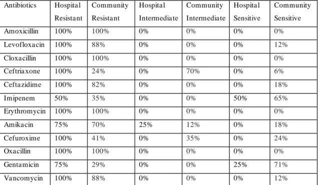
A total of 70 samples were collected from our study sampling sites in several phases from the period of June 2022 to December 2022. From the 70 samples, 21 PCR-confirmed staphylococcus aureus isolates were obtained which was 30% of the sample size. It was explored that 100% of the isolates from hospital effluents were significantly resistant to 9 antibiotics (Amoxicillin, Levofloxacin, Cloxacillin, Ceftriaxone, Ceftazidime, Erythromycin, Cefuroxime, Oxacillin, and Vancomycin), whereas 100% of the isolates from communities tap water showed resistance to 4 antibiotics (Amoxicillin, Cloxacillin, Erythromycin, and Oxacillin).
The result of our study showed the emergence of ARGs in the strains of Staphylococcus aureus in the community setting has increased significantly. These ARBs and ARGs were hypothesized to be transmitted from the hospital settings by the hospital’s untreated effluents Keywords: Staphylococcus aureus, ARGs, ARBs , MRSA, Multi-drug resistant, Hospital wastewater
6
Dedication
We would like to dedicate this thesis to our parents. Thank you so much for everything! Words can hardly describe our thanks and appreciation to you. They have been our source of inspiration, support, and guidance. they have taught us to be unique, and determined, to believe in the process, and to always persevere. To take a quote from Albert Schweitzer, “At times our own light goes out and is rekindled by a spark from another person. Each of us has cause to think with deep gratitude of those who have lighted the flame within us.” Our parents have been that spark for us when our light blew out. Thank you for your unwavering love and support along this journey we have taken.
This research is also dedicated to all the members of group “KSHJ” for their sacrifice and cooperation, for supporting one another through the ups and downs of nine long months for completing this huge task like never experienced before.
7
Acknowledgment
We would like to begin by expressing our gratitude to Almighty Allah for providing us with the opportunity and strength to complete this research. Additionally, we are grateful for His blessings on our day-to-day lives, good health, and sound mind. We acknowledge our esteem to Professor A F M Yusuf Haider, Ph.D., Professor and Chairperson of the Department of Mathematics and Natural Sciences, BRAC University, for allowing and encouraging us to complete our undergraduate thesis.
Our regards, gratitude, indebtedness, and appreciation go to our respected supervisor Akash Ahmed, Senior Lecturer, Department of Mathematics and Natural Sciences, BRAC University for his constant supervision, constructive criticism, expert guidance, enthusiastic encouragement to pursue new ideas, and never-ending inspiration throughout the entire period of our research work. We would also like to express gratitude toward Md Hasanuzzaman, Senior Lecturer, Microbiology Program, Department of Mathematics and Natural Sciences, BRAC University for his guidance and assistance.
Our sincere appreciation goes out to Laboratory Officers Md. Mahmudul Hasan, Shamim Akhter Chowdhury, and Asma Binte Afzal for their unwavering kindness and advice.
Additionally, we would like to express our gratitude to our Research assistants Mohammad Aziz Hossain and Nishat Tasnim Ananna for their exceptional kindness and assistance. We would like to express our sincere gratitude to you for guiding us through the writing of the report and occasionally offering suggestions regarding the setting of experimental designs, the interpretation of results, and subsequent directions for the entire project. Without their warm assistance, it would have been impossible to submit our report. In addition, we would like to express sincere and loving gratitude to our lab-mate Tasfia Tasnim Toma and Shamima Nasreen for all their moral support and help throughout this roller-coaster journey.
Additionally, we would like to express our appreciation to lab attendants Ashik-E Khuda, Tanzila Alam, and office attendant Nadira Yeasmin for their assistance during our nine months in the laboratory. Last but not least, we would like to express our gratitude to everyone who assisted us in the laboratory and worked alongside us.
8
Table of Contents
Declaration………2
Approval………3
Ethics Statement………...4
Abstract/ Executive Summary………5
Dedication (Optional)………..6
Acknowledgment……….….7
Table of Contents……….8
List of Tables………...11
List of Figures……….…12
List of Acronyms………....13
Chapter 1: Introduction ………..15-19 1.1 Background………..15
1.2 Transmission of antibiotic-resistant pathogens from hospital effluents to community settings………..18
Chapter 2: Literature Review………20-31 2.1 Staphylococcus aureus………....20
2.2 Virulence factors……….…21
2.3 Prevalence of MRSA (Methicillin Resistant Staphylococcus aureus)……….…...22
2.4 Hospital wastewater as a potential reservoir for ARBs and ARGs………....24
2.5 Acquisition of ARGs via horizontal gene transfer………..…26
2.5.1 Natural competence and transformation………..…27
2.5.2 Transduction……….…28
2.5.3 Conjugation mobilizes plasmid-borne ARGs………..29
2.6 Knowledge gap in the existing literature………...30
9
2.7 The novelty of Our Study……….………30
2.8 Aims, Objectives, and Hypothesis……….……...31
Chapter 3: Methods and Materials……….32-40 3.1 Sample site selection………32
3.2 Sample collection ………33
3.3 Sample processing………34
3.4 Sample enrichment and growth on selective media……….34
3.5 Colony Morphology, selection, and analysis………35
3.6 Molecular detection………..35
3.6.1 DNA extraction……….35
3.6.2 Preparation of Primers from stock solution for PCR………36
3.6.3 Preparation of controls for PCR………37
3.6.4 PCR Assay………37
3.6.5 Gel electrophoresis……….38
3.7 Antimicrobial susceptibility testing………38
3.8 Pathogenicity screening test ………..39
3.8.1 Coagulase Test……….39
3.8.2 DNase Test………..40
Chapter 4: Result and observations……….41-53 4.1 Isolation of Staphylococcus aureus ………41
4.2 PCR-based identification of Staphylococcus aureus: result interpretation…….42
4.3 Distribution of S isolates: month-wise……….43
4.4 Distribution of S. aureus: based on the sampling sites………44
4.5 Antimicrobial Susceptibility Test - Result………..45
10
4.5.1 Antimicrobial resistance pattern of total isolates ……….45
4.5.2 Antimicrobial resistance pattern in isolates of Hospital effluents ………47
4.5.3 Antimicrobial resistance pattern in isolates of Hospital adjacent communities....48
4.5.4 Comparative analysis of AMR pattern between the isolates of Hospital effluents isolates of Hospital adjacent communities ………50
4.5.5 Prevalence of Oxacillin resistant Staphylococcus aureus ………51
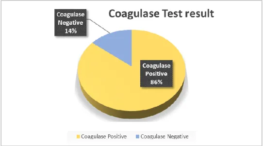
4.6 Pathogenicity Screening test result ……….51
4.6.1 Coagulase test ………..51
4.6.2 DNase test……….53
Chapter 5: Discussion………...54-57 5.1 Result analysis-based discussion ………54
5.2 Limitations of our study ……….56
Chapter 6: Conclusions………57-58 6.1 Recommendations and conclusions……….57 Chapter 7: References………..59-65
11
List of Tables
Table 1: Sequences of primers used for amplification by PCR……….22
Table 2: Antibiotics disc list used in this study with CLSI interpretation………..25
Table 3: Antimicrobial resistance pattern of total isolates……….32
Table 4: Antimicrobial resistance pattern in isolates of Hospital effluents………33
Table 5: Antimicrobial resistance pattern in isolates of Hospital adjacent communities…..35
Table 6: Comparative analysis of AMR pattern between the isolates of Hospital effluents isolates of Hospital adjacent communities………..37
12
List of Figures
Figure 1: GIS map of sampling sites………18
Figure 2: Illustration of Sample sites selection………19
Figure 3: The appearance of yellow colonies on MSA………...…….27
Figure 4: TStaG422PCR for detecting the Genus Staphylococcus, 50bp ladder was used…..28
Figure 5: Sa442 PCR for detecting the Staphylococcus aureus, 100 bp ladder was used…...28
Figure 6: Month-wise distribution of PCR-confirmed S. aureus...29
Figure 7: sampling sites-wise distribution of S. aureus……….………….…....30
Figure 8: Antibiotic susceptibility test of Staphylococcus aureus……….………..……31
Figure 9: Antimicrobial resistance pattern of total isolates………..32
Figure 10: Antimicrobial resistance pattern in isolates of Hospital effluents………...……...34
Figure 11: Antimicrobial resistance pattern in isolates of Hospital adjacent communities….36 Figure 12: Prevalence of Oxacillin resistant S.aureus………..………...37
Figure 13: Representations of coagulase test result………...……..38
Figure 14: Clot formation in plasma by coagulase enzyme………..38
Figure 15: Representations of DNase test result……….….39
13
List of Acronyms
PFT – Pore Forming Toxin ET – Exfoliative Toxin SAg – Super Antigen
MRSA – Methicillin Resistant Staphylococcus aureus PBP2a – Penicillin Binding Protein
SCCmec- Staphylococcal cassette chromosome mec MDR – Multi Drug Resistance
HA-MRSA – Health Associated Methicillin Resistant Staphylococcus aureus CDC – Centre for Disease Control and Prevention
NI – Nosocomial Infection
ARB – Antibiotic Resistant Bacteria HGT – Horizontal Gene Transfer WWTP – Waste-water Treatment Plans CNS – Central Nervous System
MHC – Major Histocompatibility Complex
CA-MRSA – Community-acquired Methicillin Resistant Staphylococcus aureus VRSA - Vancomycin Resistant Staphylococcus aureus
PCR – Polymerase Chain Reaction
MIC – Minimum Inhibitory Concentration HWWs – Hospital Wastewaters
ARGs - Antibiotic Resistance Genes AR - Antibiotic Resistance
DNA - Deoxyribonucleic Acid.
DNCC - Dhaka North City Corporation AMR – Anti-microbial Resistance
NICRH - National Institute of Cancer Research & Hospital DSH - Dhaka Shishu (Children) Hospital
TSB - Tryptic Soy Broth
14 MSA – Mannitol Salt Agar
NA – Nutrient Agar
MHA – Muller Hilton Agar LB - Luria Bertani broth
PBS - Phosphate-Buffered Saline TE – Tris – EDTA
EDTA - Ethylenediamine Tetraacetic Acid MCT - Micro-Centrifuge Tubes
TBE - Tris-borate-EDTA Bp – Base-pair
CLSI - Clinical and Laboratory Standards Institute TCTS - Tube Coagulase Test
AST – Antibiotic Susceptibility Test HAI – Healthcare Associated Infection
ORSA – Oxacillin Resistance Staphylococcus aureus UTI – Urinary Tract Infection
RNA - Ribonucleic Acid
15
Chapter 1 Introduction 1.1 Background
Staphylococcus aureus is Gram-positive cocci, facultative aerobic that has the intrinsic ability to ferment carbohydrates and forms clusters (Kluytmans & Wertheim, 2005). Gram-positive bacterium Staphylococcus aureus also known as "golden staph," is a member of the class Bacilli, Order Bacillales, family Staphylococcaceae, and genus Staphylococcus (M, 2018). It is a facultative anaerobe that can be either positive or negative for coagulase activity, and nitrate reduction activity. It is a non-motile, also non-spores former, and microscopically, it looks like a bunch of grapes (L. G. Harris et al., 2002).
They produce an extracellular cell clumping factor, are catalase and coagulase positive, and some strains produce capsules. Staphylococcus aureus is responsible for numerous pyogenic infections, food poisoning, and toxic shock syndrome, and it can produce a wide range of virulence factors. Staphylococcus aureus is rarely able to invade healthy, intact skin; the majority of the time, they only enter the body through skin breaks (Brown et al., 2005).
Pyogenic infections like breast abscesses, post-operative wound infections, folliculitis, impetigo, furuncles, septic arthritis, lung abscess, and others are caused by Staphylococcus aureus. Toxin-mediated infections include staphylococcal scalded skin syndrome, toxic shock syndrome, and septicemia, which frequently results in metastatic secondary foci (Mackie &
McCartney Practical Medical Microbiology | WorldCat.Org, n.d.).
S. aureus's arsenal of virulence factors, including its secreted toxins, contribute to its proficiency as a pathogen [13,14]. The pore-forming toxins (PFTs), the exfoliative toxins (ETs), and the superantigens (SAgs) are the three main categories into which the main S. aureus toxins fall. Hemolysin- (Hla or -toxin), leukotoxins, and phenol-soluble modulins (PSMs) are the four types of pores-forming toxins.
Before the beta-lactam antibiotic penicillin was discovered, several people died from S. aureus infections. Staphylococcal infections decreased significantly following the discovery of this antibiotic; however, within a few years, penicillin-resistant strains of S. aureus emerged.
Because these organisms produced the plasmid -encoded beta-lactamase enzyme and disrupted the beta-lactam ring, this antibiotic had no effect on these organisms. Later, a semi-synthetic antibiotic named methicillin was used to combat beta-lactamase producers and proved
16 effective. However, once more, methicillin-resistant S. aureus strains emerged shortly after its discovery in 1961(Chambers, 2001). Skin and soft tissue infections, ventilator-associated pneumonia, catheter-associated bacteremia, and numerous other infections in hospitals and communities have all been steadily increasing the mortality, morbidity, and costs in health care since its first report (Shanson, 1981; Maple et al. 1989)
Approximately about 20% of healthy individuals are persistently colonized in the nasal route with S.aureus, and 30% are colonized intermittently. Studies showed that one of the most common causes of hospital-acquired infections is Staphylococcus aureus (Kluytmans &
Wertheim, 2005). It is the most common cause of surgical site infections and infections of the lower respiratory tract, and it is the second most common cause of nosocomial bacteremia, pneumonia, and cardiovascular infections (Choo, 2017). S. aureus has a large arsenal of virulence factors, with both structural and secreted products contributing to infection pathogenesis (Choo, 2017; Thompson et al., n.d.).
The emergence of resistance to penicillin and also to newer narrow-spectrum β-lactamase–
resistant penicillin antimicrobial drugs such as methicillin, and oxacillin appeared very soon after they were introduced into medication purposes in the 1940s and 1960s, respectively (Ali et al., 2016). Initially, penicillin resistance only affected a small number of hospitalized patients. However, as penicillin use increased, resistance spread to other hospitals and then into the community. Penicillinase-resistant penicillin’s, cephalosporin’s, and a number of other groups of antibiotics that are active against Staphylococcus species have been developed to address this issue, which was brought on by penicillinase-producing Staphylococcus species (Choo, 2017). However, shortly after methicillin's introduction in 1959-1960, methicillin- resistant Staphylococcus aureus (MRSA) was identified. In 1968, a methicillin-resistant S.
aureus (MRSA) infection outbreak was observed for the first time (Sharma et al., 2013). Since then, its prevalence in hospitals has risen immensely. However, this time around, resistance was brought about by altering the penicillin-binding proteins (PBP2a), which are derived from the chromosomal mecA gene that is located on a mobile genetic element known as the Staphylococcal cassette chromosome mec (SCCmec). Resistance to all β- -lactams and their derivatives is caused by this target site alteration. Additionally, resistance to other antibiotics, such as amino-glycosides, is frequently accompanied by methicillin resistance (Kim et al., 2014).
17 After MRSA strains first appeared, it mostly infected in the hospital setting, but methicillin resistance raised exponentially in community settings also (Brown et al., 2005; Chambers, 2001). Antimicrobial resistance is particularly well-developed in healthcare-associated MRSA (HA-MRSA). Since Methicillin-resistant Staphylococcus aureus became resistant to most other structurally unrelated antibiotics, such as chloramphenicol and rifampicin, treatment of infections caused by this strain of Staphylococcus aureus became more challenging (Cosgrove et al., 2005).
Drug resistance is more common in infections acquired in hospitals than in the community.
This is because antibiotics that target these bacteria are used a lot in the hospital. The simultaneous development of resistance to multiple antibiotics is a characterist ic of these hospital strains. E. coli, S. aureus, and other bacteria that exhibit drug resistance are typical examples (Cosgrove et al., 2005). Multi-drug resistance (MDR) allows an organism that causes a disease to resist a variety of drugs or chemicals with varying structures and functions that are meant to eradicate the organism. According to the CDC, multi-drug resistance is defined as resistance to two or more antibiotics from distinct structural classes (CDC,2006). One of the most significant challenges facing global public health is multidrug resistance. Antibiotics that can be purchased without a prescription, without the assistance of a doctor or even a pharmacist, and that are used indiscriminately without regard for specific symptoms have contributed to the development of antibiotic resistance, as various studies have demonstrated.
Due to their multidrug resistance, MRSA strains are difficult to eradicate, necessitating the use of glycopeptide antibiotics like vancomycin. Vancomycin resistance in clinical Staphylococcus aureus has become a major concern since its discovery in 1988 in enterococci and it’s in vitro demonstration that its resistance genes Van A and Van B are able to transmitted to other bacterial species, including S. aureus(Chambers, 2001). The indiscriminate use of antibiotics, the prolonged stay in the hospital, the lack of awareness, the receipt of antibiotics prior to admission, and other factors increase the likelihood that MRSA will emerge and spread (McDonald et al., 1997). The spread of drug-resistant MRSA poses a serious threat to staphylococcal infection control and treatment.
18
1.2 Transmission of antibiotic-resistant pathogens from hospital effluents to community settings
The mechanism of nosocomial infection (NI) is becoming increasingly complex, making its prevention and management increasingly challenging. An increasing number of patients have a constitution that makes them susceptible, and the frequent occurrence of severe infectious diseases has also increased the cost of NI prevention and control (Pandey, 2016a). The majority of nosocomial infections occur in developing nations such as Bangladesh. Nosocomial infections are also frequently brought on by improper hospital waste management. The term
"hospital waste" refers to any solid, liquid, or other waste that is produced during either short - term or long-term healthcare, such as when a patient receives observational, diagnostic, therapeutic, and rehabilitative services for a disease or injury, as well as during research testing and vaccinations. Hospitals produce a significant amount of wastewater each day from 400 to 1200 L per bed (Khan et al., 2015; Pandey, 2016a). Microorganisms, heavy metals, harmful chemicals, and radioactive elements are all present in these effluents. Hospital waste may pose a threat to public health and the ecological balance. Leaving pathological and infectious wastes untreated could result in disease outbreaks (Pandey, 2016b; Sydnor & Perl, 2011).
Another pressing issue affecting public health in the 21st century is antibiotic resistance. The most important factor contributing to the spread of antibiotic resistance is the overuse and misuse of antibiotics (Buelow et al., 2017). In acute care hospitals, selective pressure by antimicrobial is particularly high; For instance, twenty to thirty percent of European inpatients receive antibiotic treatment during their stay (Kaur et al., 2020). Additionally, pathogens spread in hospitals, particularly through healthcare workers' hands. As a result, hospitals became the ecological niches and reservoirs for antibiotic-resistant bacteria (ARB) and contribute significantly to their emergence and spread. When effluents from healthcare facilities are directly discharged without prior treatment into the wastewater network, this situation may be exacerbated (Chonova et al., 2016). As a result, large quantities of antimicrobials are also discharged in wastewater which exerts continuous selective pressure upon the ARBs. ARBs leave hospitals on colonized patients as well as via the wastewater systems (Buelow et al., 2017). Antimicrobial selective pressure encourages the HGT (horizontal transfer) of resistance genes between and within species, as do heavy metals and disinfectants that contains antibacterial properties (Verlicchi et al., 2010).
19 ARB is found in hospital effluents all over the world, but only a few nations perform proper treatments before they are released. For instance, according to the European Directive, hospital effluents are allowed to be discharged untreated without restriction (Fekadu et al., 2015; Picão et al., 2013). As a result, these effluents are dumped into the main urban wast ewater flow, where WWTPs treat them further (Chonova et al., 2016). Wastewater treatment plants were initially constructed to safeguard surface waters from exposure to pathogenic microorganisms and were not evaluated for the specific clearance of ARB (Chonova et al., 2016). In low-income countries and developed countries like Bangladesh, wastewater may be directly released into surface water in some instances. Since people rarely drink surface water without additional treatment, the presence of pathogens in treated water poses little risk in developed nations (Fekadu et al., 2015). However, the water used for additional household and commercial purposes can never be neglected because of the possibility of human contamination with antibiotic-resistant pathogens (Fekadu et al., 2015; Picão et al., 2013; Verlicchi et al., 2010).
20
Chapter 2
LITERATURE REVIEW 2.1 Staphylococcus aureus
Gram-positive, spherical Staphylococci are typically found in grape-like clusters (Wariso et al., 2015). The Greek word staphyle, which means "bunch," is the origin of the term Staphylococcus and kokkus, which literally translates to "berry"(Mandal et al., 2015).
Staphylococci are common in nature and typically inhabit the healthy individual’s skin and also mucous membranes of birds and humans. S. aureus, which can cause both superficial and deep pyogenic infections as well as a variety of toxin-mediated illnesses, is the most important pathogenic strain. S. epidermidis, S. haemolyticus, S. hominis, S. warneri, S. capitis, S.
lugdunensis, and S. simlulans are all common Staphylococci that can be found on human skin.
All of these are opportunistic pathogens, especially in immunosuppressed patients or those with intravascular catheters or implanted prosthetic devices. The pathogenic staphylococci frequently hemolyze blood, are able to coagulate plasma, also produce a variety of toxins, and produce an extracellular enzyme (Brooks et al., 2004),
Among all of the Staphylococcus spp., Staphylococcus aureus is responsible for a wide range of infections, from minor skin infections like impetigo, boils, cellulitis, folliculitis, scalded skin syndrome, and abscesses to life-threatening conditions like osteomyelitis, meningitis, endocarditis, toxic shock syndrome, and bacteremia (Tenover & Gorwitz, 2014). A wide range of toxins also produced by Staphylococcus aureus includes Staphylococcal enterotoxin, exfoliatin toxin, toxic shock syndrome toxin, alpha toxin, and leucocidin (Steiner, 1996).
Morphologically Staphylococcus aureus has a diameter of about 1 mm, is non-motile, does not sporulate, and does not encapsulate. They do, however, have a microcapsule that can only be visualized with an electron microscope and not a light microscope (Tenover & Gorwitz, 2014).
S. aureus typically grows at temperatures between 12 and 44 °C in basic media like nutrient agar. For optimal growth, the pH and temperature should be 7.5 and 37°C, respectively. At 37°C for 24 hours, S. aureus can produce round, convex, smooth, opaque colonies with a diameter of 1-3 mm in nutrient agar. Golden yellow pigment is produced by most strains. Some strains may produce yellow or orange pigment, while others do not. The best way to observe the production of pigment is to grow the cultures aerobically at 22°C.
21
2.2 Virulence factors
A number of virulence factors allow S. aureus to adhere to surfaces, invade or evade the immune system, and cause harmful toxic effects on the host, all of which contribute to its wide range of infections (Bien et al.,2013).
Firstly, the cell gains rigidity and structural integrity from the polysaccharide peptidoglycan in its cell wall (Tenover & Gorwitz, 2014). It accounts for 50% of the weight of the cell wall and gives the cell rigidity. The peptidoglycan can induce the release of inflammatory cytokines, activate the complement, and stimulate the production of cytokines by macrophages (Jacobson et al., 2000). Secondly, teichoic acid, an antigenic component of the cell wall, helps the cocci adhere to the surface of the host cell and shields them from complement -mediated opsonization. Staphylococci adhere to mucosal cells through it (T. 0 Harris et al., 1993). Next, more than 90% of S. aureus strains contain Protein A, a surface protein covalently bound to the peptidoglycan layer (Kluytmans & Wertheim, 2005). Micrococci and Coagulase-negative Staphylococci (CNS) lack it. In addition to causing platelet damage and hypersensitivity, Protein A also possesses chemotactic, anticomplementary, and antiphagocytic biological properties. Except for IgG3, it binds to the Fc portion of IgG molecules, allowing the Fab region to combine with its particular antigen. When homologousb (test) antigen is applied to a suspension of these sensitized cells, the antigen combines with Staphylococcal cells' free Fab sites (Foster & Mcdevitt, 1994). This phenomenon, which is referred to as co-agglutination, has numerous applications in the research of cell-surface structural and immunochemical processes (Mehta et al., 2007). It has a surface-associated protein, also known as bound coagulase, which interacts with fibrinogen (Novlck & Genome, 1991). This clumping factor directly interacts with plasma and transforms it into insoluble fibrin, causing the Staphylococci to clump together or aggregate. SE-A, B, C, D, E, and G are the six antigenic types of enterotoxins (Vivek et al., 2011). When ingested, enterotoxins are responsible for Staphylococcal food poisoning and cause diarrhea and vomiting (Mehta et al., 2007). These harmful proteins are relatively heat stable and can withstand 100°C for several minutes (Mehta et al., 2007).
Staphylococcus aureus also has the ability to produce numerous toxins which are categorized based on their mode of action. The cytotoxin which has the ability of pore formation and can induce pro-inflammatory changes in mammalian cells is a type of alpha toxin with a 33kDa molecular weight (Bhakdil & Tranum-Jensen2, 1991). The resulting cellular damage may be a
22 factor in the symptoms of sepsis syndrome (Bhakdil & Tranum-Jensen2, 1991; Dinges et al., 2000; T. 0 Harris et al., 1993). The pyrogenic-toxin super-antigens are basically structurally related and they share various degrees of amino acid sequence homology (Dinges et al., 2000).
By binding to MHC class II proteins, they act as super-antigens that stimulate extensive T-cell proliferation and the release of cytokines (Cribier1992, n.d.). Toxic shock syndrome and food poisoning, two diseases brought on by these proteins, are caused by 14 distinct domains of the enterotoxin molecule (Cribier1992, n.d.; T. 0 Harris et al., 1993). The structure of toxic shock syndrome toxin 1 is very similar to that of enterotoxins B and C, despite only having a small amount of amino acid sequence homology. The gene for toxic shock syndrome toxin 1 is found in 20% of S. aureus isolates (Bhakdil & Tranum-Jensen2, 1991; Cribier1992, n.d.). In the Staphylococcal scalded skin syndrome, the exfoliative toxins, including epidermolytic toxins A and B, cause skin erythema and separation. These toxins' mechanism of action is still up for debate. Leukocytolytic toxin Panton–Valentine leukocidin has been linked to severe cutaneous infections in epidemiological studies (Cribier1992, n.d.).
2.3 Prevalence of MRSA (Methicillin Resistant Staphylococcus aureus)
Staphylococcus aureus strains that are resistant to methicillin and related beta-lactam antibiotics (like penicillin, oxacillin, and others) are also referred as methicillin-resistant Staphylococcus aureus (MRSA)(Otto, 2012). Commonly used antimicrobials like aminoglycosides, chloramphenicol, ciprofloxacin, erythromycin, and tetracycline are frequently multiple-resistant in MRSA isolates (Otto, 2012). Methicillin-resis tant Staphylococcus aureus (MRSA) first emerged as a nosocomial pathogen in the early 1960s (Boucher & Corey, 2008). They are of great concern to public health and have been frequently observed in clinical samples taken from human patients (Thompson et al., n.d.).
S. aureus strains that are resistant to virtually all antibiotics, with the exception of Vancomycin, have emerged in recent years. These have been referred to as "multiple resistant S. aureus"
strains instead of "methicillin-resistant Staphylococcus aureus" strains due to the fact that many MRSA strains are also resistant to macrolides, tetracycline, lincosamides, fluoroquinolones, and aminoglycosides. MRSA strains are also increasingly exhibiting resistance to trimethoprim/sulfamethoxazole (Mandal et al., 2015; Saroglou et al., n.d.). MRSA accounts for nearly 90% of S. aureus isolates found in some hospitals (Hsueh et al., 2004). Different strains of methicillin-resistant Staphylococcus aureus have also emerged in the community, making them a problem outside of hospitals. Both community settings and healthcare facilities
23 have been infected with CA-MRSA strains (Choo, 2017). Hospital-acquired MRSA (HA- MRSA) and community-acquired MRSA (CA-MRSA) may cross-contaminate healthcare workers who work at the hospital-community interface (Hsueh et al., 2004).
A study conducted in Iran's Department of Microbiology and Department of Pathology found that, out of 175 S. aureus strains, 53 were found to be resistant to methicillin using the E-test, while 52 were found to be resistant to methicillin using the disk diffusion method with oxacillin or cefoxitin (Boubaker et al., 2004).
Using the disc diffusion method, 78 out of 300 S. aureus strains were found to be Methicillin- resistant in another study conducted at the B.P. Koirala Institute of Health Science, Dharan (Baral et al., 2011).
Another study found that, using the Kirby-Bauer disc diffusion method, 19 strains of methicillin were identified in 65 of 210 different clinical samples that were collected and analyzed (Kayastha, n.d.).
S. aureus strains with varying degrees of methicillin resistance have been discovered in India.
In 2008 and 2009, 5864 and 5133 of 13975 S. aureus isolates were MRSA, respectively, according to a study (Joshi et al., 2013).
A study was conducted in Hospitals in Chittagong, Bangladesh which found 66 S. aureus isolates identified from the 100 samples. 53 of them (80.30 percent) had a positive oxacillin test result; however, in order to avoid false positives, the 53 oxacillin-positive isolates were tested again with cefoxitin; 43 of them (65.15 percent) were resistant to cefoxitin and were therefore confirmed as MRSA; and the remaining 23 isolates (34.85%) were confirmed as methicillin-sensitive S. aureus (T. Islam et al., 2018).
Another study conducted in a tertiary care hospital Dhaka, Bangladesh that found out of the 44 S. aureus strains that were isolated, 15 were MRSA (two were VRSA), and 29 were S. aureus that was sensitive to methicillin. Oxacillin was highly ineffective against all MRSA isolates.
The oxacillin disc diffusion method had a sensitivity of 93.33 percent and a specificity of 100 percent when compared to polymerase chain reaction (PCR). Both the sensitivity and specificity of the cefoxitin disc diffusion method and the MIC of oxacillin were 100 percent (T. A. B. Islam & Shamsuzzaman, 2015).
Another study conducted with hospital patients in Dhaka found that MRSA, CA-MRSA, and HA-MRSA infections are more prevalent (46.15 percent) in individuals between the ages of 51
24 and 60. Females are 32.30 percent more likely to contract MRSA. 53 of the 65 isolates were phenotypically identified as S. aureus. In addition, 38 of 53 isolates tested positive, or 72%, of which 33 were phenotypically classified as MRSA (Parvez et al., 2018).
2.4 Hospital wastewater as a potential reservoir for ARBs and ARGs
Hospital wastewater can cause a serious threat to humans in terms of contagiousness and drastic spread of infectious diseases in healthcare settings, community settings, hospital employees, and the environment (Galvin et al., 2010; Lien et al., 2017). In hospitals, various solvents, pharmaceuticals, and radionuclides are utilized for research, diagnostics, and disinfection purposes. Some drugs remain in the body un-metabolized or partially assimilated after consumption, resulting in excretion into wastewater (Lien et al., 2017). The HWW also contains residual quantities of disinfectants used to treat skin microbial infections and disinfect hospital instruments and surfaces, which increases the population of pathogenic microbes (Lax
& Gilbert, 2015). The pathogenic micro-flora that is found in HWWs also come from medical devices, the environment, and the water that is used in hospital practice. Patients' excrement is the primary source of the pathogens that are released. As a result, HWW is made up of a variety of pathogenic microbes, such as bacteriophages, yeasts, algae, viruses, protozoa, parasites, and fungi. Most of the time, hospital effluent is discharged and treated with household wastewater without being treated first (Galvin et al., 2010; Lax & Gilbert, 2015). If the pathogens in the receiving water are not treated, they can persist for a considerable amount of time in soil or water before entering the food chain and posing a health risk to humans and the spread of infectious diseases (Wang et al., 2017).
One of the most effective and significant medications used in therapeutic applications is antibiotics, and the indiscriminate use of these substances has allowed them to enter the environment. ARGs (antibiotic resistance genes) and ARB (antibiotic-resistant bacteria), which compromise or decrease the effectiveness of antibiotic compounds by becoming resistant to multiple drugs, have emerged as a result of the overuse and misuse of antibiotics in human therapeutics, veterinary, agricultural, and aquaculture applications (Kaur et al., 2020).
Sub-inhibitory concentrations, which are comparable to concentrations found in some aquatic and soil environments, can also promote resistance among bacteria, according to some studies (Kümmerer, 2009). These studies have demonstrated that resistant bacteria can also be present in environments with low antibiotic concentrations. Genes encoding for -lactamase have been found in remote Alaskan soil, suggesting that the environment is a reservoir of ARGs (Allen et
25 al., 2009). ARGs have been found in locations with little human activity, such as genes encoding for β-lactamase in remote Alaskan soil, indicating that the environment serves as a reservoir for ARGs (Allen et al., 2009). Lechuguilla Cave in New Mexico, which has been isolated for more than 4 million years, has been reported to have antibiotic-resistant bacteria, including some strains that were resistant to over 14 different antibiotics and in ancient permafrost samples where communities had resistance mechanisms at least 5000 years ago (Bhullar et al., 2012; Perron et al., 2015). Some strains of these bacteria were resistant to over 14 different antibiotics. These kinds of studies help us learn more about how common resistance genes were in environments long before antibiotics were used by humans (Allen et al., 2009).
Evidence from recent years has demonstrated a link between environmental and clinical resistance and the mobilization of these resistance genes from the environment into pat hogenic bacteria that pose health risks to humans and animals (Marti et al., 2014a). Additionally, the environment has become a reactor for ARB and ARGs as a result of the introduction of antibiotics from diverse human activities, which contributes to the evolution and spread of resistance genes. As a result of the introduction and spread of ARGs, at least one class of antibiotics is now resistant to at least 70% of hospital-acquired illnesses (Berglund, 2015).
HWW is a significant source of ARGs and ARB, and its infectious and toxic characteristics make it extremely hazardous. In the past, resistance was thought to be a health issue; however, it is now known that the environment outside of the clinic plays a significant role in the spread of resistance genes (Berglund, 2015). WWTPs receive a wide variety of antibiotics and ARGs from hospital wastewater and urban wastewater (Marti et al., 2014b).
This has made a web of resistance between humans, animals, and the environment. Even after they are treated, they can still be there and help spread them around. In addition, there is still a largely untapped pool of genes that have the potential to be transmitted to pathogenic bacteria and used as ARGs. Therefore, antibiotic resistance development must be ad dressed because it has a wider impact on human health and the environment than just a local health problem. To identify the proliferation of ARGs and antibiotics, advanced biological risk assessment evaluations are required because the current risk assessment is inadequate (Kaur et al., 2020).
This includes limiting the spread of resistance throughout the environment and among humans.
This requires defining resistance in environmental samples and standardizing testing in those samples, both of which will necessitate the creation of databases that are easier to understand and can combine environmental and clinical metadata. It would enhance the risk assessment of
26 ARGs and ARBs in order to further develop control strategies and help to comprehend relationships between resistomes from various settings (Berendonk et al., 2015) .
2.5 Acquisition of ARGs via horizontal gene transfer
Essential and most effective clinical tools are antibiotics that kill or inhibit infectious diseases;
however, resistance continues to emerge, diversify, and rapidly spread in the recent era.
Antimicrobial-resistant infections cause at least 700,000 deaths annually worldwide and resistant infections are anticipated to kill 10 million people annually within 30 years, significantly more than cancer deaths1(O’Neill, 2014). By 2050, it is anticipated that the greatest obstacle in healthcare will be this apocalypse of resistance. Routine surgical procedures and hospital stays are already becoming increasingly risky as a result of bacterial antibiotic resistance (AR). Over 25% of nosocomial infections are caused by antibiotic- resistant bacteria, making the epidemic which can be problematic in long-term acute healthcare facilities (O’Neill, 2014). Antibiotics cause selective pressures that favor resistance, which results in the spread of resistant bacterial populations.
This public health issue has emerged because microbial species can acquire new genetic material from outside their clonal lineage through horizontal gene transfer (HGT). Microbes are able to sample and share a large gene pool through HGT, which may encode traits useful to their local environment (Sørensen et al., 2005). For instance, when bacteria are subjected to strong selective pressures, such as the presence of antibiotics, horizontal acquisition of antibiotic resistance genes (ARG) makes it possible for genome diversification and the potential for rapid fitness gains. Indeed, HGT can provide genes that serve the necessary for survival more quickly than spontaneous mutations (Charpentier et al., 2012). By transferring pathogenic traits like virulence genes and the capacity to form biofilms, HGT also contributes to outbreaks and infections (Hiller et al., 2010). Through conjugation, transduction, and natural transformation, plasmids, bacteriophages, extracellular DNA and are the three primary drivers of HGT, respectively. Bacterial plasmids and bacteriophages are common genetic features.
Each of the three mechanisms favors gene transfer between organisms that are closely related, but it can also occur between organisms that are phylo-genetically distant (Wiedenbeck &
Cohan, 2011).
27
2.5.1 Natural competence and transformation
Natural transformation is the ability of competent bacteria to take in free DNA (Chen &
Dubnau, 2004). The uptake of DNA can be used as a source of nutrients, repair DNA or genetic innovation. Homologous recombination, homology-facilitated illegitimate recombination, or the formation of an autonomously replicating element can follow the uptake of DNA into the bacterial genome (de Vries & Wackernagel, 1998). A means of gene transfer is provided by natural transformation, which enables competent bacteria to use the DNA in their immediate environment to generate genetic variation (de Vries & Wackernagel, 1998). The uptake and stable integration of captured DNA, the development of competence, and the availability of free DNA are prerequisites for natural transformation. Acinetobacter, Haemophilus, Neisseria, Pseudomonas, Staphylococcus, and Streptococcus are among the clinically relevant antibiotic- resistant pathogens that are capable of natural DNA transformation (Traglia et al., 2014). The majority of community and hospital-acquired antibiotic-resistant infections are caused by Escherichia and Klebsiella, it is anticipated that both are naturally competent in nature (Cameron & Redfield, 2006). This prediction also holds true for all other Enterobacteriaceae, increasing the spread of ARGs in numerous priority pathogens is aided by natural transformation (Cameron & Redfield, 2006).
S. aureus was shown to have the ability to become naturally competent. An alternative sigma factor known as SigH is responsible for regulating competence gene expression in S. aureus.
Normally, SigH is not expressed, but SigH accumulates to the point where the natural transformation of S. aureus cells with both chromosomal and plasmid DNA, including the SCCmecII element, is possible (Haaber et al., 2017) . Antibiotics that act on the cell-219 wall have also been shown to alter SigH expression in recent years.
28
2.5.2 Transduction
Non-pathogenic DNA can be transferred via infectious or noninfectious virus particles from an infected host bacterium to a new host via transduction, a DNA acquisition mechanism (Heuer
& Smalla, 2007). When the phage particle is produced, the host DNA is erroneously packed into the empty head. Phage particles that are defective and released from lysed host cells have the ability to adsorb onto new host cells and deliver the DNA that is contained in the capsid into the new host. The recipient's genome can incorporate the injected bacterial DNA (Lerminiaux & Cameron, 2019). Even though the majority of bacteriophages only infect a small number of hosts, this method of gene transfer has the advantages of being quite long- lasting in the environment, not requiring cell-to-cell contact, and protecting DNA in transducing phage particles (Wommack & Colwell, 2000). Transduction is recognized as a potential factor in the spread of ARGs, particularly among members of the same species (Hens and co. 2006). When viral particles transfer bacterial genes, this is called transduction. Bacterial DNA can be accidentally packaged in a bacteriophage capsid after infection with the virus (Wommack & Colwell, 2000). A capsid that contains bacterial DNA is fully capable of injecting that foreign DNA into a recipient cell by binding to it. Transduction has taken place if the transferred bacterial DNA is recombined into the genome of the recipient cell (Lerminiaux & Cameron, 2019).
In addition to plasmids, S. aureus strains typically contain one to four prophages—functional bacteriophages embedded in the genome—that have long been recognized as being necessary for human colonization and the evolution of S. aureus strains(Xia & Wolz, 2014) . Prophages can excise either naturally or through induction by activating the bacterial SOS response, which is triggered when DNA damage is caused by, for example, oxidative stress or antibiotic exposure. Both plasmid and chromosomal DNA can be transferred as 45 kbp of bacterial D NA, or roughly 1.5% of the S. aureus genome, is packaged into the transducing particles during transduction (Xia & Wolz, 2014). Generalized transduction in S. aureus was previously thought to be restricted to temperate phages, with serotype B and F phages receiving the majority of research (Stanczak-Mrozek et al., 2017) . The serotype B phages 11 and 80 have been extensively utilized as tools for the transfer of mutations and genes between strains in molecular biology. Phages that do not belong to serotypes B or F have recently undergone transduction. Among S. aureus strains, 187 only bind and transduce ST395 strains, which have an unusual teichoic acid in the cell wall. Surprisingly, it also binds and transduces other
29 staphylococcal species other than S. aureus and Listeria monocytogenes, where the structure is similar (Haaber et al., 2017).
2.5.3 Conjugation mobilizes plasmid-borne ARGs
Conjugation is the process whereby a DNA molecule (plasmid or conjugative transposon) is transferred from the donor to a physically attached recipient cell via the conjugation apparatus (Mihajlovic et al., 2009). The majority of conjugative systems, such as the synthesis of conjugative pili, share some mechanistic principles, but Gram-negative and Gram-positive bacteria have remarkably different conjugative systems. Plasmids transfer better on surfaces, such as in biofilms or between planktonic cells, depending on the shape and characteristics of the plasmid-encoded pili (Pukall et al., 1996). By mobilizing plasmids, phages, or transformation, plasmids that do not contain the complete set of genes encoding proteins required for the conjugative transfer apparatus can still be transferred to recipient cells (Mihajlovic et al., 2009). Extra-chromosomal genetic elements that have the ability to replicate independently of chromosomes are known as plasmids. When these selfish genetic elements carry genes that are beneficial to the host cell, like ARGs in the presence of antibiotics, their persistence is enhanced. As a result, numerous ARGs can be found on plasmids (Heuer &
Smalla, 2007). Primarily through conjugation, plasmids spread through bacterial populations.
The formation of a bridge that facilitates the transfer of a plasmid from a donor cell to a recipient cell is the first step in conjugation, which requires physical contact between two cells in the same environment (Sørensen et al., 2005).
pSK41 belongs to one of the most well-known conjugative plasmid families in S. aureus(Berg et al., 1998) . They typically confer resistance to aminoglycosides, but they may also confer resistance to penicillin, mupirocin, tetracycline, macrolides, vancomycin, and other antibiotics.
In addition to genes for conjugation, stability, and replication, the 46.4 kilobase pSK41 plasmid also contains transposon-like structures and co-integrated plasmids flanked by copies of the resistance-containing insertion sequence IS257 (Berg et al., 1998; Stanczak-Mrozek et al., 2017). Due to the fact that only about 5% of S. aureus plasmids encode all of the products necessary for autonomous transfer, conjugation was previously thought t o be a relatively uncommon event (Haaber et al., 2017; Xia & Wolz, 2014). However, conjugative plasmids, as we now know, facilitate the transfer of other plasmids. The mobilizable plasmids contain an oriT and a relaxase, but do not contain the genes necessary for mating pore formation or the coupling protein. Importantly, half of all sequenced S. aureus plasmids, including the large
30 plasmids pIB485, pMW2, and the USA300 p03 family, contain sequences that are similar to the oriT of pWBG749, suggesting that this type of transfer may be common in S. aureus (Haaber et al., 2017; Pukall et al., 1996).
2.6 Knowledge gap in the existing literature
The bacteriological analysis of different aquatic systems and food ecological systems was studied heavily by researchers on large scale; however, a big knowledge gap still exists in these previous studies.
Hospital wastewater effluents were largely studied in European, Asian, and other sub- continental regions, however, studies regarding wastewater from hospital effluents in Bangladesh remain understudied in many stances. The effluent management systems were evaluated as the screening and surveillance methods to study the burden of diseases and the spreading of antibiotic resistance. These types of studies were found to be carried out frequently in other continents and as well as in the south Asian region. However, Bangladesh remained in the dark when it comes to these types of studies to determine the burden of ARBs and ARGs that is transmitted through the hospital effluents.
Moreover, there was huge unawareness regarding hospitals’ effluent management and its burden on public health at the policy level in Bangladesh. This has resulted in the more contagious spreading of hospital pathogens to the environment which creates the ultimate burden on public health. Due to the lack of studies carried out in this field, it remains out of focus in designing the controls and prevention strategies to reduce the burdens of ARGs and ARBs.
2.7 The novelty of Our Study
The transmission and spread of hospital pathogens to the environment have created a major public health concern since the establishment of hospital concepts. This issue has become more concerning when microorganisms started to become resistant to antibiotics and share their ARGs among species by horizontal gene transfer.
Since most of the studies carried out in Bangladesh focused on analyzing the microbiological burden on foods or aquatic systems, the reservoirs from where it actually transmitted were unidentified. Our study focuses to create the baseline for analyzing the burden of antibiotic- resistant Staphylococcus aureus transmitted to the environment from hospitals and creating awareness in individuals and at national policy levels.
31
2.8 Aims, Objectives, and Hypothesis
Considering all the above facts and issues, the general objective of our study was to determine the burden of antibiotic-resistant Staphylococcus aureus in hospitals and their adjacent communities in Dhaka North City Corporation. The study also aims to perform a comparative evaluation of the AMR pattern of Staphylococcus aureus and analyze the ARGs’ burden spread from hospitals to the environment.
The study started with a hypothesis that the burden of ARBs and ARGs was potentially transmitted from hospitals to the environment by the means of untreated wastewater effluents from hospitals.
32
Chapter 3
Methods and Materials 3.1 Sample site selection
The sample collection site for this study was selected in the Dhaka Metropolitan area, under the Dhaka North City Corporation. The study was conducted from June 2022 to December 2022 focusing on major three government hospitals and their adjacent community households located in DNCC. During this period, samples have been collected every month from each study site. Selected Hospitals and their adjacent area as study sites are DNCC Dedicated Covid- 19 Hospital, Mohakhali, Dhaka-1212, National Institute of Cancer Research & Hospital (NICRH), and Dhaka Shishu (Children) Hospital, Shaymoli-1207. The samples were mainly Hospital effluents (wastewater) and tap water from the adjacent community. Considering the Hospital area as the center, a quadrant of community sampling point has been determined within the range of 300m. Emphasizing the issue that hospital effluents are released into the environment without proper treatment and it may create severe disease outbreaks. Furthermore, the targeted areas are often crowded with mass people, mostly patients, and their attendants.
Therefore, these sites were fit for conducting this study.
Figure 1: GIS map of sampling sites
33 Figure 2: Illustration of Sample sites selection
3.2 Sample collection
Samples were collected from hospital effluents and tap water from all possible quadrants of the community surrounding the hospital. Samples were targeted to collect in the morning. Five samples from the sampling sites have been obtained in each phase of sample collection. From every hospital, one effluent wastewater and four community tap water were collected every month. Before the sample collection, all the equipment and utilities were autoclaved properly.
To avoid cross-contamination while collecting samples, one pair of gloves, sterile sample collection bottles (500ml), and sterile falcon tubes (50ml) were used. To carry the samples from sampling sites to the lab, sterile ice boxes and ice packs were used. A laboratory apron and gloves were worn as a safety protocol.
Wastewater samples from hospital effluents were collected in a 50 ml sterile falcon tube. Tap water from the hospital’s surrounding communities was collected in separated sterile water collection bottles (500ml) and marking them with unique identifiers. The falcon tubes and water collection bottles were sealed with caps and transferred into the icebox. The lid of the icebox was then closed. After the sample has been collected, the used pair of gloves was kept in a separate Ziplock bag, and then the hands were sanitized with 70% ethanol. Samples were then immediately transferred to the lab for further processing within 2 hrs.
34
3.3 Sample processing
For processing the sample filter apparatus, test tubes (10 ml), falcon tubes (50 ml), sterile filter paper (0.45ul), modified TSB (containing 15% NaCl), and normal physiological saline (0.9%
NaCl), modified MSA (containing 7.5% NaCl) and pair of gloves were used. Modified TSB was transferred into sterile falcon tubes under laminar airflow according to the community sample number. From all the community tap water samples, approximately 50ml of water was poured into the filter apparatus and filtered with 0.45uL filter paper. The filter papers were then transferred with the sterile tweezer to the falcon tubes containing modified TSB accordingly.
After that, all the falcon tubes containing enriching samples were placed in a beaker and sealed with foil paper, incubated in the shaker incubator at 37°C and observed for 48 hours. After 48 hours when growth was observed by determining the turbidity of TSB broth, each sample was diluted up to 8-fold in normal physiological saline (0.9% NaCl). Then 0.1 ml from each sample 10-2, 10-4, 10-6 was poured on MSA agar media plates and spread evenly. The media plates containing samples were then stacked together and labeled with masking tape and placed in the incubator at 37°C for 18-24 hours.
At the same time, wastewater from the hospital effluent was serially diluted in normal saline up to 8 folds inside the laminar. From the diluted hospital samples, 0.1 ml of raw, 10-2, 10-4, and 10-6 were then poured and spread evenly on modified MSA media plates. The media plates were then stacked and labeled with masking tape and then placed in the incubator at 37°C for 48 hours to observe the growth. All the equipment, utilities, and remaining samples were discarded after autoclaving to avoid the burden on the environment.
3.4 Sample enrichment and growth on selective media
The study was focused on the isolation and characterization of Staphylococcus aureus from environmental samples, mainly from hospital effluents and community tap water. Therefore, Mannitol Salt Agar (HiMedia), Tryptic Soy Broth (HiMedia), and Nutrient Agar (HiMedia), Luria Bertani broth (HiMedia) were used in this study. The community tap water samples were enriched in modified TSB (containing 15% NaCl) broth as bacterial loads are significantly low in community tap water. Additional sodium chloride was used for the growth preferences of Staphylococci spp.(Chapman, n.d.). Mannitol salt agar was used as it is he selective media for Staphylococci spp. which can differentiate among Staphylococci spp. was used for presumptive identification of S. aureus. On top of that, sodium chloride was also added to the MSA media make it effective for the isolation of S. aureus (Chapman, n.d.). Additional salt concentration
35 was determined by several trial-and-error processes during the protocol establishment period.
Furthermore, Nutrient agar media and Luria-Bertani broth were also used frequently for routine microbiological works.
3.5 Colony Morphology, selection, and analysis
After the incubation period, all MSA media plates were brought out from the incubat or, and bacterial growth and morphology were observed. The pink MSA plates turned yellow which indicates the mannitol fermenting ability of Staphylococcus aureus. The total colony-forming unit/ml was counted by following the Standard plate count methods. From every sample, 6 to 8 yellow/white (surrounded by yellow zone), golden yellow, and pink colonies were presumptively selected for streaking on MSA plates and incubated at 37°C for 18-24 hours.
These presumptive Staphylococcus aureus colonies were stored for further processing.
3.6 Molecular detection 3.6.1 DNA extraction
Genomic DNA isolation is a critical and vital step for the molecular detection of any bacteria.
For the isolation of genomic DNA, the boiling method of DNA extraction was used in this study (Shin et al., n.d.). A pure culture from each isolate was grown in Luria-Bertani broth by incubating overnight at 37°C. After the incubation period, 700uL of grown cell culture broth from each isolate was transferred into the separate sterile micro-centrifuge tubes, and then centrifuged at 13000 rpm for 5 minutes. Then, the supernatant was discarded very carefully and the pellet was retained. The cell pellets were then vigorously washed with 300uL of 1x PBS (phosphate-buffered saline). In this step, the vortex machine was used for a homogeneous mixture of cell pellets with PBS. The solution was centrifuged again at 14000 rpm for 5 minutes. The cell supernatant was discarded again and pellets were retained. The pellet was then re-suspended with 200uL of 1x TE (Tris-EDTA) buffer with gentle re-pipetting. Once the cell pellet was mixed evenly with TE buffer, it was placed in a water bath setting the temperature at 100°C to boil for 15 minutes. After boiling, MCTs were brought out from the water bath with extra precautions and placed on ice to chill for 10 minutes. Then, the cells were subjected to centrifuging at 13000 rpm for 5 minutes. After centrifugation was completed, the supernatant that contains genomic DNA was transferred to a new set of micro-centrifuge tubes and stored at -20°C.
36
3.6.2 Preparation of Primers from stock solution for PCR:
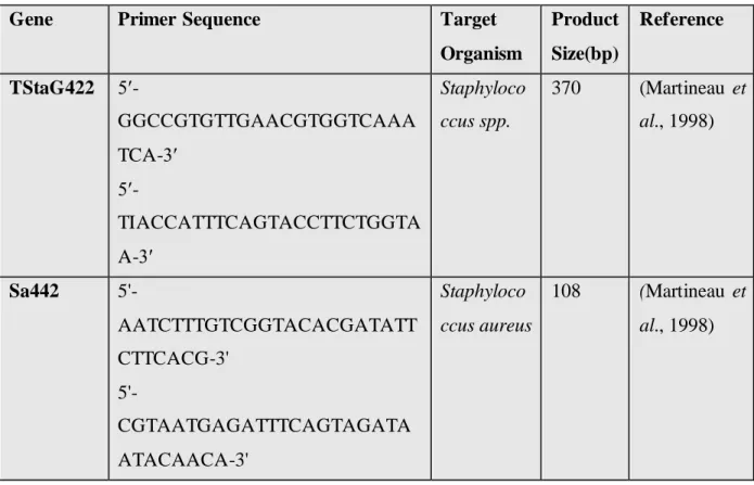
Two sets of primers were used in this study for the molecular detection of Staphylococcus aureus. The TStaG422 primer was used for detection at the genus level which is Staphylococci specific, and the Sa442 primer was used to detect the species level such as S. aureus (Martineau et al., 1998). The stock solutions of both of these primers TStaG422 and Sa442 were available in the lab.
For the preparation of the 100 µl of working solutions ((10µM) of TStaG422 primer from 100µM, 10µl of100µM forward and 10µl of 100µM reverse primers were taken in two different MCTs. The remaining 90 µl was then filled with molecular-grade nuclease-free water in each tube. A gentle re-pipetting and short spin for 20 seconds were followed after adding the nuclease-free water. The same process was followed while preparing the working solution of Sa442 primers.
Table 1: Sequences of primers used for amplification by PCR Gene Primer Sequence Target
Organism
Product Size(bp)
Reference
TStaG422 5′-
GGCCGTGTTGAACGTGGTCAAA TCA-3′
5′-
TIACCATTTCAGTACCTTCTGGTA A-3′
Staphyloco ccus spp.
370 (Martineau et al., 1998)
Sa442 5'-
AATCTTTGTCGGTACACGATATT CTTCACG-3'
5'-
CGTAATGAGATTTCAGTAGATA ATACAACA-3'
Staphyloco ccus aureus
108 (Martineau et al., 1998)
37
3.6.3 Preparation of controls for PCR
For performing the polymerase chain reaction (PCR), a positive control was used each time, which serves as quality control of all the processes of molecular detection of Staphylococcus aureus. Laboratory standard true positive isolate of Staphylococcus aureus was available at the laboratory and was used as positive control throughout the study, and negative control containing nuclease-free water with master mix was also used.
3.6.4 PCR assay
The amplification of certain genes by polymerase chain reaction under sets of conditions helps to detect the bacterial isolates at the molecular level effectively. The PCR-based detection of Staphylococcus aureus by amplifying TStaG422 and Sa442 genes was performed frequently in this study as samples were obtained each week from the selected sites.
PCR assay was performed in PCR tubes and the PCR mixtures were in a 15 µl volume, which comprised of 3.9 µl Nuclease free water, 0.8µl of each set of primers (10µM), 7.5 µl of 2X emerald PCR Master Mix (Takara Bio), and 2 µl of DNA template. Gentle re-pipetting and spinning were performed very carefully for proper mixing and to avoid bubbles forming. The PCR was performed in an Applied Bio-system (Thermo-Fischer) thermal cycle and the program (modified) was set as follows: initial denaturation of 94°C fo