CHARACTERIZATION OF METHICILLIN RESISTANT
Staphylococcus aureus
(MRSA) ISOLATED FROM HUMAN AND DIARY COWS
SYARIFUDDIN TATO 1, SITI ISRINA OKTAVIA SALASIA2, SUGIYONO2, KURNIASIH3
1
Pharmacology Dept., 2 Clinical Pathology Dept., Pathology Dept.3 Faculty of Veterinary Medicine, Gadjah Mada University, Yogyakarta
ABSTRACT
Staphylococcus aureus is the causative agent of clinical or subclinical
mastitis in dairy cows. Staphylococcus aureus cause also pathogenic diseases in human. Several hospitals reported the increase of methicillin resistant S. aureus (MRSA) frequency, because this srtain was resistant to several antibiotics. The aim of research was to characterize S. aureus, distribution of resistant factor to methicillin and several other antibiotics.
The isolates of S.aureus originated from dairy cows (11 isolates) and from patients of Sarjito hospital (10 isolates). Bacterial identification based on the growth in PAD media, Gram staining, mannitol salt agar (MS) fermentation, catalase, coagulase, clumping factor tests and the resistancy/sensitivity to methicillin, penicillin G, ampicillin, oxacillin, tetracyclin, gentamycin and erythromycin. It was also identified to 23S rRNA and mecA genes for the resistancy screening to methicillin (MRSA) by using polymerise chain reaction (PCR).
All isolates from milk and human used in this study were S.aureus based on phenotypic and genotypic identifications. Staphylococcus aureus were resistant to penicillin group (20-100%), and to tetracyclin, gentamycin and erythromicin (9-40%). By PCR amplification could be observed for 90% methicillin resistant (mecA) genes for human isoates and 45% for milk isolates. The resistancy of
S.aureus to methicillin in the present study might help to understand the
distribution of methicillin resistant S. aureus (MRSA) among human and bovine isolates and might help to control S. aureus infecions in Indonesia.
Key words : Staphylococcus aureus, methicillin resistant, mecA
INTRODUCTION
Staphylococcus aureus is the causative agent of clinical or subclinical
mastitis in dairy cows. Staphylococcus aureus cause also pathogenic diseases in
human. Several hospitals reported the increase of methicillin resistant S. aureus
(MRSA) frequency, because this srtain was resistant to several antibiotics. The
primary reservoir of Staphylococcus aureus is located in the infected quarter of
(Akineden et al., 2001). Intra-mammae infection can cause a high economical
loss because of the decrease of milking production between 10 – 25% from total
dairy milk production (Han et al., 2000).. Staphylococcus aureus is the primary
pathogen to human that it cause several diseases with clinical signs such as
impetigo, scalded skin syndrome, pneumonia, osteomyelitis, pioarthrosis,
endocarditis, metastasis, food poison, toxic shock syndrome (TSS), meningitis
and sepsis (Marrack & Kappler, 1990; Dinges et al., 2000; Omoe et al., 2002).
Toxic shock syndrome oxin-1 (TSST-1) produced by Staphylococcus aureus is a
toxin, especially, it is related to menstruation case in woman. Several strain
Staphylococcus aureus can also produce one or two exfoliatif ETA or ETB that it
has connection with impetigo disease or scalded skin syndrome (Lee et al., 1987;
Marrack & Kappler, 1990; Akineden et al., 2001). Staphylococcal infection is
often known as nosokomial infection in the hospital that is caused from lesion of
post surgery, infection during hemodialisis, bacteriaemia and pneumonia (Na’was
et al., 1997).
The primary problem to solve Staphylococcus aureus infection is the
antibiotic resistancy. Several strains of Staphylococcus aureus has been reported
resistant to almost all commercial antibiotics (Todar, 2002). It is reported from
several hospitals that it occured the frequency increase of methicillin resistant
Staphylococcus aureus (MRSA) and it is usually resistant to multiple antibiotics
(Na’was et al., 1997; Todar, 2005).
MATERIALS AND METHODS
Staphylococcus aureuswas isolated from several dairy cattle farm in DIY
and Central Java (11 isolates). Staphylococcus aureus from human was isolated
from patient of Sarjito hospital which was collected from Laboratorium
Microbiology, Faculty of Medicine, GMV (10 isolates).
Samples from milk of cattle and skin of human was selected in blood agar
plate, THB, Gram stain, annitol salt agar, Catalase, Koagulase tests. Clumping
factor, Biochemistry test (Bruchler et al, 1994). Hemolysa production used agar
Antibiotic sensitivity test used. Mϋller – Hinton agar to Methicillin (Met),
Tetracyclin(TE), Penicillin G(P), Ampicillin (AMP), Erythromycin(E),
Gentamisin(GM), Oxacillin (OX). The inhibition zone was evaluated based on,
Anonim (1998). The diameter zone of inhibition was compared with the standart
of Kirby Baner (Jang et al, 1978).
Samples from cattle and human were rRNA extracted using Qiamp tissue
kit (Qiagen, Germany), then were amplified in 23S region with species primers.
rRNA extraction product was then digested with CoU Hind III (invitrogen)
and 5 µM spermidine trihydrochloride (Sigma – Aldrich, USA) for 16 hours, 5 µl
of digested DNA was ligated with 0,2 µg adapter oligonucleotide (ADH-1 ACG
GTA TGC GAC AG and ADH-2 AGC TCT GTC GCA TAC CGT GAG), IU T4
DNA ligase (invitrogen) until 20 µl of volume, was incubated at 200
C for 4
hours. They were heated at 800C for 10 minutes for inactivation of T4 ligase then
soluted in 1/5 sterile water. The amplification reaction used 50 µl of total volume
containing 2,5 µl of template DNA, 200 µM of dNTPs, 1 µl of primer HI-X
(GGT ATG CGA CAG AGC TTX (X=A, T, G or C) ( 100 pmol/µl), and 1 µl
(5U) of Taq DNA polymerase in 1x PCR buffer.
Each HI-X primer (derivative) was used in 4 different PCR reaction. The
product of PCR was examined with gel electrophoresis 1,5 % W/V agarose.
RESULT AND DISCUSSION
The result of of sensitivity test to several antibiotics showed that all
antibiotics produced the inhibition zone to S. aureus with diameter variation
(Table 1).
Table 1. Inhibition zone diameters of several antibiotics to Staphylococcus
aureus.
No Isolate
code
Isolate origin Inhibition zone diameter (mm)
MET
5
TE
30 P
10
AMP
10
GM
10 E
15 OX
5
Dairy milk origin
1. SU 2 Milk, yogya 6 18 6 13 13 6 6
3. SU 16 Milk, yogya 17 28 30 25 20 25 10
4. SU 24 Milk, yogya 12 20 8 7 20 20 6
5. SU 25 Milk, yogya 13 18 8 10 20 20 6
6. SU 28 Milk, yogya 20 20 8 7 22 28 6
7. SU 34 Milk, Solo 17 22 23 21 20 25 14
8. SU 39 Milk, Solo 12 17 7 7 18 20 6
9. BY 5 Milk, Boyolali 17 25 7 6 17 20 18
10. BY 7 Milk, Boyolali 20 20 7 8 15 20 13
11. Y 7 Milk, Yogya 20 21 6 6 15 20 6
Human skin infection
12. 169 Human skin,
Yogya
22 20 7 7 17 25 7
13 179 Human skin,
Yogya
20 25 7 7 17 20 15
14. 198 Human skin,
Yogya
22 8 9 9 15 20 20
15. 199 Human skin,
Yogya
6 6 6 15 12 6 6
16. 255 Human skin,
Yogya
20 25 10 10 16 20 15
17. 262 Human skin,
Yogya
25 20 9 9 20 6 6
18. 274 Human skin,
Yogya
28 30 12 10 12 20 25
19. 870 Human skin,
Yogya
20 16 16 30 17 15 24
20. 979 Human skin,
Yogya
22 20 20 7 20 27 8
21. 1091 Human skin,
Yogya
7 7 7 7 7 7 7
All inhibition zones were then compared with standar zones of
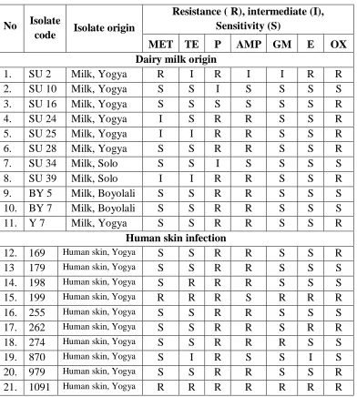
Table 2. Sensitivity of isolates S.aureus from dairy cows and human skin to
several antibiotics.
No Isolate
code Isolate origin
Resistance ( R), intermediate (I), Sensitivity (S)
MET TE P AMP GM E OX
Dairy milk origin
1. SU 2 Milk, Yogya R I R I I R R
2. SU 10 Milk, Yogya S S I S S S S
3. SU 16 Milk, Yogya S S S S S S R
4. SU 24 Milk, Yogya I S R R S S R
5. SU 25 Milk, Yogya I I R R S S R
6. SU 28 Milk, Yogya S S R R S S R
7. SU 34 Milk, Solo S S I S S S S
8. SU 39 Milk, Solo I I R R S S R
9. BY 5 Milk, Boyolali S S R R S S S
10. BY 7 Milk, Boyolali S S R R S S S
11. Y 7 Milk, Yogya S S R R S S R
Human skin infection
12. 169 Human skin, Yogya S S R R S S R
13 179 Human skin, Yogya S S R R S S S
14. 198 Human skin, Yogya S R R R S S S
15. 199 Human skin, Yogya R R R S R R R
16. 255 Human skin, Yogya S S R R S S S
17. 262 Human skin, Yogya S S R R S R R
18. 274 Human skin, Yogya S S R R R S S
19. 870 Human skin, Yogya S I R S S I S
20. 979 Human skin, Yogya S S R R S S R
21. 1091 Human skin, Yogya R R R R R R R
Sensitivity of S.aureus to several antibiotics showed that isolates from
milk (36,4%) and isolates from human (20%) were resistant to methicillin.
Isolates from milk (90,9%) and isolates from human (100%) were resistant to
Table 3. Sensitivity percentage of S. aureus isolates from dairy cows and human
skin to several antibiotics (%).
Antibiotics Milk Origin Isolate Human isolate
Resistance(R) Sensitivity(S) Resistance(R) Sensitivity(S)
Methicillin 36,4 63,6 20 80
Penicillin G 90,9 9,1 100 0
Ampicillin 72,7 27,3 80 20
Oxacillin 63,6 36,4 50 50
Tetracyclin 27,3 72,7 40 60
Gentamycin 9,1 90,9 30 70
Erythromycin 9,1 90,9 40 60
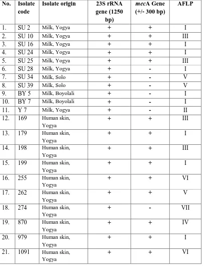
Based on the presence of gene which had responsible to methicillin,
isolates from human positive 9 (90%) had methicillin resistancy (mecA gene), and
it was only 1 (10%) was negative. Staphylococcus aureus from dairy cows were
5 isolates (45,5%) which was positive contained mecA gene (Table 4). MecA gene
of S. aureus from dairy cows and human in this research showed the same result
(300 bp).
It was found the derivate of Penicillin (methicillin) that was effective to
penicillin resistant of microorganism (Black, 1999). However, it has been reported
from many hospitals that the frequency of methicillin resistant S.aureus (MRSA)
was increase to multiple antibiotics (Na’was et al., 1997; Todar, 2002).
The presence of MRSA in dairy cows in Indonesia has not been reported.
This research showed that 45,5% of S. aureus isolates from dairy cows had
MRSA. The infection of MRSA might be caused by the resistancy of S. aureus to
Table 4. Amplification result of isolates from dairy cows and human skin in 23S
of rRNA gene region and mecA gene.
No. Isolate code
Isolate origin 23S rRNA gene (1250
bp)
mecA Gene
(+/- 300 bp)
AFLP
1. SU 2 Milk, Yogya + + I
2. SU 10 Milk, Yogya + + III
3. SU 16 Milk, Yogya + + I
4. SU 24 Milk, Yogya + + I
5. SU 25 Milk, Yogya + + III
6. SU 28 Milk, Yogya + - I
7. SU 34 Milk, Solo + - V
8. SU 39 Milk, Solo + - V
9. BY 5 Milk, Boyolali + - I
10. BY 7 Milk, Boyolali + - I
11. Y 7 Milk, Yogya + - II
12. 169 Human skin,
Yogya
+ + III
13. 179 Human skin,
Yogya
+ + I
14. 198 Human skin,
Yogya
+ + III
15. 199 Human skin,
Yogya
+ + I
16. 255 Human skin,
Yogya
+ + VI
17. 262 Human skin,
Yogya
+ + V
18. 274 Human skin,
Yogya
+ - VII
19. 870 Human skin,
Yogya
+ + IV
20. 979 Human skin,
Yogya
+ + I
21. 1091 Human skin, Yogya
CONCLUSION
Isolates from dairy cows (11 isolates) and from human skin (10 isolates)
were positive Staphylococcus aureus which was resistant to penicillin (20-100%)
and to tetracycline, gentamycine, erythromycine (9-40%). Isolates of S.aureus
from human (90%) and isolates from dairy cow (45,5%) contained mecA gene.
Isolates of Staphylococcus aureus from human (60%) was similar to isolates from
dairy cows (30%)
REFFERENCES
Akineden, Ö., Annemüller, C., Hassan, A. A., Lämmler, Ch., Wolter, W.,
Zschöck, M., 2001. Toxin genes and other characteristics of Staphylococcus
aureus isolates from milk of cows with mastitis. Clin.Diagn. Lab. Immunol.
8: 959-964.
Brückler, J., Schwarz, S. and Untermann, F., 1994. Staphylokokken-Infektionen
und Enterotoxine, Band.II/1, In Blobel, H. und Schlieβer (Herausber),
Handbuch der bakteriellen Infektionen bei Tieren, 2 Auflage. Gustav
Fischer Verlag Jena, Stuttgart.
Han, H.-R, S.-I.Park, S.-W.Kang, W.-S.Jong and C.-J.Youn, 2000.Capsular
polissacharide typing of domestic mastitis-causing Staphylococcus aureus
strains and its potential exploration of bovine mastitis vaccine development.
I. Capsular polyssacharide typing, isolation and purification of the strain. J.
Vet. Sci. 1: 53-63.
Jang, S.S., Biberstein E. L. and Hirsh D.C., 1978. A Diagnostic Manual of
Veterinary Clinical Bacteriology And Micology. UNESCO/CIDA Regional
training Course in Veterinary Diagnostic Microbiology, Peradeniya, 67-69.
Lee, J. C., S.L. Xu, A. Albus, and P.J. Livolsi, 1994.Genetic analysis of type 5
capsular polyssacharide expression by Staphylococcus aureus.J. Bacteriol.
27: 4883-4889.
Marrack, P. and J. Kappler, 1990.The staphylococcal enterotoxin and their
relatives.Science, 248: 705-711.
Na’was,T., A. Hawwari, E. Hendrix, J. Hebden, R. Edelman, M. Martin, W.
genotypic characterization of nosocomial Staphylococcus aureus isolates
from trauma patients. J. Clin. Microbiol.36(2): 414-420.
Omoe, K., Ishikawa, M., Shimoda, Y., Hu, D. –L., Ueda, Shinagawa, K., 2002.
Detection of seg, she, and sei genes in isoaltes and determination of the
enterotoxin productivities of S. aureus isolates harbouring seg, she, and sei
genes. J. Clin.
Microbiol.40: 857-862.
Skalka, B., mola, J. and Pillich, J., 1979. A Simple method of detecting
staphylococcal hemolysin.Zbl. Bacteriol.Hyg. I. Abt. Orig. A. 245: 283-286.
Todar,K., 2002. Staphylococcus.Bacteriology at UW-Bacteriology 330 Home
Page.1-7.
Todar,K.,2005.Todar”s online textbook of bacteriology.
Staphylococcus.University of Winconsin- Madison Department of
Bacteriology.www.textbookofbacteriology.net/staph.html. Tanggal
download :1 November 2007.
Woods, G. I., and Washington, J. A. 1995. Manual of clinical Microbiology, 6thed,