Stem Cells as Therapeutic Carrier in Cancer Treatment
By
Mysha Yeasmin Anika 16346027
A thesis submitted to the Department of Pharmacy in partial fulfillment of the requirements for the degree of
Bachelors of Pharmacy (Hons.)
Department of Pharmacy Brac University
July 2021
© [2021]. Brac University All rights reserved
Declaration
It is hereby declared that
1. The thesis submitted is my own original work while completing degree at Brac University.
2. The thesis does not contain material previously published or written by a third party, except where this is appropriately cited through full and accurate referencing.
3. The thesis does not contain material which has been accepted, or submitted, for any other degree or diploma at a university or other institution.
4. I have acknowledged all main sources of help.
Student’s Full Name & Signature:
__________________________
Mysha Yeasmin Anika 16346027
Approval
The thesis titled “Stem Cells as Therapeutic carrier in Cancer Treatment” submitted by Mysha Yeasmin Anika (16346027) of Summer 2016, has been accepted as satisfactory in partial fulfillment of the requirement for the degree of Bachelors of Pharmacy on 27thJuly, 2021.
Examining Committee:
Supervisor: _______________________________
Dr. Hasina Yasmin
Professor, Department of Pharmacy Brac University
Academic Coordinator: _______________________________
Dr. Hasina Yasmin
Professor, Department of Pharmacy Brac University
Departmental Head: _______________________________
Dr. Eva Rahman Kabir Professor, Department of Pharmacy
Brac University
27.7.2021
Ethics Statement
The study does not involve any kind of animal or human trial.
Abstract
Cancer is one of the leading causes of death worldwide. Traditional surgery or chemotherapy are not sufficient to remove cancer completely. Researchers are trying to find out individual pathways for targeting tumor niches with precise drug amounts to eradicate cancer. The latest framework of stem cell therapy focuses on an optimistic instrument for treating cancer. Modified stem cells are utilized as therapeutic carriers with oncolytic viruses, nanoparticles, exosomes at particular tumor niches in the human body. However, therapeutic application of stem cell therapy can be improved by targeting malignancy and screening of anticancer medications. Additionally, mesenchymal stem cells, neural stem cells and hematopoietic stem cells transplantation have possible remedial viability in different cancer treatment. This review article highlights the use of different types of stem cells as therapeutic carriers and examines the future role of this therapy in various cancers.
Key words:Cancer, stem cells, therapeutic carrier, stem cell therapy, tumor niches.
Dedication
I want to dedicate this project to my respectable supervisor Dr. Hasina Yasmin, Professor of Department of Pharmacy, Brac University for her continuous guidance throughout my project.
Acknowledgements
I would like to proceed by thanking the Almighty who is the source of our strength, knowledge, patience which have enabled me to complete this project with full diligence. It would be impossible to complete my project work without His mercy.
I would like to express my deepest gratitude and appreciation to my project supervisor, Dr.
Hasina Yasmin (Professor of Department of Pharmacy, Brac University) whose expertise, ample time spent and consistent guidance in every step providing me with her continuous support and guidance since the very first day of this journey. I would like to thank her for great advice and patient behavior throughout this phase whenever I encountered difficulty.
Moreover, I would like to thank Professor Dr. Eva Rahman Kabir, Chairperson of Department of Pharmacy, Brac University, for giving me the opportunity to perform this project work individually and giving me the chance to work with Dr. Hasina Yasmin madam.
Subsequently, I would also like to thank my parents for their support and words of encouragement which motivated me to work harder to overcome the difficulties.
Table of Contents
Declaration... ii
Approval...iii
Ethics Statement... iv
Abstract... v
Dedication...vi
Acknowledgements...vii
List of Tables...x
List of Figures... xi
List of Acronyms...xii
Chapter 1... 1
Introduction...1
1.1 Cancer...1
1.2 Stem cell therapy...3
1.3 Purpose of the study... 4
Chapter 2... 5
Stem cells...5
2.1 Definition and properties...5
2.2 Discovery and development of stem cells...7
2.3 Sources of stem cells... 8
2.4 Classification of stem cells...10
Chapter 3... 18
Stem cells as therapeutic carrier in malignancy... 18
3.1 Introduction... 18
3.2 Genetically modified stem cells to discharge anticancer proteins... 19
3.3 Stem cell-mediated suicide therapy...20
3.4 Stem cells as nanoparticle carriers... 22
3.5 Stem cells loaded with oncolytic virus...24
3.6 Stem cell-derived exosomes...28
Chapter 4... 30
Conclusion and Future Prospects...30
Reference... 31
List of Tables
Table 1: Various conventional treatment options for cancer...2
Table 2: Discovery and development of stem cells... 7
Table 3: Sources of stem cells... 9
Table 4: MSCs-based anti-cancer drug carrier techniques... 29
List of Figures
Figure 1: Lineage potential of stem cells (Berdasco & Esteller, 2011)...6 Figure 2: Classification of stem cells (Hawsawi et al., 2018)... 11 Figure 3: Formation of embryonic stem cells and their differentiation (Zakrzewski et al., 2019).
... 12 Figure 4: Using optimized reprogramming of induced pluripotent stem cells (Hockemeyer &
Jaenisch, 2016)... 16 Figure 5: Effect of cancer stem cells on tumor development (Houghton et al., 2007)...17 Figure 6: Modified stem cells to assist tumor cell destruction (Stuckey & Shah, 2014)... 22 Figure 7: Conveyance of MSCs loaded with PTX-PLGA NPs in orthotopic glioma model (Wang et al., 2018).... 24 Figure 8: The utilization of stem cells to transmit oncolytic viruses to malignant cells as suitable vectors (Zendedel et al., 2019)... 25 Figure 9: Benefits of systemic injection with oncolytic virus protected by MSCs (Subramani et al., 2009)... 27
List of Acronyms
RM Regenerative medication
GVHD Graft-Versus-Host Disease
ESCs Embryonic stem cells
HSCs Hematopoietic stem cells
MSCs Mesenchymal stem cells
NSCs Neural stem cells
ESCs Endothelial stem cells
iPSCs Induced pluripotent stem cells
CSCs Cancer stem cells
PTX Paclitaxel
GDEPT Gene directed enzyme prodrug therapy SCNT Somatic cell nuclear transfer
CD Cytosine deaminase
5-FC 5-fluorocytosine
DR4 Death receptor 4
EGFR Epidermal growth factor receptor
MYXV Myxoma virus
PTX-NPs Paclitaxel loaded nanoparticles
IFN-α Interferon-α
IFN-β Interferon-β
TSP1 Thrombospondin 1
OVs Oncolytic viruses
Chapter 1
Introduction 1.1 Cancer
Cancer is one of the life-threating diseases due to its driving causes of mortality and morbidity throughout the world (Yang et al., 2020). In reference to WHO, more than nineteen million new cases and about ten million cancer deaths were estimated in 2020 across the world. Among the different types the most commonly diagnosed cancer is the breast cancer. The next most common type is lung cancer (11.7%), followed by colorectal (10.0 %), prostate (7.3%), and stomach (5.6%) cancers (Sung et al., 2021).
Various strategies have been established for cancer treatment such as-surgery, radiotherapy, chemotherapy and targeted therapy (Shown in table 1). But the efficacies of various therapeutic options are restricted due to metastasis, relapse, heterogeneity, resistance to drugs and radiotherapy, and lack of immunological monitoring that hold up cancer treatment (C. L. Zhang et al., 2017a). The lack of tumor specificity is the major hindrance in effectiveness of conventional therapies for cancer treatment (Mitku et al., 2017).
The available anticancer agents at present do not show target specificity. They can affect all types of cells because of the similarity between normal and abnormal cells. Anticancer agents are toxic to normal cells and can cause various side effects (Stringer & Snyder, 2018). Efficacy of an anti-cancer drug is dependent on its therapeutic concentrations at the tumor site. Variable hypoxia, intra-tumoral pressure gradients, abnormal vasculature within the tumor etc. results in less concentration of drugs at the solid tumor site. This is difficult to reach in most solid tumors (Blakeley, 2008). Stem cells are engineered with various cytotoxic agents so that they can
decrease tumor volumes and extend survival rates in preclinical animal models (C. L. Zhang et al., 2017a).
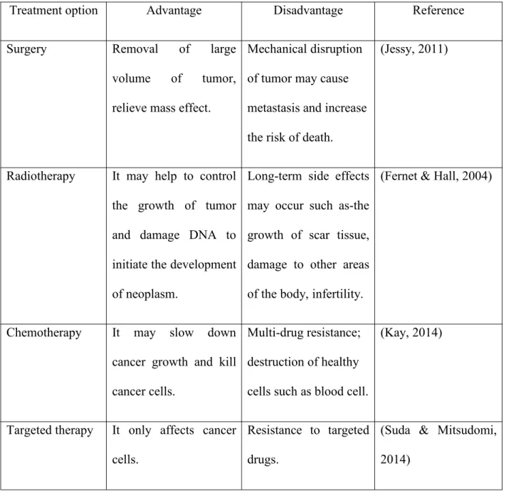
Table 1: Various conventional treatment options for cancer
Treatment option Advantage Disadvantage Reference
Surgery Removal of large
volume of tumor, relieve mass effect.
Mechanical disruption of tumor may cause metastasis and increase the risk of death.
(Jessy, 2011)
Radiotherapy It may help to control the growth of tumor and damage DNA to initiate the development of neoplasm.
Long-term side effects may occur such as-the growth of scar tissue, damage to other areas of the body, infertility.
(Fernet & Hall, 2004)
Chemotherapy It may slow down cancer growth and kill cancer cells.
Multi-drug resistance;
destruction of healthy cells such as blood cell.
(Kay, 2014)
Targeted therapy It only affects cancer cells.
Resistance to targeted drugs.
(Suda & Mitsudomi, 2014)
Stem cell therapy has provided a hopeful option in combating cancer. Therapeutic efficacy of stem cell therapy may be increased due to its greater target-specificity on tumors (Chu et al., 2020a).
1.2 Stem cell therapy
Stem cell therapy is the form of regenerative medicine that promotes repairing of diseased, dysfunctional or injured tissue of specific patients' suffering from severe wounds or chronic disease conditions. This therapy is useful when patients' self-regenerative responses are inadequate (Mahla, 2016). Stem cells are undifferentiated cells and they have the ability to extensively proliferate (self-renewal), usually arise from a single cell, and differentiate into different types of specialized cells and tissue with varying potency (Kolios & Moodley, 2012).
These properties of stem cells are responsible for making stem cell therapy as a viable alternative treatment for various therapeutic conditions. Stem cells can be used in treatment of various diseases including lymphoblastic leukemia, thalassemia, multiple myeloma, myeloid leukemia, and sickle cell anemia, Parkinson's disease, liver, lungs and heart diseases, diabetes, renal disease, chronic wounds, graft-versus-host disease, sepsis, cancer etc. (Larijani et al., 2012).
Stem cells can be used as therapeutic carriers for targeting tissues or organs of interest. Stem- cell-mediated delivery of genes, proteins or small molecules as therapeutic agents has the potential to enhance local or systemic repair processes and take advantage of the innate capability of stem cells to migrate to injury sites (Labusca et al., 2018). Additionally, stem cells can be applied in regenerative medicine, immunotherapy, cancer stem cell-targeted therapy, and
anticancer drug screening applications. Currently, this therapy is a highly advanced treatment choice for cancer as a targeted cell therapy. Because stem cells can accumulate inside tumors after systemic injection, and hence can be applied as carriers for tumor specific therapy (Takayama et al., 2020).
Advantages of stem cell therapy include- less post-procedural recovery time, no risk of rejection because this treatment uses the patient's biologic extracts, no risk of transmission of communicable disease. This therapy does not require any general anesthesia (Rahman et al, 2020).
1.3 Purpose of the study The purpose of the study is-
to gain information about the types of stem cells that can be utilized as therapeutic carrier in treatment of cancer.
to attain current scenario and future perspective of stem cell therapy in cancer treatment.
Chapter 2 Stem cells
2.1 Definition and properties
Stem cells are immature cells that are thought to be progenitor of more than 200 cell types existing in human adult body (Deepak & Vishnupriya, 2020). They exist in different stages of life including embryonic, fetal, and adult stages (Kolios & Moodley, 2012).
Stem cells are defined by their ability to have three main properties:
Multi-lineage differentiation (capacity to distinguish into any mature cell type);
self-renewal (ability to experience various cycles of cell proliferation while keeping pace with the undifferentiated state); and
homeostatic control (Palomeras et al., 2018).
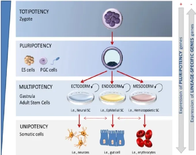
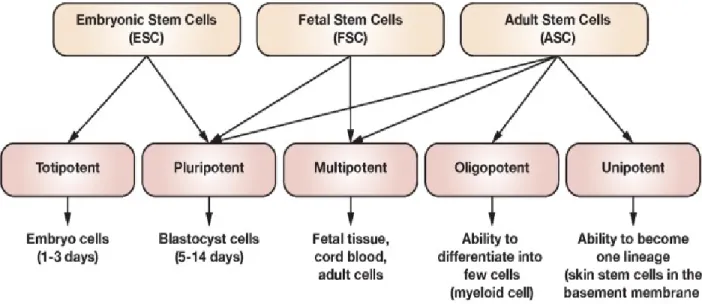
Another property incorporates potency of the stem cells. These cells may be either totipotent, pluripotent, multipotent, or unipotent. The lineage potential of stem cells is shown in figure 1.
Totipotent cells can produce all kinds of cells such as embryonic and extra-embryonic tissues (placenta). The zygote is the singular mammalian cell prepared for conveying all cells and tissues of a living organism (Mitku et al., 2017).
Pluripotent cells can make cells of the embryo including germ cells and cells from any of the germ layers.
Multipotent cells simply produce cells of a particular germ layer. For example- blood cell (mesoderm).
Unipotent cells can differentiate into one lineage. An example includes germ cell, that matures to become egg or sperm by one lineage differentiation, but not other cell types (Shihadeh, 2015).
Figure 1: Lineage potential of stem cells (Berdasco & Esteller, 2011).
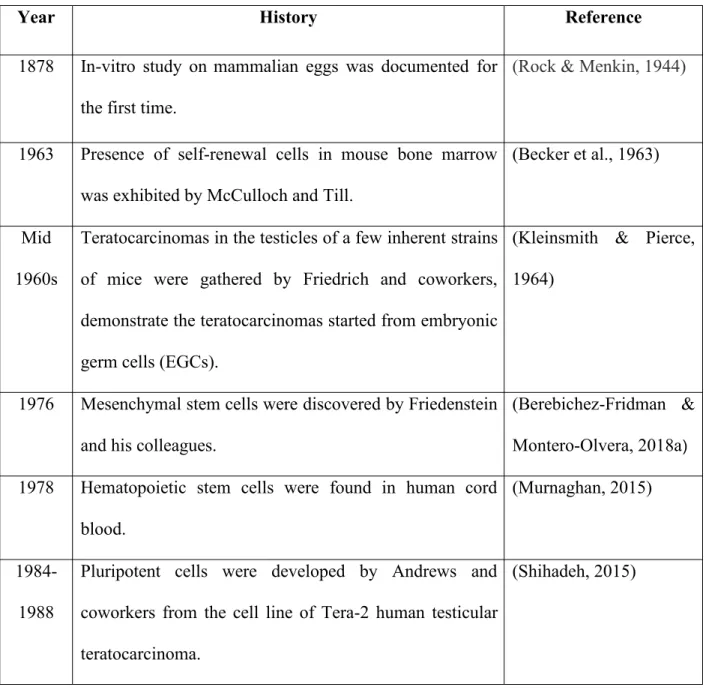
2.2 Discovery and development of stem cells
The development of stem cells was a progressing effort of several decades. They were not recognized by any single researcher or group of researchers (Shihadeh, 2015). The major events toward developing stem cells are given below in table 2.
Table 2: Discovery and development of stem cells
Year History Reference
1878 In-vitro study on mammalian eggs was documented for the first time.
(Rock & Menkin, 1944)
1963 Presence of self-renewal cells in mouse bone marrow was exhibited by McCulloch and Till.
(Becker et al., 1963)
Mid 1960s
Teratocarcinomas in the testicles of a few inherent strains of mice were gathered by Friedrich and coworkers, demonstrate the teratocarcinomas started from embryonic germ cells (EGCs).
(Kleinsmith & Pierce, 1964)
1976 Mesenchymal stem cells were discovered by Friedenstein and his colleagues.
(Berebichez-Fridman &
Montero-Olvera, 2018a)
1978 Hematopoietic stem cells were found in human cord blood.
(Murnaghan, 2015)
1984- 1988
Pluripotent cells were developed by Andrews and coworkers from the cell line of Tera-2 human testicular teratocarcinoma.
(Shihadeh, 2015)
1997 From Bonnet and Dick's evidence, Leukemia derived from hematopoietic stem cells was the primary confirmation of cancer stem cells (CSCs). The first cloning of artificial animals (Dolly) was discovered.
(Murnaghan, 2015;
(Kumar et al., 2010)
2001 The first human embryo was cloned at the early stage of 4-6 cells.
(Shihadeh, 2015)
2006 Induced pluripotent stem cells (iPSCs) were discovered. (Robinton & Daley, 2012)
2007 Fetal stem cells (FSCs) were discovered, isolating from amniotic fluid.
(Robinton & Daley, 2012)
2010 The trial of human embryonic stem cells took place in U.S. for the first time.
(Ebert et al., 2009)
2.3 Sources of stem cells
Stem cells from several sources exhibit different growth, movement, and separation abilities that determine their use in anti-tumor therapy. They can be found from two areas like- allogeneic and autologous sources (Chu et al., 2020a).
Allogeneic stem cells are separated from healthy persons and are available with better quality and quantities. They can be packed away and treated as "off-the-shelf" products, making
Autologous stem cells are gained from the tissue of patients themselves. There is less risk of host immune rejection, yet adequate time is required to achieve adequate quantities of cells of viable character (Öztürk et al., 2020).
In other words, two essential stem cell sources are accessible: adult stem cells and embryonic stem cells (showed in table 2). Induced pluripotent stem cells (iPSCs) have currently been artificially constructed by genetic modification of somatic cells (Takahashi & Yamanaka, 2006). Embryonic stem cells and induced pluripotent stem cells are alluded to as pluripotent stem cells since they can develop from each of the three germinal layers into all cell types. Interestingly, most adult stem cells are multi-potent and they can separate into a predetermined number of cell types (Dds et al., 2012).
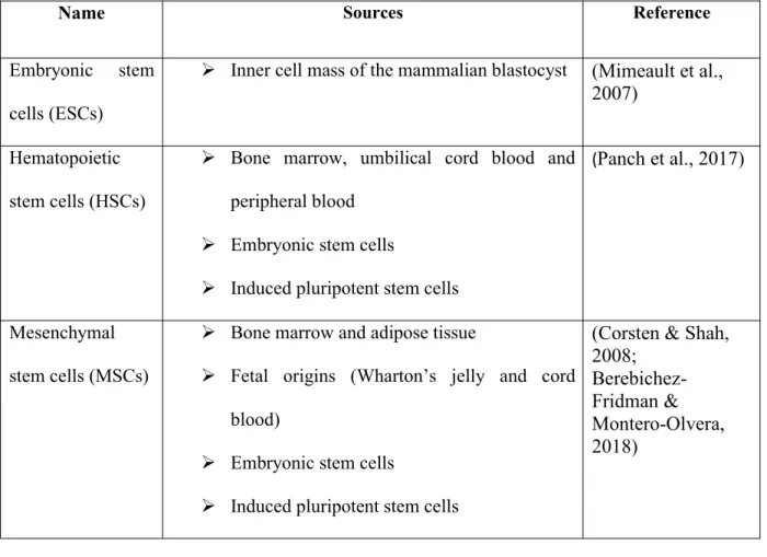
Table 3: Sources of stem cells
Name Sources Reference
Embryonic stem cells (ESCs)
Inner cell mass of the mammalian blastocyst (Mimeault et al., 2007)
Hematopoietic stem cells (HSCs)
Bone marrow, umbilical cord blood and peripheral blood
Embryonic stem cells
Induced pluripotent stem cells
(Panch et al., 2017)
Mesenchymal stem cells (MSCs)
Bone marrow and adipose tissue
Fetal origins (Wharton’s jelly and cord blood)
Embryonic stem cells
Induced pluripotent stem cells
(Corsten & Shah, 2008;
Berebichez- Fridman &
Montero-Olvera, 2018)
Neural stem cells (NSCs)
Brain, spinal cord and retina
Embryonic stem cells
Induced pluripotent stem cells
(Gottlieb, 2002)
Endothelial stem cells (ESCs)
Bone marrow
Embryonic stem cells
Induced pluripotent stem cells
(S. Kim & Von Recum, 2008)
Fetal stem cells (FSCs)
fetal tissues
Extra-embryonic segments such as amniotic liquid, placenta
(Goodarzi et al., 2019)
Induced
pluripotent stem cells (iPSCs)
obtained from somatic cells using reprogrammed technologies
(Hockemeyer &
Jaenisch, 2016)
2.4 Classification of stem cells
Stem cells can be mainly classified as ‘embryonic stem cells’ (ESCs) or ‘somatic stem cells’
(SSCs) based on their source and differentiating capacity (C. L. Zhang et al., 2017a). The other kind of cell is fetal stem cells (FSCs) which emerges from the placenta, amniotic liquid and fetal tissues (Hawsawi et al., 2018). The types of stem cells are showed in figure 2.
Figure 2: Classification of stem cells (Hawsawi et al., 2018).
2.4.1 Embryonic stem cells (ESCs)
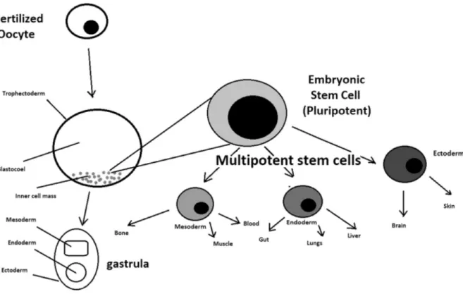
Human embryonic stem cells are obtained from embryos that are four or five days old and they form a hollow microscopic ball of cells which is called blastocyst. The cells of the inner cell masses are intended to distinguish into three germ layers such as- ectoderm, mesoderm and endoderm (Mimeault et al., 2007). These cells might have incompatibility with immune reaction between recipients and donors (Dds et al., 2012).
There is an ethical discussion about utilizing embryonic stem cells for therapeutic purposes (Henon, 2003). Tumorigenicity of embryonic stem cells after transplantation is a vital issue that ought to be appropriately addressed before ESCs transplantation in clinical trials. These cells may cause tumor development after transplantation (Newman et al., 2005). The last issue is that they are allogeneic and express elevated levels of complex-I proteins with histocompatibility and can be turned off during transplantation. Due to this issue, embryonic stem cells could not be
treated as the principal alternative in clinical trial until 2006 (Mitku et al., 2017). The formation of embryonic stem cells and their differentiation are showed in figure 3.
Figure 3: Formation of embryonic stem cells and their differentiation (Zakrzewski et al., 2019).
2.4.2 Adult Stem Cells (ASCs)
Adult stem cells also known as somatic stem cells (Dds et al., 2012). These cells have been studied widely and approved by the US Food and Drug Administration for human diseases, for example, Parkinson's disease, juvenile diabetes (Corsten & Shah, 2008). They are established
hematopoietic stem cells (HSCs), mesenchymal stem cells (MSCs), and neural stem cells (NSCs).
In disease treatment, they are often considered as most ideal option due to their utility.
Hematopoietic stem cells (HSCs) are available in bone marrow and they can establish all types of blood cell in body. The implantation of this type of cells inferred from cord blood, used for treating particular leukemia, and few sorts of blood related diseases (C. L. Zhang et al., 2017a).
Mesenchymal stem cells (MSCs) are found from bone marrow. This cells can also be used as host cells for replication, transportation, and local discharge of intact oncolytic adenoviruses (Corsten & Shah, 2008). In vitro, they can multiply and develop many specific kinds of cells, for example, osteocytes, adipocytes, and chondrocytes, rapidly (Chu et al., 2020b). MSCs are connected broadly within the treatment of distinctive cancers. They have become popular candidates for cellular therapy due to easy handling purpose and clinical safety issue. MSCs have therapeutic advantages in different malignancies that is the great chance for numerous clinical trials (Parfejevs et al., 2020).
Neural stem cells, initially present in the central nervous system (CNS), can regenerate by own and form new neurons and glial cells. This type of cells is generally tested to treat breast, lung, and prostate malignancies in mice. They can separate into three significant kinds of CNS cells, for example, (i) neurons; (ii) astrocytes; and (iii) oligodendrocytes. They have great potential to treat neurogenerative diseases in research purpose (Ahmed et al., 2010).2.4.3 Fetal stem cells (FSCs)
Fetal stem cells (FSCs) can be derived not only from a few fetal tissues but also from extra- embryonic segments, for example, amniotic liquid and placenta, which are enriched source of fetal stem cells with less moral concerns (Goodarzi et al., 2019). They are less potent than embryonic stem cells (Ilic & Polak, 2011). They can be present in fetal tissues such as blood, liver, bone marrow, pancreas, lung, spleen and kidney. Their potential properties, development capacity and absence of tumorigenicity make them as a great choice in cell therapy (Abdulrazzak et al., 2010).
In addition, fetal blood derived mesenchymal stem cells exhibit greater capacity for differentiation, increased ability for homing and engraftment, and diminished immunogenic reactions compared with adult stem cells. The development rate and self-renewal capability of fetal stem cells are better than adult stem cells. Fetal mesenchymal stem cells are able to more effectively bind to ligands of the extracellular matrix that come up with their homing to the tumor site and reduce tumor burden. Fetal liver derived stem cells have been appeared to have ten folds capacity for engraftment than adult stem cells of the bone marrow (Goodarzi et al., 2019).
Other types of stem cells are also available such as induced pluripotent stem cells (iPSCs) and
2.4.4 Other types of stem cells
Induced pluripotent stem cells (iPSCs)
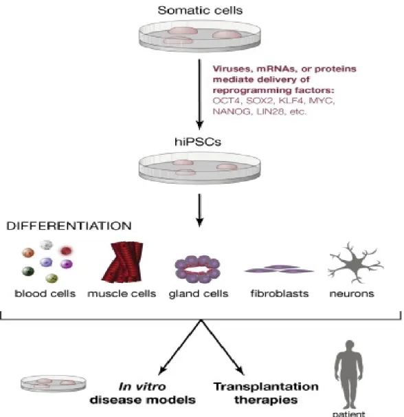
Induced pluripotent stem cells are artificially produced from non-pluripotent stem cells (Mitku et al., 2017). The capacity to reconstruct somatic cells to induced pluripotent stem cells (iPSCs) gives a chance to make cell lines (Malik & Rao, 2013). This cells are reinvented with identical characteristics from undeveloped stem cells, for example, self-repair ability and separation into various cell types (Yoshida & Yamanaka, 2017). This process of reprogramming is called retro- differentiation and is performed by transfection into non pluripotent cells (showed in figure 4).
Viral vectors, including retroviruses, adenoviruses and non-viral vectors, can be utilized for the transfection (Mitku et al., 2017).
Many years before Yamanaka's research, John Gurdon had appeared that the epigenetic profile of a totally separated cell can be reconstructed to a pluripotent state. Many years after the fact, the cloning by somatic cell nuclear transfer (SCNT), sheep "Dolly" revealed with the Gurdon's outcome. The reconstructing effectiveness of induced pluripotent stem cells was at first exceptionally low, however it has been essentially upgraded. The cultural environment improves the reprogramming efficiency, for example, hypoxic development (Yoshida & Yamanaka, 2017).
By utilizing advanced reinventing microRNAs, patient cells may be reprogrammed into induced pluripotent stem cells (Hockemeyer & Jaenisch, 2016).
Figure 4: Using optimized reprogramming of induced pluripotent stem cells (Hockemeyer & Jaenisch, 2016).
Cancer Stem Cells (CSCs)
Cancer stem cells are considered as potential targets for malignancy since they were identified in
accessible inside tumor tissues that act basic parts in malignancy improvement, metastasis, and recurrence of malignancy. In this way, focusing on cancer stem cells can yield an extraordinary chance to treat different kinds of tumors (Chu et al., 2020b). The base of cancer stem cells is fully unknown but numerous pathways involved in carcinogenesis (Soltanian & Matin, 2011).
Figure 5: Effect of cancer stem cells on tumor development (Houghton et al., 2007)
(A)A group of cells within a tumor are capable to replicate and assist tumor growth (showed in figure 5). These cancer stem cells give indistinguishable immortal daughter cells (red) and transient amplifying cells (TA-white) which is capable for the bulk of tumor cell expansion.
(B) The conventional therapy decreases tumor bulk but cause tumor recurrence but targeting tumor stem cells result in apoptosis (Houghton et al., 2007).
Chapter 3
Stem cells as therapeutic carrier in malignancy 3.1 Introduction
Tissue engineering based on stem cells is intended to create transplantable bio-replacements and to serve as an effective substitute for tissue and organ transplantation (Mandpe et al., 2020). The reasons for using stem cells as carrier in malignancy are:
to protect therapeutic agents from fast natural deterioration;
to limit adverse effect and
to extend local therapeutic levels because of inherent tumor-targeting effect of stem cells.
The amount of stem cells present in tumor niches determines anti-tumor efficacy (Chu et al., 2020a). Because of stem cell-secreted factors and physical interactions between stem cells and tumor cells, unmodified stem cells may have an inherent anti-tumor effect. Besides, stem cells have been updated to treat malignancy in several ways (Stuckey & Shah, 2014).
Mesenchymal stem cells and neural stem cells, may be modified for prospective use in the treatment of malignancy through various mechanisms. The enzyme/prodrug system, nanoparticle or oncolytic virus or exosomes transportation at the tumor site through stem cells are common modifications for therapeutic applications in malignancy (showed in figure 6) (C. L. Zhang et al., 2017b).
3.2 Genetically modified stem cells to discharge anticancer proteins
Stem cells are usually modified to exhibit transgenes by releasing proteins through viral transduction but non-viral techniques have some advantages, for example, poor immunogenicity of the host. The apoptotic proteins are immediate effectors that binds with death receptor 4 (DR4, also known as TRAILR1) and (DR5, also known as TRAILR2), specially expressed on malignant cells and actuates apoptosis(Stuckey & Shah, 2013; Sasportas et al., 2009). Another method is to hinder multiplication pathways in malignant cells to utilize proteins that can beat the endogenous substance to their related receptors. For example, in different preclinical cancer models, stem cell based biological components bind to the epidermal growth factor receptor (EGFR) and cytokines, for example, interferon-α (IFN-α) and interferon-β (IFN-β) have been appeared to adversely control tumor development (Dembinski et al., 2013 ;Changchun Ren et al., 2008).
Indirect effector such as anti-angiogenic thrombospondin 1 (TSP1) inhibit tumor-related vasculature development, building up a threatening micro-environment that is not permissive (Van Eekelen et al., 2010). It has experimented the emission of immunomodulatory particles, for example, interleukins (ILs). It is possible to transform the immunosuppressive tumor climate that coordinates an immune reaction against malignancy. Human MSCs have been developed to discharge IL-12 or IL-18 and have been experimented in mice with renal cell carcinoma, cervical tumors and glioblastoma (Stuckey & Shah, 2014).
In addition, neural stem cells (NSCs), utilized as a transporter to persistently deliver TRAIL, have been appeared to limit the metastasis of brain tumor, hence survival rate was found to be increased in the treated mice (Kauer et al., 2012). In a clinical trial (NCT03298763), this framework has been utilized for treating human lung adenocarcinoma. Co-expression in stem
cells of suicide gene (i.e., cytosine deaminase, CD) and cytotoxic gene (i.e., IFN-β, inhibit tumor growth and trigger apoptosis) demonstrates a possibility to build the adequacy of treatment (Yi et al., 2014).
3.3 Stem cell-mediated suicide therapy
The secretion of soluble factors such as prodrug-converting enzymes or tumor-toxic chemokines can be increased by this technique (Sage et al., 2016). Stem cells mediated suicide gene therapy is a technique by which stem cells are programmed inserting a virus or bacterial gene to exhibit an enzyme that changes after administration of a non-toxic drug into a cytotoxic medication (Duarte et al., 2012). The advantage of this therapy is that there is no concern about long term fate of stem cells because they are removed after therapeutic effect (Stuckey & Shah, 2014).
An experiment was performed in October 2018 (ClinicalTrials.gov identifier: NCT02015819) utilizing modified neural stem cells to treat glioma. In this treatment, Herpes simplex virus thymidine kinase (HSV-TK) has additionally been utilized (C. L. Zhang et al., 2017b). This is the most common form of gene that codes the prodrug activating enzyme and can be isolated from the Herpes simplex virus or E. coli. This is well characterized suicide gene. The herpes simplex virus thymidine kinase (HSV-TK) changes over ganciclovir (GCV) to GCV- monophosphate, which is further phosphorylated to GCV-triphosphate, which prevents DNA synthesis (Kosaka et al., 2012). This triphosphate metabolite is a purine analog and incorporation of this analog hinders DNA polymerase during DNA synthesis. Numerous clinical fields, for
Another study established that neural stem cells thymidine kinase (NSCs-TK) infused into the cerebrum moved to the contralateral hemisphere, co-situated with U87 cells, and gave ganciclovir-treated mice with long term survival (C. L. Zhang et al., 2017b). A few stage I/II clinical trials are tested on the gene directed enzyme prodrug (GDEPT) technique (Chu et al., 2020b). This technique is also potentiated by mesenchymal stem cells (MSCs) which has arisen as a potential cancer therapy. Mesenchymal stem cells act to convey anti-malignant component directly to tumor niches due to the intrinsic tumor tropism. Other clinical trials for example, ClinicalTrials.gov Identifier: NCT03298763, NCT02530047 and NCT02079324 and clinical trials with MSC-based gene directed enzyme prodrug treatment have given hopeful outcomes for further advancement (von Einem et al., 2017).
MSCs have been found in a wide view in treatment of malignancy including breast cancer and glioblastoma. An important protein that is utilized in prodrug treatment is cytosine deaminase (CD). It changes 5-fluorocytosine (5-FC) to 5-fluorouracil, a harmful antimetabolite (Stuckey &
Shah, 2014). Cytosine deaminase/5-fluocytosine (CD/5FC) has been demonstrated to be powerful in preclinical investigations, where 4% of CD positive cells in tumor mass are adequate to completely eliminate the tumor (Ho et al., 2020). However, when this therapy is utilized in conjunction with new therapeutic strategies, recent investigations on pre-clinical cancer models show the immense capability of this procedure (Duarte et al., 2012).
The modification of stem cells is shown in figure 6 that assist tumor cell destruction.
Figure 6: Modified stem cells to assist tumor cell destruction (Stuckey & Shah, 2014).
3.4 Stem cells as nanoparticle carriers
Stem cells can be used as nanoparticle (NP) carrier that can resolve the inability to target tumor
requires the modification of the surface of a nanoparticle by chemical / biological components to tumor. Physical targeting consists of controlling nanoparticles with the help of external magnetic field to tumor cells (Subramani et al., 2009). The surface of nanoparticles can be altered by changing properties such as stability, solubility and targeting technique. NPs can be targeted against tumors, generally by association with cell surface functional groups of amine or thiol (Chu et al., 2020b). Stem cells can be given with nanoparticles in vivo, where they can transfer to malignant site and store the nanoparticles close to the tumor (Stuckey & Shah, 2014). From one investigation, MSCs loaded with doxorubicin-containing permeable silica have been appeared to move more effectively to initiate apoptosis in intracranial tumors than doxorubicin alone. More investigations are necessary for nanoparticle-based therapeutics with stem cells in malignancy (L. Li et al., 2011).
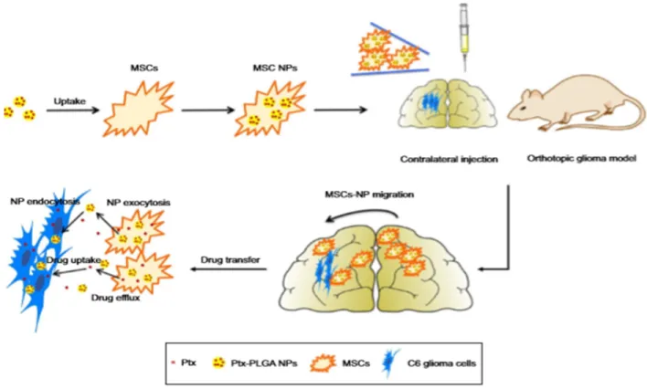
From another investigation, it was showed that mesenchymal stem cells (MSCs) based photosensitizer loaded silica nanoparticle (PS loaded SiO2NP) did not cause toxic effects against MSCs. Photodynamic therapy inhibited the tumor growth by injecting PS loaded MSCs (Kidd et al., 2009). The retention ability of PS loaded MSCs resulted in deposition of PS in tumor and successfully destructed the tumor. The usage of mesenchymal stem cells (MSCs) is a hopeful target therapy for delivering drugs to tumor niches. Another analysis demonstrated that intravenous administration of paclitaxel loaded mesenchymal stem cells (PTX-NPs loaded MSCs) deposited more nanoparticles in orthotopic lung tumor and local drug depots were produced in mice (Cao et al., 2014). MSCs carried paclitaxel-polylactic-co-glycolic acid nanoparticles (PTX- PLGA NPs) to the brain tumor in mice model of human gliomas (shown in figure 7).
Interestingly, tumor growth in mice were significantly inhibited by these NP-loaded MSCs. It
was discovered that because of their tumor-tropic impact, MSCs were first adhered in the lung parenchymal tissue and afterward moved to lung tumor niches (Roger et al., 2010).
Figure 7: Conveyance of MSCs loaded with PTX-PLGA NPs in orthotopic glioma model (Wang et al., 2018).
3.5 Stem cells loaded with oncolytic virus
Oncolytic viruses (OVs) replicate restrictively in tumor cells. Tumor cells have the ability to house OV-transduced neural stem cells (NSCs). NSC-delivered oncolytic viruses have demonstrated more antitumor activity than viruses against glioblastomas (C. L. Zhang et al., 2017b). Various types of stem cells are utilized as host cells for the vehicle in murine models of
Figure 8: The utilization of stem cells to transmit oncolytic viruses to malignant cells as suitable vectors (Zendedel et al., 2019).
Oncoviruses specifically attack and effectively destroy the neoplastic cells. The remedial capability of oncolytic viral treatment relies upon the activity against the modified cells (shown in figure 8). Moreover, the anti-malignant activity of these viruses are noticed when oncolytic viruses are expected to impart immune‐stimulatory particles (Zendedel et al., 2019). Oncolytic Herpes simplexvirus (HSV1716) was used for clinical assessment in patients with gliomas in the USA and the UK. The usage of Talimogene laherparepvec (T-VEC) genetically engineered from oncolytic Herpes Simplex Virus Type-1 was approved by FDA to treat patients with melanoma in the USA in 2015 (Q. Zhang & Liu, 2020).
From another analysis, the therapy was investigated with breast cancer stem cells and oncolytic Herpes Simplex viruses. The outcome of the investigation indicated that onco-HSV vector G47δ was powerful against breast cancer stem cells (J. Li et al., 2012).
Mesenchymal stem cells (MSCs) synergistically work with oncolytic viruses and deliver cytokines. MSCs generate a tumor-tolerogenic niches through prostaglandins and interleukins to diminish immune cell activity. Allogeneic MSCs have been documented to prompt immune reaction systemically and intratumorally penetration of leukocytes in mice. The adequacy of mesenchymal stem cells was illustrated in stage I clinical trial. (J. Kim et al., 2015).
A study showed that mesenchymal stem cells loaded with Myxoma virus (MYXV) experimentally develops with actuated injuries of pulmonary melanoma to immunocompetent mice. Myxoma viruses (MYXV) carrying mesenchymal stem cells transport MYXV from the site of infusion to the tumor site and tumor homing has been reported (Weng et al., 2014). No unfavorable neurotic impacts were discovered related to MSC-mediated MYXV oncolytic treatment. Mesenchymal stem cells (MSCs) permit Myxoma viruses (MYXV) to be easily moved to the micro-environment of pulmonary melanoma, causing effective response (Jazowiecka-Rakus et al., 2020).
When unshielded myxoma viruses are administered intravenously, antiviral reaction causes no oncolytic activity. On the other hand, shielded viruses having defensive carrier like- mesenchymal stem cells (MSCs) that permit powerful transportation to tumor site and give oncolytic activity (shown in figure 9). Mesenchymal stem cells (MSCs) loaded with oncolytic viruses improve oncolysis and show antitumor activity (Hadryś et al., 2020).
Figure 9: Benefits of systemic injection with oncolytic virus protected by MSCs (Subramani et al., 2009).
3.6 Stem cell-derived exosomes
Various therapeutic drugs, proteins and mi-RNAs are encapsulated by exosomes. Compared with other engineered nano-particles, these natural carriers have various benefits, including unique biocompatibility, durability, high loading capacity and greater internalization into tumor cells (Fuhrmann et al., 2015). Special proteins or ligands can be effectively functionalized to improve the targeting impact on the tumor niches (Kooijmans et al., 2016). The conventional transfection technique has successfully packaged genetic products such as mRNAs or siRNAs into exosomes derived from stem cells. From one study, exosomes were directly injected into the malignant site which showed noticeable decrease in glioma xenograft development in rat with primary brain tumor (Katakowski et al., 2013). In another study, miR-122-expressing MSCs derived exosomes significantly increased antitumor effect of sorafenib was observed when applied on hepatocellular tumor model (Lou et al., 2015).
Two approaches could encapsulate small molecule drugs into exosomes. First, it was found that stem cells may pick up and pack certain agents into exosomes after priming with exogenous materials and release them to the culture medium by exocytosis. Moreover, these exosomes have been found to inhibit the development of leukemia and myeloma cell lines in tumor (Pessina et al., 2013; Bonomi et al., 2017). MSCs were used for loading other medicines, including doxorubicin, gemcitabine, and cisplatin (Coccè et al., 2017). Post-loading method can be used for loading therapeutic drugs into exosomes. Drugs were encapsulated into exosomes by using
Table 4: MSCs-based anti-cancer drug carrier techniques
SC-loaded agents for tumor site
Example Benefits Reference
Oncolytic viruses Adenovirus;
Measles virus;
Herpes simplex virus
Anti-tumor activity with multiple infections;
Selective replication in the cells of tumors
(Stoff-Khalili et al., 2007;
Castleton et al., 2014; Duebgen et al., 2014)
Tumor/tissue‐
specific prodrugs
CD + 5‐5‐FU;
HSV‐tk+
Ganciclovir
Selective activation of drugs in tumor niches
(Kucerova et al., 2007; von Einem et al., 2017)
Immunomodulatory agents
IL‐2;
IL‐12;
Interferon‐β
Directly and indirectly powerful impact on malignancy development;
Synergy with other forms of immunotherapy
(Stagg et al., 2004; Ren et al., 2008)
Apoptosis‐inducing agents
TRAIL;
EGF agonist;
IFN-α; IFN-β
Endogenous signaling agent; recently in clinical trial
(Luetzkendorf et al., 2010; Sage et al., 2016)
Cytotoxic chemotherapy
Paclitaxel;
Doxorubicin
Chemotherapeutic drugs approved by Food and Drug Administration
(Pessina et al., 2011)
Chapter 4
Conclusion and Future Prospects
Cancer is the uncontrolled growth of abnormal cells in the body. It is a leading cause of death worldwide with an estimated 10 million deaths in 2020. Various conventional treatment options are available for treating cancer such as chemotherapy, radiotherapy, surgery and targeted therapy. The main goal of these therapies is to cure cancer patients or prolong their lives. The effectiveness of these therapies reduces due to the lack of tumor specificity, metastasis, recurrence, heterogeneity, resistance to chemotherapy. But the use of stem cell therapy in cancer treatment can overcome the drawbacks of the conventional therapies. It can be used as a therapeutic carrier to target tumor niches. There is a lot of study about how stem cells can be utilized for different types of cancers. Stem cell therapy is planned to be used in the future to cure multiple forms of cancer for which no successful therapies are available now. However, embryonic stem cells /induced pluripotent stem cells-based vaccines can be applied to treat malignant growths. The standardization of the procedure for the production of exosomes as well as anti-tumor vaccines are necessary to gain stable effects. Laws and standards should be carefully considered to ensure the ethical integrity of embryonic stem cells application and to remove the barriers for the purpose of research and therapy with embryonic stem cells. More studies are required for the development of stem cell therapy to treat cancer. Though stem cell technologies are found to be highly potent for cancer treatment, further researches are required to
Reference
Abdulrazzak, H., Moschidou, D., Jones, G., & Guillot, P. V. (2010). Biological characteristics of stem cells from foetal, cord blood and extraembryonic tissues. In Journal of the Royal Society Interface (Vol. 7, Issue SUPPL. 6, p. S689). Royal Society.
https://doi.org/10.1098/rsif.2010.0347.focus
Ahmed, A. U., Alexiades, N. G., & Lesniak, M. S. (2010). The use of neural stem cells in cancer gene therapy: Predicting the path to the clinic. In Current Opinion in Molecular Therapeutics.
Auffinger, B., Morshed, R., Tobias, A., Cheng, Y., Ahmed, A. U., & Lesniak, M. S. (2013).
Drug-loaded nanoparticle systems and adult stem cells: A potential marriage for the
treatment of malignant glioma? Oncotarget, 4(3), 378–396.
https://doi.org/10.18632/oncotarget.937
Becker, A. J., McCulloch, E. A., & Till, J. E. (1963). Cytological demonstration of the clonal nature of spleen colonies derived from transplanted mouse marrow cells.Nature,197(4866), 452–454. https://doi.org/10.1038/197452a0
Berdasco, M., & Esteller, M. (2011). DNA methylation in stem cell renewal and multipotency.
In Stem Cell Research and Therapy (Vol. 2, Issue 5, p. 42). BioMed Central.
https://doi.org/10.1186/scrt83
Berebichez-Fridman, R., & Montero-Olvera, P. R. (2018a). Sources and Clinical Applications of Mesenchymal Stem Cells: State-of-the-art review. Sultan Qaboos University Medical Journal,18(3), e264. https://doi.org/10.18295/SQUMJ.2018.18.03.002
Berebichez-Fridman, R., & Montero-Olvera, P. R. (2018b). Sources and clinical applications of mesenchymal stem cells state-of-the-art review. Sultan Qaboos University Medical Journal, 18(3), e264–e277. https://doi.org/10.18295/squmj.2018.18.03.002
Blakeley, J. (2008). Drug delivery to brain tumors. In Current Neurology and Neuroscience Reports(Vol. 8, Issue 3, pp. 235–241). NIH Public Access. https://doi.org/10.1007/s11910- 008-0036-8
Bonomi, A., Steimberg, N., Benetti, A., Berenzi, A., Alessandri, G., Pascucci, L., Boniotti, J., Coccè, V., Sordi, V., Pessina, A., & Mazzoleni, G. (2017). Paclitaxel-releasing mesenchymal stromal cells inhibit the growth of multiple myeloma cells in a dynamic 3D culture system.Hematological Oncology,35(4), 693–702. https://doi.org/10.1002/hon.2306 Cao, B., Yang, M., Zhu, Y., Qu, X., & Mao, C. (2014). Stem Cells Loaded with Nanoparticles as
a Drug Carrier for In Vivo Breast Cancer Therapy.Advanced Materials,26(27), 4627–4631.
https://doi.org/10.1002/adma.201401550
Castleton, A., Dey, A., Beaton, B., Patel, B., Aucher, A., Davis, D. M., & Fielding, A. K. (2014).
Human mesenchymal stromal cells deliver systemic oncolytic measles virus to treat acute lymphoblastic leukemia in the presence of humoral immunity. Blood, 123(9), 1327–1335.
https://doi.org/10.1182/blood-2013-09-528851
Chu, D.-T., Nguyen, T. T., Tien, N. L. B., Tran, D.-K., Jeong, J.-H., Anh, P. G., Thanh, V. Van,
Truong, D. T., & Dinh, T. C. (2020b). Recent Progress of Stem Cell Therapy in Cancer Treatment: Molecular Mechanisms and Potential Applications. Cells, 9(3), 563.
https://doi.org/10.3390/cells9030563
Coccè, V., Farronato, D., Brini, A. T., Masia, C., Giannì, A. B., Piovani, G., Sisto, F., Alessandri, G., Angiero, F., & Pessina, A. (2017). Drug Loaded Gingival Mesenchymal Stromal Cells (GinPa-MSCs) Inhibit In Vitro Proliferation of Oral Squamous Cell Carcinoma. Scientific Reports,7(1). https://doi.org/10.1038/s41598-017-09175-4
Corsten, M. F., & Shah, K. (2008). Therapeutic stem-cells for cancer treatment: hopes and hurdles in tactical warfare. In The Lancet Oncology (Vol. 9, Issue 4, pp. 376–384).
https://doi.org/10.1016/S1470-2045(08)70099-8
Dds, H. E., Dds, W. S., Dds, M. N., Dds, I. A., & Dds, K. A. (2012). Stem cells in dentistry – Part I : Stem cell sources. Journal of Prosthodontic Research, 56(3), 151–165.
https://doi.org/10.1016/j.jpor.2012.06.001
Deepak, A., & Vishnupriya, V. (2020). Stem Cell Therapy for Breast Cancer : A Review.8(6), 533–535.
Dembinski, J. L., Wilson, S. M., Spaeth, E. L., Studeny, M., Zompetta, C., Samudio, I., Roby, K., Andreeff, M., & Marini, F. C. (2013). Tumor stroma engraftment of gene-modified mesenchymal stem cells as anti-tumor therapy against ovarian cancer. Cytotherapy, 15(1), 20. https://doi.org/10.1016/j.jcyt.2012.10.003
Duarte, S., Carle, G., Faneca, H., Lima, M. C. P. de, & Pierrefite-Carle, V. (2012). Suicide gene therapy in cancer: Where do we stand now? InCancer Letters(Vol. 324, Issue 2, pp. 160–
170). Elsevier Ireland Ltd. https://doi.org/10.1016/j.canlet.2012.05.023
Duebgen, M., Martinez-Quintanilla, J., Tamura, K., Hingtgen, S., Redjal, N., Wakimoto, H., &
Shah, K. (2014). Stem cells loaded with multimechanistic oncolytic herpes simplex virus variants for brain tumor therapy. Journal of the National Cancer Institute, 106(6).
https://doi.org/10.1093/jnci/dju090
Ebert, A. D., Yu, J., Rose, F. F., Mattis, V. B., Lorson, C. L., Thomson, J. A., & Svendsen, C. N.
(2009). Induced pluripotent stem cells from a spinal muscular atrophy patient. Nature, 457(7227), 277–280. https://doi.org/10.1038/nature07677
Fernet, M., & Hall, J. (2004). Genetic biomarkers of therapeutic radiation sensitivity. In DNA Repair (Vol. 3, Issues 8–9, pp. 1237–1243). Elsevier.
https://doi.org/10.1016/j.dnarep.2004.03.019
Fuhrmann, G., Serio, A., Mazo, M., Nair, R., & Stevens, M. M. (2015). Active loading into extracellular vesicles significantly improves the cellular uptake and photodynamic effect of
porphyrins. Journal of Controlled Release, 205, 35–44.
https://doi.org/10.1016/j.jconrel.2014.11.029
Goodarzi, P., Falahzadeh, K., Aghayan, H., Payab, M., Larijani, B., Alavi-Moghadam, S., Tayanloo-Beik, A., Adibi, H., Gilany, K., & Arjmand, B. (2019). Therapeutic abortion and ectopic pregnancy: alternative sources for fetal stem cell research and therapy in Iran as an Islamic country. In Cell and Tissue Banking (Vol. 20, Issue 1, pp. 11–24). Springer
cells as carriers for systemic delivery of oncolytic viruses. In European Journal of
Pharmacology (Vol. 874, p. 172991). Elsevier B.V.
https://doi.org/10.1016/j.ejphar.2020.172991
Hawsawi, Y. M., Al-Zahrani, F., Mavromatis, C. H., Baghdadi, M. A., Saggu, S., & Oyouni, A.
A. A. (2018). Stem cell applications for treatment of cancer and autoimmune diseases: Its promises, obstacles, and future perspectives. In Technology in Cancer Research and Treatment(Vol. 17). SAGE Publications Inc. https://doi.org/10.1177/1533033818806910 Henon, P. R. (2003). Human embryonic or adult stem cells: An overview on ethics and
perspectives for tissue engineering. In Advances in Experimental Medicine and Biology (Vol. 534, pp. 27–45). Springer Science and Business Media Deutschland GmbH.
https://doi.org/10.1007/978-1-4615-0063-6_3
Ho, Y. K., Woo, J. Y., Tu, G. X. E., Deng, L. W., & Too, H. P. (2020). A highly efficient non- viral process for programming mesenchymal stem cells for gene directed enzyme prodrug cancer therapy.Scientific Reports,10(1), 14257. https://doi.org/10.1038/s41598-020-71224- 2
Hockemeyer, D., & Jaenisch, R. (2016). Induced pluripotent stem cells meet genome editing. In Cell Stem Cell (Vol. 18, Issue 5, pp. 573–586). Cell Press.
https://doi.org/10.1016/j.stem.2016.04.013
Houghton, J. M., Morozov, A., Smirnova, I., & Wang, T. C. (2007). Stem cells and cancer. In Seminars in Cancer Biology (Vol. 17, Issue 3, pp. 191–203). Academic Press.
https://doi.org/10.1016/j.semcancer.2006.04.003
Huang, R., & Rofstad, E. K. (2017). Cancer stem cells (CSCs), cervical CSCs and targeted
therapies. In Oncotarget (Vol. 8, Issue 21, pp. 35351–35367). Impact Journals LLC.
https://doi.org/10.18632/oncotarget.10169
Ilic, D., & Polak, J. M. (2011). Stem cells in regenerative medicine: Introduction: In British Medical Bulletin (Vol. 98, Issue 1, pp. 117–126). Oxford Academic.
https://doi.org/10.1093/bmb/ldr012
Jazowiecka-Rakus, J., Sochanik, A., Rusin, A., Hadryś, A., Fidyk, W., Villa, N., Rahman, M. M., Chmielik, E., Franco, L. S., & McFadden, G. (2020). Myxoma Virus-Loaded Mesenchymal Stem Cells in Experimental Oncolytic Therapy of Murine Pulmonary Melanoma.Molecular Therapy - Oncolytics,18, 335–350. https://doi.org/10.1016/j.omto.2020.07.003
Jessy, T. (2011). Immunity over inability: The spontaneous regression of cancer. Journal of Natural Science, Biology and Medicine,2(1), 43. https://doi.org/10.4103/0976-9668.82318
Katakowski, M., Buller, B., Zheng, X., Lu, Y., Rogers, T., Osobamiro, O., Shu, W., Jiang, F., &
Chopp, M. (2013). Exosomes from marrow stromal cells expressing miR-146b inhibit
glioma growth. Cancer Letters, 335(1), 201–204.
https://doi.org/10.1016/j.canlet.2013.02.019
Kauer, T. M., Figueiredo, J. L., Hingtgen, S., & Shah, K. (2012). Encapsulated therapeutic stem cells implanted in the tumor resection cavity induce cell death in gliomas. Nature Neuroscience,15(2), 197–204. https://doi.org/10.1038/nn.3019
advantages-and-limitations-to-existing-conventional-treatment
Kidd, S., Spaeth, E., Dembinski, J. L., Dietrich, M., Watson, K., Klopp, A., Battula, V. L., Weil, M., Andreeff, M., & Marini, F. C. (2009). Direct evidence of mesenchymal stem cell tropism for tumor and wounding microenvironments using in vivo bioluminescent imaging.
Stem Cells,27(10), 2614–2623. https://doi.org/10.1002/stem.187
Kim, J., Hall, R. R., Lesniak, M. S., & Ahmed, A. U. (2015). Stem cell-based cell carrier for targeted oncolytic virotherapy: Translational opportunity and open questions. In Viruses (Vol. 7, Issue 12, pp. 6200–6217). MDPI AG. https://doi.org/10.3390/v7122921
Kim, S., & Von Recum, H. (2008). Endothelial stem cells and precursors for tissue engineering:
Cell source, differentiation, selection, and application. In Tissue Engineering - Part B:
Reviews(Vol. 14, Issue 1, pp. 133–147). https://doi.org/10.1089/teb.2007.0304
Kleinsmith & Pierce. (n.d.). MULTIPOTENTIALITY OF SINGLE EMBRYONAL CARCINOMA
CELLS - PubMed. Retrieved May 2, 2021, from
https://pubmed.ncbi.nlm.nih.gov/14234000/
Kolios, G., & Moodley, Y. (2012). Introduction to stem cells and regenerative medicine. In Respiration(Vol. 85, Issue 1, pp. 3–10). Respiration. https://doi.org/10.1159/000345615 Kooijmans, S. A. A., Schiffelers, R. M., Zarovni, N., & Vago, R. (2016). Modulation of tissue
tropism and biological activity of exosomes and other extracellular vesicles: New nanotools for cancer treatment. Pharmacological Research, 111, 487–500.
https://doi.org/10.1016/j.phrs.2016.07.006
Kosaka, H., Ichikawa, T., Kurozumi, K., Kambara, H., Inoue, S., Maruo, T., Nakamura, K.,
Hamada, H., & Date, I. (2012). Therapeutic effect of suicide gene-transferred mesenchymal stem cells in a rat model of glioma. Cancer Gene Therapy, 19(8), 572–578.
https://doi.org/10.1038/cgt.2012.35
Kucerova, L., Altanerova, V., Matuskova, M., Tyciakova, S., & Altaner, C. (2007). Adipose tissue-derived human mesenchymal stem cells mediated prodrug cancer gene therapy.
Cancer Research,67(13), 6304–6313. https://doi.org/10.1158/0008-5472.CAN-06-4024
Kumar, R., Sharma, A., Pattnaik, A., & Varadwaj, P. (2010). Stem cells: An overview with respect to cardiovascular and renal disease. Journal of Natural Science, Biology and Medicine,1(1), 43. https://doi.org/10.4103/0976-9668.71674
Labusca, L., Herea, D. D., & Mashayekhi, K. (2018). Stem cells as delivery vehicles for regenerative medicinechallenges and perspectives. In World Journal of Stem Cells(Vol. 10,
Issue 5, pp. 43–56). Baishideng Publishing Group Co.
https://doi.org/10.4252/wjsc.v10.i5.43
Larijani, B., Esfahani, E. N., Amini, P., Nikbin, B., Alimoghaddam, K., Amiri, S., Malekzadeh, R., Yazdi, N. M., Ghodsi, M., Dowlati, Y., Sahraian, M. A., & Ghavamzadeh, A. (2012). A r c h i v e o f S I D Stem Cell Therapy in Treatment of Different Diseases. In Acta Medica Iranica(Vol. 50, Issue 2). ACTA MEDICA IRANICA. http://journals.tums.ac.ir/
Li, J., Zeng, W., Huang, Y., Zhang, Q., Hu, P., Rabkin, S. D., & Liu, R. (2012). Treatment of
mesenchymal stem cells for tumor-tropic therapy. ACS Nano, 5(9), 7462–7470.
https://doi.org/10.1021/nn202399w
Lou, G., Song, X., Yang, F., Wu, S., Wang, J., Chen, Z., & Liu, Y. (2015). Exosomes derived from MIR-122-modified adipose tissue-derived MSCs increase chemosensitivity of hepatocellular carcinoma. Journal of Hematology and Oncology, 8(1).
https://doi.org/10.1186/s13045-015-0220-7
Luetzkendorf, J., Mueller, L. P., Mueller, T., Caysa, H., Nerger, K., & Schmoll, H. J. (2010).
Growth inhibition of colorectal carcinoma by lentiviral TRAIL-transgenic human mesenchymal stem cells requires their substantial intratumoral presence.Journal of Cellular and Molecular Medicine, 14(9), 2292–2304. https://doi.org/10.1111/j.1582- 4934.2009.00794.x
Mahla, R. S. (2016). Stem cells applications in regenerative medicine and disease therapeutics.
In International Journal of Cell Biology (Vol. 2016). Hindawi Limited.
https://doi.org/10.1155/2016/6940283
Malik, N., & Rao, M. S. (2013). A review of the methods for human iPSC derivation.Methods in Molecular Biology,997, 23–33. https://doi.org/10.1007/978-1-62703-348-0_3
Mandpe, P., Prabhakar, B., & Shende, P. (2020). Role of Liposomes-Based Stem Cell for Multimodal Cancer Therapy. In Stem Cell Reviews and Reports(Vol. 16, Issue 1, pp. 103–
117). Springer. https://doi.org/10.1007/s12015-019-09933-z
Marcus, A. J., & Woodbury, D. (2008). Fetal stem cells from extra-embryonic tissues: Do not discard: Stem Cells Review Series. Journal of Cellular and Molecular Medicine, 12(3), 730–742. https://doi.org/10.1111/j.1582-4934.2008.00221.x
Mimeault, M., Hauke, R., & Batra, S. K. (2007). Stem Cells: A Revolution in Therapeutics—
Recent Advances in Stem Cell Biology and Their Therapeutic Applications in Regenerative Medicine and Cancer Therapies. Clinical Pharmacology & Therapeutics, 82(3), 252–264.
https://doi.org/10.1038/sj.clpt.6100301
Mitku, W., Tesfaye, W., & Wubshet, A. (2017). Review on Stem Cell Therapy and their Role in Cancer Treatment.International Journal of Biotechnology and Bioengineering,3(4), 71–79.
https://doi.org/10.25141/2475-3432-2017-4.0071
Murnaghan. (n.d.). History of Stem Cell Research. Retrieved May 3, 2021, from http://www.explorestemcells.co.uk/HistoryStemCellResearch.html
Newman, M. B., Misiuta, I., Willing, A. E., Zigova, T., Karl, R. C., Borlongan, C. V., & Sanberg, P. R. (2005). Tumorigenicity issues of embryonic carcinoma-derived stem cells: Relevance to surgical trials using NT2 and hNT neural cells. In Stem Cells and Development (Vol. 14, Issue 1, pp. 29–43). Mary Ann Liebert Inc. https://doi.org/10.1089/scd.2005.14.29
Ong, H. T., Federspiel, M. J., Guo, C. M., Ooi, L. L., Russell, S. J., Peng, K. W., & Hui, K. M.
(2013). Systemically delivered measles virus-infected mesenchymal stem cells can evade host immunity to inhibit liver cancer growth. Journal of Hepatology, 59(5), 999–1006.
https://doi.org/10.1016/j.jhep.2013.07.010
Öztürk, S., Elçin, A. E., Koca, A., & Elçin, Y. M. (2020). Therapeutic Applications of Stem
https://doi.org/10.3390/molecules23092193
Panch, S. R., Szymanski, J., Savani, B. N., & Stroncek, D. F. (2017). Sources of Hematopoietic Stem and Progenitor Cells and Methods to Optimize Yields for Clinical Cell Therapy. In Biology of Blood and Marrow Transplantation (Vol. 23, Issue 8, pp. 1241–1249). Elsevier Inc. https://doi.org/10.1016/j.bbmt.2017.05.003
Parfejevs, V., Sagini, K., Buss, A., Sobolevska, K., Llorente, A., Riekstina, U., & Abols, A.
(2020). Adult Stem Cell-Derived Extracellular Vesicles in Cancer Treatment: Opportunities and Challenges.Cells,9(5), 1171. https://doi.org/10.3390/cells9051171
Pessina, A., Bonomi, A., Coccè, V., Invernici, G., Navone, S., Cavicchini, L., Sisto, F., Ferrari, M., Viganò, L., Locatelli, A., Ciusani, E., Cappelletti, G., Cartelli, D., Arnaldo, C., Parati, E., Marfia, G., Pallini, R., Falchetti, M. L., & Alessandri, G. (2011). Mesenchymal stromal cells primed with paclitaxel provide a new approach for cancer therapy. PLoS ONE,6(12).
https://doi.org/10.1371/journal.pone.0028321
Pessina, A., Coccè, V., Pascucci, L., Bonomi, A., Cavicchini, L., Sisto, F., Ferrari, M., Ciusani, E., Crovace, A., Falchetti, M. L., Zicari, S., Caruso, A., Navone, S., Marfia, G., Benetti, A., Ceccarelli, P., Parati, E., & Alessandri, G. (2013). Mesenchymal stromal cells primed with Paclitaxel attract and kill leukaemia cells, inhibit angiogenesis and improve survival of leukaemia-bearing mice. British Journal of Haematology, 160(6), 766–778.
https://doi.org/10.1111/bjh.12196
Rahman et al. (n.d.). Stem Cell Therapy in Treatment of Non-communicable Diseases:
Possibilities and Challenges | Asian Journal of Immunology. Retrieved November 12, 2020, from https://journalaji.com/index.php/AJI/article/view/30124
Ren, C., Kumar, S., Chanda, D., Kallman, L., Chen, J., Mountz, J. D., & Ponnazhagan, S. (2008).
Cancer gene therapy using mesenchymal stem cells expressing interferon-β in a mouse prostate cancer lung metastasis model. Gene Therapy, 15(21), 1446–1453.
https://doi.org/10.1038/gt.2008.101
Ren, Changchun, Kumar, S., Chanda, D., Chen, J., Mountz, J. D., & Ponnazhagan, S. (2008).
Therapeutic Potential of Mesenchymal Stem Cells Producing Interferon-α in a Mouse Melanoma Lung Metastasis Model. Stem Cells, 26(9), 2332–2338.
https://doi.org/10.1634/stemcells.2008-0084
Robinton, D. A., & Daley, G. Q. (2012). The promise of induced pluripotent stem cells in research and therapy. In Nature (Vol. 481, Issue 7381, pp. 295–305). Nature.
https://doi.org/10.1038/nature10761
Rock, J., & Menkin, M. F. (1944). In vitro fertilization and cleavage of human ovarian eggs.
Science,100(2588), 105–107. https://doi.org/10.1126/science.100.2588.105
Sage, E. K., Thakrar, R. M., & Janes, S. M. (2016). Genetically modified mesenchymal stromal cells in cancer therapy. In Cytotherapy (Vol. 18, Issue 11, pp. 1435–1445). Elsevier B.V.
https://doi.org/10.1016/j.jcyt.2016.09.003
Sasportas, L. S., Kasmieh, R., Wakimoto, H., Hingtgen, S., Van De Water, J. A. J. M., Mohapatra, G., Figueiredo, J. L., Martuza, R. L., Weissleder, R., & Shah, K. (2009).
Shihadeh, H. (2015). History and Recent Advances of Stem Cell Biology and the Implications for Human Health. InHealth. http://digitalcommons.uri.edu/srhonorsprog/421
Soltanian, S., & Matin, M. M. (2011). Cancer stem cells and cancer therapy. In Tumor Biology (Vol. 32, Issue 3, pp. 425–440). Springer. https://doi.org/10.1007/s13277-011-0155-8
Stagg, J., Lejeune, L., Paquin, A., & Galipeau, J. (2004). Marrow stromal cells for interleukin-2 delivery in cancer immunotherapy. Human Gene Therapy, 15(6), 597–608.
https://doi.org/10.1089/104303404323142042
Stoff-Khalili, M. A., Rivera, A. A., Mathis, J. M., Banerjee, N. S., Moon, A. S., Hess, A., Rocconi, R. P., Numnum, T. M., Everts, M., Chow, L. T., Douglas, J. T., Siegal, G. P., Zhu, Z. B., Bender, H. G., Dall, P., Stoff, A., Pereboeva, L., & Curiel, D. T. (2007).
Mesenchymal stem cells as a vehicle for targeted delivery of CRAds to lung metastases of breast carcinoma. Breast Cancer Research and Treatment, 105(2), 157–167.
https://doi.org/10.1007/s10549-006-9449-8
Stringer, & Snyder. (n.d.). Anticancer drug | pharmacology | Britannica. Retrieved March 12, 2021, from https://www.britannica.com/science/anticancer-drug
Stuckey, D. W., & Shah, K. (2013). TRAIL on trial: Preclinical advances in cancer therapy. In Trends in Molecular Medicine (Vol. 19, Issue 11, pp. 685–694). Trends Mol Med.
https://doi.org/10.1016/j.molmed.2013.08.007
Stuckey, D. W., & Shah, K. (2014). Stem cell-based therapies for cancer treatment: separating hope from hype.Nature Publishing Group,September, 1–9. https://doi.org/10.1038/nrc3798 Subramani, K., Hosseinkhani, H., Khraisat, A., Hosseinkhani, M., & Pathak, Y. (2009).
Targeting Nanoparticles as Drug Delivery Systems for Cancer Treatment. Current Nanoscience,5(2), 135–140. https://doi.org/10.2174/157341309788185406
Suda, K., & Mitsudomi, T. (2014). Successes and Limitations of Targeted Cancer Therapy in Lung Cancer. In Progress in tumor research (Vol. 41, pp. 62–77). Karger Publishers.
https://doi.org/10.1159/000355902
Sung, H., Ferlay, J., Siegel, R. L., Laversanne, M., Soerjomataram, I., Jemal, A., & Bray, F.
(2021). Global cancer statistics 2020: GLOBOCAN estimates of incidence and mortality worldwide for 36 cancers in 185 countries. CA: A Cancer Journal for Clinicians, caac.21660. https://doi.org/10.3322/caac.21660
Takahashi, K., & Yamanaka, S. (2006). Induction of Pluripotent Stem Cells from Mouse Embryonic and Adult Fibroblast Cultures by Defined Factors. Cell, 126(4), 663–676.
https://doi.org/10.1016/j.cell.2006.07.024
Takayama, Y., Kusamori, K., Tsukimori, C., Shimizu, Y., Hayashi, M., Kiyama, I., Katsumi, H., Sakane, T., Yamamoto, A., & Nishikawa, M. (2020). Anticancer drug-loaded mesenchymal stem cells for targeted cancer therapy. Journal of Controlled Release, 329.
https://doi.org/10.1016/j.jconrel.2020.10.037
Van Eekelen, M., Sasportas, L. S., Kasmieh, R., Yip, S., Figueiredo, J. L., Louis, D. N., Weissleder, R., & Shah, K. (2010). Human stem cells expressing novel TSP-1 variant have
V. (2017). Treatment of advanced gastrointestinal cancer with genetically modified autologous mesenchymal stem cells - TREAT-ME-1 - A phase I, first in human, first in class trial.Oncotarget,8(46), 80156–80166. https://doi.org/10.18632/oncotarget.20964 Wang, X., Gao, J., Ouyang, X., Wang, J., Sun, X., & Lv, Y. (2018). Mesenchymal stem cells
loaded with paclitaxel– poly(Lactic-co-glycolic acid) nanoparticles for glioma-targeting therapy. International Journal of Nanomedicine, 13, 5231–5248.
https://doi.org/10.2147/IJN.S167142
Weng, M. Z., Zhang, M. Di, Qin, Y. Y., Gong, W., Tang, Z. H., Quan, Z. W., & Wu, K. J.
(2014). Targeting gallbladder carcinoma: Bone marrow-derived stem cells as therapeutic delivery vehicles of myxoma virus. Chinese Medical Journal, 127(12), 2350–2356.
https://doi.org/10.3760/cma.j.issn.0366-6999.20132704
Yang, L., Shi, P., Zhao, G., Xu, J., Peng, W., Zhang, J., Zhang, G., Wang, X., Dong, Z., Chen, F.,
& Cui, H. (2020). Targeting cancer stem cell pathways for cancer therapy. In Signal Transduction and Targeted Therapy (Vol. 5, Issue 1). Springer Nature.
https://doi.org/10.1038/s41392-020-0110-5
Yi, B. R., Hwang, K. A., Aboody, K. S., Jeung, E. B., Kim, S. U., & Choi, K. C. (2014).
Selective antitumor effect of neural stem cells expressing cytosine deaminase and interferon-beta against ductal breast cancer cells in cellular and xenograft models.Stem Cell Research,12(1), 36–48. https://doi.org/10.1016/j.scr.2013.09.010
Yoshida, Y., & Yamanaka, S. (2017). Induced Pluripotent Stem Cells 10 Years Later. In Circulation Research(Vol. 120, Issue 12, pp. 1958–1968). Lippincott Williams and Wilkins.
https://doi.org/10.1161/CIRCRESAHA.117.311080
Zakrzewski, W., Dobrzyński, M., Szymonowicz, M., & Rybak, Z. (2019). Stem cells: Past, present, and future. In Stem Cell Research and Therapy(Vol. 10, Issue 1). BioMed Central Ltd. https://doi.org/10.1186/s13287-019-1165-5
Zendedel, E., Atkin, S. L., & Sahebkar, A. (2019). Use of stem cells as carriers of oncolytic viruses for cancer treatment. Journal of Cellular Physiology, 234(9), 14906–14913.
https://doi.org/10.1002/jcp.28320
Zhang, C. L., Huang, T., Wu, B. L., He, W. X., & Liu, D. (2017a). Stem cells in cancer therapy:
Opportunities and challenges. In Oncotarget (Vol. 8, Issue 43, pp. 75756–75766). Impact Journals LLC. https://doi.org/10.18632/oncotarget.20798
Zhang, C. L., Huang, T., Wu, B. L., He, W. X., & Liu, D. (2017b). Stem cells in cancer therapy:
Opportunities and challenges. In Oncotarget (Vol. 8, Issue 43, pp. 75756–75766). Impact Journals LLC. https://doi.org/10.18632/oncotarget.20798
Zhang, Q., & Liu, F. (2020). Advances and potential pitfalls of oncolytic viruses expressing immunomodulatory transgene therapy for malignant gliomas. In Cell Death and Disease (Vol. 11, Issue 6, pp. 1–11). Springer Nature. https://doi.org/10.1038/s41419-020-2696-5