Application of a modiied method for stem cell isolation from lipoaspirates in
a basic lab
Caroline T. Sardjono1,2, Melina Setiawan1, Frisca1, Virgi Saputra3, Gwendy Aniko4, Ferry Sandra1
1 Stem Cell and Cancer Institute
2 Microbiology Department, Faculty of Medicine, Maranatha Christian University, Bandung, Indonesia 3 Kalbe Farma Tbk
4 Mitra Keluarga Kemayoran
Abstrak
Tujuan Lipoaspirate mengandung jumlah sel punca mesenkimal yang banyak, sehingga lipoaspirate kini menjadi sumber sel punca mesenkimal yang sangat potensial bagi riset maupun untuk aplikasi klinis. Metode sederhana isolasi sel punca mesenkimal yang dapat diaplikasikan pada laboratorium dasar akan memfasilitasi perkembangan riset sel punca di negara berkembang. Diharapkan, hasil studi ini dapat meningkatkan pengembangan riset sel punca di Indonesia.
Metode Lipoaspirate dicerna dengan enzim collagenase type I kemudian dilakukan iltrasi. Pemurnian sel punca
mesenkimal dilakukan dengan mengkultur sel selama 2-3 hari disusul dengan pembuangan supernatan. Konirmasi populasi yang homogen dilakukan melalui analisis sel dengan metode lowcytometry sesuai dengan kriteria dari Mesenchymal and Tissue Stem Cell Committee of the International Society of Cell Therapy.
Hasil Sel punca mesenkimal yang dapat diperoleh dengan menggunakan prosedur ini adalah sebanyak 16,41 ± 8,22 x 10⁸ sel per 120 ml lipoaspirate. Sel hasil kultur menunjukan morfologi ibroblastik, sesuai dengan karakteristik sel punca mesenkimal dan berhasil dipuriikasi dari sel lainnya. Hal ini dikonirmasi dengan analisis lowcytometry yang menunjukan ekpresi CD105, tanpa adanya ekspresi HLA-Class II, CD 45, CD 34, CD14, and CD19.
Kesimpulan Studi ini menunjukan bahwa sel punca mesenkimal dapat diisolasi dari lipoaspirate secara sederhana.
Prosedur ini sangat memungkinkan untuk dilakukan di laboratorium dasar. (Med J Indones 2009; 18:91-6)
Abstract
Aim Lipoaspirate, a wasted by product from liposuction procedure recently has been shown to contain abundant
mesenchymal stem cells (MSCs). MSCs have been studied in many research areas to regenerate many cell lineages including, myogenic, cardiomyogenic, and angiogenic lineages. The large quantity of MSCs in lipoaspirate, makes it an attractive source for stem cells used in research and clinical applications. A simpliied method which is suitable to be performed in a basic laboratory will facilitate development of stem cell research in developing countries. Therefore the outcomes from this study are expected to encourage the progress of stem cell research in Indonesia.
Methods Lipoaspirate was digested using collagenase type I, followed by a basic iltration method. Puriication of MSCs was done by cell culture for 2-3 days followed by supernatant removal. To conirm the homogenous population of MSCs, an analysis using lowcytometry was performed based on the MSCs minimal criteria developed by Mesenchymal and Tissue Stem Cell Committee of the International Society of Cell Therapy.
Resuts MSCs were able to be obtained at 16.41 ± 8.22 x 10⁸ cells per 120 ml lipoaspirate. The cultured cells showed ibroblastic morphology which is characteristic for MSCs and were able to be puriied from non-MSCs cells. This was conirmed by lowcytometry assay showing expression of CD105 and the absence of HLA-Class II, CD 45, CD 34, CD14, and CD19.
Conclusions This study has shown that it was feasible to isolate messenchymal stem cell from human lipoaspirate. The procedure was practicable to be performed within a basic laboratory. (Med J Indones 2009; 18: 91-6)
92 Sardjono et al. Med J Indones
Liposuction is one of the most popular cosmetic surgi-cal procedures worldwide with an estimated one mil-lion liposuctions performed annually.1 Lipoaspirate, which often wasted as by product from liposuction procedure recently has been shown to contain abundant mesenchymal stem cells (MSCs).2
The use of lipoaspirate as a source for stem cells of-fers a far less invasive procedure for cell sampling than the aspiration of bone marrow. Moreover, the numbers of stem cells obtained from lipoaspirate are reportedly higher in lipoaspirate than its bone marrow counter-part.3 The average of MSCs occurrence in lipoaspirate was 1.2 ± 0.3 per 100 Adipose-derived Cells, much
higher compared to 1 MSC per 50,000 bone marrow
nucleated cells.4
The clinical utilizations of MSCs from adipose tissue have been reported in the regenerative-reconstructive surgical procedures such as calvarial repair, tracheal
repair, istula healing in Crohn’s disease, osteogen -esis imperfecta, breast reconstructions, and tissue re-construction of post-radiotherapy damage.5-8 Recently, MSC therapies have been extended into treatment for cardiovascular diseases including acute myocardial in-farction and chronic heart failure.9 Moreover, several
types of medical device have been developed in order
to fulill the high demand of procedures that can cater
quick isolation with high yield of MSCs from adipose tissue.6,10 Moreover, there have been several providers offering an option for patients undergo liposuction to be able to store their stem cells in cryopreservation fa-cility.11 This option allows the individuals to take
ben-eits from their own stem cells for future applications.
Mesenchymal stem cells (MSCs), also known as mar-row stromal cells or mesenchymal progenitor cells, are
deined as self-renewable, multipotent progenitor cells
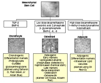
with the capacity to differentiate into several distinct mesenchymal lineages. MSCs can be induced to differ-entiate into adipogenic, chondrogenic, and osteogenic linages in the laboratory using appropriate cocktails of growth factors and supplements (Figure 1).12 Fur-thermore, MSCs have been studied in many research area to regenerate cell linages including, myogenic, cardiomyogenic lines, and also differentiate into neu-rogenic, angiogenic and hepatic lineages.10,13,14 Despite their capability to differentiate in vivo and in vitro into mesenchymal lineages, MSCs have also been shown to have an important role in the maintenance of hae-matopoiesis.13
Figures and legends
Figure 1. Multilineages Differentiation of Me
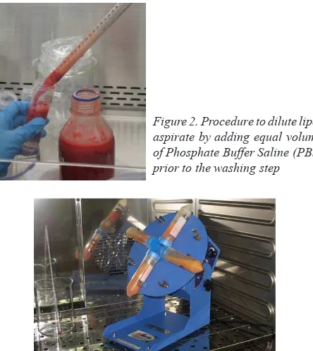
Figure 2. Procedure to dilute lipoaspirate by adding equal volume of Phosphate Buffer
Saline (PBS) prior to the washing step
Chondrocyte Osteoblast Adipocyte (stained von Kossa, Alizarin
Red) (stained von Kossa, Alizarin
Red)
Many publications have shown MSCs present and able to be isolated from many tissue types, such as bone marrow, trabecular bone, periosteum, synovial mem-brane, muscle, dermis, pericytes, blood, and adipose tissue.15-17 These demonstrate the potential of MSCs in addition to their plasticity and their usefulness in cell therapy. It is also of interest that neither autolo-gous nor allogeneic MSCs induce any immunoreacti-vity in the host upon local transplantation or systemic administration.18,19 Finally, the large quantity of MSCs in lipoaspirates, makes it an attractive source for stem cells isolation for research and clinical applications.14,17 An optimized method which is suitable to be performed in a basic research laboratory clearly will facilitate de-velopment for basic laboratory to conduct stem cell re-search. Therefore the aim of this study was to modify the method of Zuk et al so that it would perform well in a minimal laboratory setting.
Methods
Mesenchymal stem cell isolation from lipoaspirates
All protocols were reviewed and approved by the Stem Cell and Cancer Institute Institutional Review
Board prior to the study (Proposal number 13/IRB/ SCI/KF/2008). Lipoaspirates were obtained with in -formed consent from individuals undergoing tumes-cent liposuction surgery in hospitals collaborated with Stem Cell and Cancer Institute. Lipoaspirates were stored at 2–8°C for no longer than 24 hours before they were used. Methods used to isolate the mesenchymal stem cells from lipoaspirate were adapted from meth-ods in Zuk et al.17 The raw lipoaspirates (120 ml) were
diluted with equal volume of Phosphate Buffer Saline (PBS) and were divided evenly in 50ml-tubes (Figure
2). The diluted lipo aspirates were centrifuged at 430 × g for 10 minutes continously at 20oC. After centrifu-gation, the target cell–containing lipid phase was re-moved from the top and transferred in to new tubes and
diluted with an equal volume of PBS. This washing
step was repeated twice followed by further an equal volume dilution of cell-containing lipid fraction with
pre-warmed (37oC) 0.075% collagenase type I (Sigma) in PBS. Enzyme digestion was done by incubation at 37°C for 30 minutes on an orbital shaker (Figure 3).
After digestion, enzyme activity was neutralized by
adding equal volume of Dulbecco’s modiied Eagle’s medium (DMEM) containing 10% Fetal Bovine Serum (FBS). Digested product then subjected to centrifuga
-tion at 600×g for 10 minutes. Pellet was resuspended in DMEM with 10% PBS then iltered through a 100-µm
strainer mesh attached to a vacuum-pump to remove
cellular debris (Figure 4). Collected cells after iltration
were then ready for utilization. An aliquot was taken for cell count using hemocytometer under a light mi-croscope to determined cell yields. Counts and viability were determined with a hemocytometer and the trypan
blue dye exclusion technique. Briely, 10 l trypan blue
stock solution (0.4% w/v) was mixed with 10 l cells suspension, incubated for 3 minutes at room tempera-ture then cells were counted in a hemocytometer cham-ber. With this method, dead cells appear blue and were therefore distinguishable from viable cells.
Figure 3. Procedure to isolate mesenchymal stem cells from lipo aspirate using collagenase was done by incubation at 37oC for 30 minutes on a rotating shaker
Figure 4. Filtration procedure by using a 100-m strainer mesh attached to a vacuum pump to remove cellular debris
Figures and legends
Figure 1. Multilineages Differentiation of Me
Figure 2. Procedure to dilute lipoaspirate by adding equal volume of Phosphate Buffer Saline (PBS) prior to the washing step
E
E E
E PP
Figure 3. Procedure to isolate mesenchymal collagenase was done by incubation at 37
Figure 3. Procedure to isolate mesenchymal collagenase was done by incubation at 37
94 Sardjono et al. Med J Indones
Mesenchymal stem cell culture
The simplest technique to purify MSC from other contaminating cells was done by allowing the cells to adhere on plastic-surfaced disc. Cells were seeded
at 1.3x10⁵cells/cm2 in M-199 medium (Gibco 11150)
supplemented with 20 % Fetal Bovine Serum (Invit
-rogen 26140-079), 100 unit/ml penicillin and 0.1 mg/ ml streptomycin antibiotics (Sigma P0781), then kept in 37oC, 5% CO2. After 2-3 days unwanted cells (non-MSC cells and debri) were removed by two washes
with medium and expanded to reach 80% conluence. In another 6-7 days, adherent cells were detached us
-ing 0.25% trypsin EDTA solution (Sigma T4049), then M-199 medium + 20% FBS was used to inactivate trypsin. Detached cells with ibroblast-like morphol
-ogy were cultured in another lask with cell density of 1.2x10⁵ cells/cm2 for 1 week or until conluence was achieved.
Flowcytometry assay to determine MSC homog-enous population
To conirm the simpliied protocols were able to isolate
good quality MSCs, several assays were conducted.
MSCs have been known to express CD105 and de
-icient in the expression of HLA Class II, CD34 and CD45.
Expression HLA Class II molecules were determined using PE luorescein conjugated monoclonal antibod
-ies against HLA Class II (abcam 23901). Antibod-ies directed against CD45 were conjugated with FITC (BD 555482) and CD34 with FITC (BD 348053) incubated at 4oC in the dark for 15 minutes. Mouse IgG1-FITC (BD 349041) isotype matched negative controls were used to deine background staining. The cells then anal
-ysed by low analysis using FACSCalibur 3 argon laser 488nm (Becton Dickinson).
Results
Mesenchymal stem cell isolation and culture
Using the methods described, MSCs were able to be obtained at 16.41 ± 8.22 x 10⁸ cells per 120 ml lipo-aspirate which have provided an abundant number of
cells for further used (Table 1). The simpliied method
has proven to be gentle enough for the cells as shown
by the high viability cells obtained (viable cells 92.57% ± 4.5%).
Table 1. Lipoaspirate donors and cell yield Donor No. Lipoaspirate
volume (ml)
Cell yield (x 108)
Viability (%) 1
2 3 4
120 120 120 120
4.27 20.55
22.2 18.6
90.05 91.95 89.16 99.12
average 120 16.405 92.57
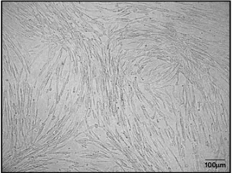
In cell culture, the isolated cells showed a ibroblast-like morphology which is characteristic for MSCs (Figure 5).
Figure 5. Homogenous MSCs showed ibroblast-like morphology during cell culture
Flowcytometry analysis
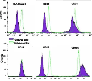
Cell surface molecules expressions were evaluated by
lowcytometry assay on the harvested cell after a 14-day culture. Based on standard criteria for deining
multi-potent mesenchymal stem cells, we characterized the cultured cells by using several markers. MSCs should
express CD105 (known as endoglin). CD105 is a type I
membrane glycoprotein located on cell surfaces and is part of the TGF beta receptor complex. Moreover, the cultured cells should be excluded from the non-MSC cells most likely to be found in MSC cultures such as
leukocytes (detected by CD45 expression), hemato
-poetic stem cells (expressing CD34), monocytes (expressing CD14), and B cell lymphocytes (CD19). HLA-DR (HLA Class II) molecules are not expressed on MSC unless stimulated, e.g. by IFN-. We conirmed
our isolated and cultured cells were MSCs as they were
mainly expressed CD105 and only a small proportion beared HLA-Class II, CD 45, CD 34, CD14, and CD19
(Figure 6).
Figure 5. Homogenous MSCs showed fibr
MSCs, i.e. lacked of HLA class II expression, CD45, CD34, CD 14 and CD19; with high expression of CD 105.
+/$&ODVV,, &' &'
&'
&' &'
&XOWXUHGFHOOV ,VRW\SHFRQWURO
95
Vol.18, No.2, April - June 2009 Stem cell isolation from lipoaspirates
DISCuSSIon
Stem cell research clearly is needed to be initiated in the developing countries including Indonesia. This is
also due to the requirements to ind a good strategy for stem cell as treatment in the regenerative medicine. De -veloping country has been getting an increase in the de-generative diseases cases each year. It is estimated by
2020, 7 out of 10 deaths in developing countries will be
attributable to non-communicable diseases 20.
More-over, developing countries now account for 80% of
global cardiovascular disease-related deaths with
two-thirds of the 171 million people worldwide affected by
diabetes reside in developing countries20. This report clearly has shown the urge for stem cell research to be conducted in Indonesia as soon as possible to support the progress in the stem cell-based therapy. The concern about starting a stem cell research also partly caused by
the lack of experience in the possible modiication of
protocols for cell isolation, processing and analysis to suit the local laboratory conditions.
This study has demonstrated that it is possible to isolate mesenchymal stem cells from the lipoaspirate using a
modiication of Zuk’s method. Lipoaspirate is truly rich
with Mesenchymal Stem cell. Compared to the MSCs collected from bone marrow, the MSCs from lipoaspi-rate obviously is easier to obtain with the yields above the yield often found in the isolation of stem cell from
bone marrow. Due to the unique property of MSCs,
the international society for Mesenchymal and Tissue Stem Cell Committee of the ISCT21 proposes minimal
criteria to deine human MSCs. First, MSCs must be
plastic-adherent when maintained in standard culture
conditions. Second, MSCs express CD105, CD73, or CD90, and lack expression of CD45, CD34, CD14 or CD11b, CD79α or CD19, or HLA-DR (HLA Class II).
Third, MSCs should demonstrate their ability for trilin-eage mesenchymal differentiation i.e. should be able to differentiate into osteoblasts, adipocytes and chondro-blasts using standard in vitro tissue
culture-differentiat-ing conditions. This study has been able to fulill two
out of the three criteria. A further study for MSCs dif-ferentiation using media and additives shown in Figure 1 would be done in a subsequent study.
Further puriication of MSCs from unwanted cells such
as white blood cells, endothelial cells, and smooth mus-cle cells, can be achieve using standard culture method Figure 6. Flowcytometry analysis of molecules expression on MSC from lipoaspirate. Flowcytometry assay showed the cultured cells were consistent with the characteristic of MSCs, i.e. lacked of HLA class II expression, CD45, CD34, CD14 and CD19; with high expression of CD 105.
Figure 5. Homogenous MSCs showed fibr
MSCs, i.e. lacked of HLA class II expression, CD45, CD34, CD 14 and CD19; with high expression of CD 105.
101 102 103 104
100
101 102 103 104
100
0 200
160
120
80
40
101 102 103 104
100
+/$&ODVV,, &' &'
101 102 103 104
100
0 200
160
120
80
40
101 102 103 104
100
101
102
103
104
&'
&' &'
&XOWXUHGFHOOV ,VRW\SHFRQWURO
100
Counts
96 Sardjono et al. Med J Indones
using in plastic-surfaced discs. MSCs were identiied as cell population with ibroblast-like morphology.21 The MSCs isolated from above protocol were able to be used in further experiments including differentiation into osteogenic, adipogenic, and chondrogenic
progen-itor cells. In this study, the lowcytometry assay was used to conirm that the cells were mainly CD105 bear -ing cells that were most likely MSC. This fact strongly suggests that the protocol is very well adapted and able to isolate nearly homogenous MSCs population in a minimal laboratory setting. For the application of this method in a circumstances where the experiments should be done without cell analysis equipments,
an-other method such as detection of mRNA for CD105, CD73, or CD90, with the absence of CD34, CD 45 or HLA-DR (HLA Class II) can be pursued. It is impor -tant to notice though, that the grade of cells obtained using this method was designated to be used in research and should not be attempted for clinical uses.
This study has shown that by using simple equipments it was feasible to isolate messenchymal stem cell from human lipoaspirate an otherwise discarded material. The procedure was practicable to be performed within a basic laboratory with the good yield of cell. The out-comes from this study are expected to encourage the progress of stem cell research in Indonesia.
Acknowledgements
The authors wish to thank Prof. Santoso Cornain, dr, DSc. for sharing his invaluable expertise, to Boenjamin Setiawan, dr., PhD who delivered the idea of stem cell
research, to the donors for donating their lipoaspirate, and the collaborated hospital staffs who have coordi-nated the material to be used in this study.
ReFeRenCeS
1. Moseley TA, Zhu M, Hedrick MH. Adipose-derived stem and progenitor cells as illers in plastic and reconstructive surgery. Plast Reconstr Surg. 2006;118(3 Suppl):S121-8. 2. Rebelatto CK, Aguiar AM, Moretao MP, Senegaglia AC, Han
-sen P, Barchiki F, et al. Dissimilar differentiation of me-senchy -mal stem cells from bone marrow, umbilical cord blood, and adipose tissue. Exp Biol Med. 2008;233(7):901-13.
3. Kern S, Eichler H, Stoeve J, Kluter H, Bieback K. Com -parative Analysis of Mesenchymal Stem Cells from Bone Marrow, Umbilical Cord Blood, or Adipose Tissue Stem Cells. 2006;24:1294-1301
4. Fraser JK, Schreiber R, Strem B, Zhu M, Alfonso Z, Wulur I, et al. Plasticity of human adipose stem cells toward en
-dothelial cells and cardiomyocytes. Nat Clin Pract Cardio -vasc Med. 2006;3 Suppl 1:S33-7.
5. Meliga E, Strem BM, Duckers HJ, and Serruys PW. Adipose-Derived Cells. Cell Transplantation. 2007;16:963–70. 6. Garcia-Olmo D, Garcia-Arranz M, Herreros D, Pascual I,
Peiro C, Rodriguez-Montes JA. A phase I clinical trial of the treatment of Crohn’s istula by adipose mesenchymal stem cell transplantation. Dis. Colon Rectum. 2005;48(7):1416–23. 7. Horwitz EM, Gordon PL, Koo WK, Marx JC, Neel MD,
McNall RY, et al. Isolated allogeneic bone marrow-derived mesenchymal cells engraft and stimulate growth in children with osteogenesis imperfecta: Implications for cell therapy of bone. Proc. Natl. Acad. Sci. USA 2002;99:8932–7. 8. Yoshimura K, Matsumoto D, Gonda KA. Clinical trial of
soft-tissue augmentation by lipoinjection with adipose-derived stromal cells (ASCs). International Fat Applied Technology Society (IFATS). 2005;9–10.
9. Psaltis PJ, Zannettino A, Worthley SG, Gronthos S. Mesen chymal Stromal Cells – Potential for Cardiovascular Repair. Stem Cells. 2008;26(9):2201-10
10. Lin K, Matsubara Y, Masuda Y, Togashi K, Ohno T, Ta -mura T, et al. Characterization of adipose tissue-derived cells isolated with the CelutionTM system. Cytotherapy. 2008;10(4):417-26.
11. Ilic D. Explores the latest developments in the ield of stem cell research and regenerative medicine. Regenerative Medicine 2008;3(2):137-43.
12. Marion NW and Mao JJ. Mesenchymal Stem Cells and Tis -sue Engineering. In: Klimanskaya I and Lanza R, editors. Methods in enzymology. Pasadena, California: Elsevier; 2006. p. 339-61.
13. Alhadlaq A, Mao JJ. Mesenchymal Stem Cells: Isolation and Therapeutics. Stem cells develop. 2004; 13:436-48. 14. Meliga E, Strem BM, Duckers HJ, and Serruys PW. Adi
-pose-Derived Cells. Cell Transplantation. 2007;16:963–70 15. Asakura A. Stem cells in adult skeletal muscle. Trends Car
-diovasc Med 2003;13:123-8.
16. Belicchi M, Pisati F, Lopa R, Porretti L, Fortunato F, Sironi M, et al. Human skin-derived stem cells migrate through -out forebrain and differentiate into astrocytes after injection into adult mouse brain. J Neurosci Res. 2004;77:475-86. 17. Zuk PA, Zhu M, Mizuno H, Huang J, Futrell W, Katz AJ et al.
Multilineage cells from human adipose tissue: implications for cell-based therapies. Tissue Eng. 2001;7:211-28. 18. Le Blanc K dan Ringden. Immunobiology of human
me-senchymal stem cells and future use in hematopoietic stem cell transplantation. Biology of Blood and Marrow Trans -plantation. 2005;11:321-4.
19. Yanez R, Lamana ML, García-Castro J, Colmenero I, Ramírez M, and Bueren JA. Adipose tissue-derived mesen-chymal stem cells (ADMSCS) have in vivo immuno-suppressive properties applicable for the control of the graft-versus-host disease. Stem Cells. 2006;24(11):2582-91. 20. Greenwood HL, Thorsteinsdottir H, Perry G, Renihan J,
Singer PA, and Daar AS. Regenerative medicine: new oppor-tunities for developing countries. Int. J. Biotechnology. 2006;8(12):60-77.