Suppl
I
- 1998
Clinical features anclpathogenesis
219Analysis of the
interaction
of-Salmonellas
\ilith
CP.2
macrophages from
different
host
species.
P.R.Watson,
A.V.
Gautier,
S.M. Paulin,
P.W. Jones, T.S.Wallis
Abstrak
Dasar molekuler
tlari
spesifisitas pejamu Sctlmonella masih belwn jelas. Serotilt Salmonella dublirL S. choleraesuis dan S. abor-tusovis metnperLihatkan fenotip pejarnu spesffik untuk berrurut-turut sapi, babi dan elomba. Berbecla dengan serotip tersebut, S. ty-phimurium metnpunyai pejamu yang beranel<n ragant. Inleraksi serotip-serotip tersebut tlengan makrofag tlari tnencit, sttpi, babi dandomba tehh dibandingkan. Hasil dari interaksi tergantung pcttla serotilt tlan jenis hospes tetapi tidak berkorelasi clengan kespesifikctn
peiamu clari serotip-serotip tersebut. Galur S. dlblin dan S. typhimurium ntengintluksi lebih besar lisis pacla makrofag sapi clan babi clibantlingkan dengan galur S. choleraesuis. HaI yang sanw clitemukan pada nmkrofag domba jika dibanclingkan clengan grzlrlr S.
chol-eraesuis r/an S. abortusovis. Sebal.ihtya keentpat serotip tersebLrt mengincluksi lisis tlari nmkrofag peritorrinn ntencit pacla tingkat yang sebanding. Makrofag sapi, dotnba tlan mencit lebihsensitif daripada makrofag babi untuk dilisis oleh SaLntonella. Lisis makrofag tklak berhubungan dengan gambaran karakteristik apoptosis. Dengan nikroskop elektron îransmisi ticlak
terljlnt
adanl,a pen.ingkcttan. gant-baran morfologik yang berhublmgan dengan apoptosis pcrcla nnkrofag monolayer terinfeksi. Juga tklak acla peningkntan laddering of DNA ;,61ag klns dari sel yang dilisis oleh Salntonello dibandingkan clengan sel monolayer kontrol. Lisis nnkrofery tidak cliperantarai oleh TNE tetapi memerlukan aelanya salmonella ekstrasel hiclup. Gangguan sistenr sekresi tipeIII
yang dikotle oleh operon invlsparnelenyapkan kemampuan salmonella melisis makrofag. Hnsil ini memperliharkan balwa inlerpretasi hasil interaksi Sal.ntonel!.ct/nakro-fery dalam uji protektif gentalnisin harus dilakukan clengan lnti-hati. Karena nnkrofag babi lebih resistan terhadap lisis oleh salntoneLla,
persistensi salmonelLa intrasel serta procluksi sitokin pro inflatninatorl, setelclr infeksi ol.eh galur S. dublin, S. choleraesuis dtut S. ty-phimurium telalr lcanù teliti dan lusiltq,a akan karni diskusikan.
Abstract
Tlte ntolecular basis of Salntonella host-specfficily retnains unclear The serol-ttpes Salmonella dublin, S. choleraesuis anrl S.
abor-tusovis s/row a host specific phenotype
for
cattle, pigs and slrcep respectively. In contrast to these serotypes, S. typhimiu4um has tt broarl host range.The inreraclions of these serolypes witlt macrophages from nice,cattle, pigs and slrcep were compared. The outcome of theinteractions was serotype-ancl host-tlependent but dicl not correlate to the host specific phenotypes of these serorypes. S. dublin nnd S. typhimurium strains inclucecl grealer lysis of both bovine ctnd porcine nncrophuges îhctn S. choleraesuis strains. Sittri/arll' S. dublin anrl
S. typhimurium stains incluced gre(tter lysis of ottine macrophages compared witlr S. choleraesuis anrl S. abortusovis strains. In conlrast all four serotypes irtclucetl comparabl.e leveh of lysis of nturhrc peritoneaL macroplrages. Boyine, ovine, cutcl murine macrophages were
more sensitive thart porcine macroplnges to SahnoneLlct inducecl l1'sis. lysrs of macrophages was not associatedwithfeatures
chttrctc-teristic of apoptosis. Infected macroplnge monolayers showetl no increase in rhe morplnlogical featnres associated with apoptosis when
exanined by transmission electron microscolty. Sùnilarly llrcre was no increase in the characteristic laeldering of DNA isolatetl front cells
untlergoing Salmonel.la-incluced h,sis cotnparecl u,ith uninfected conîtol monolayers. Macropltage lysis was not mecliatecl by TNFa, but requiretl the presence ofviable extracellular Salntonellas. Disruplion ofthe type
III
secretion system encoded by the inv/spa operon abol-islrcd Sahnonella-induced nncrophage lysis. These resulls tlemonslrate that extretne cautiottis
required when interpretingSalnto-nella/macropltage interactions in gentamicin prolection assays. As porcine nncrophages were relatively resistant to Sctlmonellct-inchrcecl lysis the intracellular persistance of Salntonella and the production of pro-ùdlamnntory cytokines was assessecl fotlowittg infection with
S. dublin, S. choleraesuis and S. typhimurium strarns,' the result of tlrcse experintents
will
be discrlssecl.INTRODUCTION
Macrophages
may potentially
have
akey role in
thepathogenesis
of
enteric and systemic
salmonellosis.
Their distribution throughout
the
body
andtheir
bac-Inslitttte for Animal Health, Compton, Newbury, Berkhire,
RG2O 7NN. U.K.
tericidal
properties
could influence
the successful
es-tablishment
of
a Salmonella infection and their
im-muno-regulatory properties could influence the
host
response
to Salmonella infections.
natural Salmonella
serotype
and host
cell
combina-tions.
Therefore, we
studied the
interaction of
S.dub-lin and
S. choleraesais strains
in bovine
and porcine
macrophages.
S.dublin is
associatedwith
severe dis-easein
calves and S. choleraesuis
is
associatedwith
severe disease
in
pigs. The outcome
of
theinteraction
of Salmonella
serotypes
with
macrophages
was
as-sessedwith
respect
to
the
effect on both
the bacteria
(persistence)
and the
macrophage
(amount
of
lysis,
mechanism
of
lysis
andproduction
of
cytokines).
MATERIALS AND
METHODS
Bacterial
strains
S.
dublin strains 5D2229
and
SD3246 were
isolated
from
casesof
salmonellosis
in cattle.
S.choleraesuis
var
kunzendoy' strains SCSA5O and
SCS14/74
were
isolated
from
casesof
salmonellosis
in
pigs.
Preparation
of
conditioned
culture supernatants
from macrophages
Bovine
and
porcine alveolar
macrophages were
iso-lated
asdescribed befores. Macrophages were
seededinto 24
well
tissue
culture
plates
at 5x 10s cellsper ml
in
Dulbecco's
modified Eagle's medium
and
Ham's
nutrient mix
F-12
containing
107oFCS and
100gml-t
gentamicin and incubated overnight
at
37"C
in
5Vo COz.Two
hours before
infection,
theculture
medium
was replaced
with DME/F12 medium
containing
5VoFCS
and
no antibiotics. Bacterial cultures
were
pre-pared as described above and diluted
in
DMEÆ12
medium to give
aratio
of infection of
5bacteria
to
1macrophage.
The overgrowth
of
bacteria
in
the
cul-ture medium
of the monolayers incubated
for
24
or
48 hours
was prevented
by
washing the
monolayers
after
t
hour
and
adding medium containing
57o FCSand
100
gml-t gentamicin, followed
by
washing
themonolayers after a further hour
and
adding medium
containing
57oFCS and
10pgml-l
gentamicin.
After
the appropriate
incubation time,
the monolayers were
centrifuged
(300
x g,
10min,
4'C)
and theculture
su-pernatants, termed conditioned macrophage
super-natants,
were aliquoted
into
sterile eppendorf
tubesheld on ice. The aliquots were
stored at
-70"C
until
assay.
Macrophage
damage wasestimated
by
measuring
theamount
of
lactate dehydrogenase released
by the
macrophages
into
the
supernatantsusing the
Cytotox
96
Non-radioactive
cytotoxicity assay
(Promega,
Madison,
USA). The number
of
bacteria
associatedwith
the macrophage monolayers at each
of
the
time
points,
and also
at
one
hour after infection, was
de-termined by viable count
asdescribed previouslys.
Characterisation
of DNA
from macrophages
DNA from
macrophages
was
extracted
using
the
method
of
Zychlinsky
et al6.
Actinomycin
D
manni-tol
(1
pgml-l)
was added as a positive control
for
apoptosis.
Macrophage monolayers were infected
ata
ratio
of
infection
of
5 bacteria
to
1
macrophage.
They
were
either
incubated
for
3 h without the
addi-tion of
gentamicin
or
t
hour after
infection
the
mono-layers
were
washed
once
with
prewarmed medium
and incubated
for
afurther
18hours
in medium
con-taining
57oFCS and
100Fgml-t
gentamicin.
Bioassay
for
ll--l-like
activity
L-l-like
activity
was
measuredusing the A375 cell
line,
whose
growth
is
inhibited in
the presenceof
hu-man IL-17.
Aliquots
of
the conditioned
macrophage
supernatants
were thawed and serially diluted
in
96
well
tissue cultur"eplates and
1x
10+A375 cells
were
added.The
assayplates
were
incubated
for
96
hours
at37"C in
5Vo COz.Following incubation,
theculture
medium was removed and
the wells
were
washedonce with
PBS.
The
remaining
cells
were
stained
with crystal violet
stainfor
2h
atroom
temp
andthen
the
excessstain
wasremoved
by
washing
thoroughly
with
PBS.
The
amount
of
retained
crystal violet
wasmeasured
by
spectrophotometry
using
a
wavelength
of
595 nm
following solubilisation of
the
crystal
vio-let in
I00Vo methanol.
Bioassay
for ll--6-like
activity
Il-6like
activity
was measured using
the TTD1
cell
line,
whose
growth is
dependent
on the
presence
of
IL-69.
Aliquots
of
theconditioned
macrophage
super-natants
were thawed and serially diluted
in 96 well
tissue culture plates
and
I
x
1047TD1 cells
were
added.The
assayplates
were
incubated
for
96
hoursat 37"C in
5VoCO1 The
assayplates
were
incubated
for
72 hours at 37"C in an
atmosphere
of
5VoCOz.
Growth
of the 7TD1 cells
was estimated
by
measur-ing
DNA
synthesis.An aliquot of FÉI1,1'tt-idine
con-taining
0.015
MBq
of
radioactivity
was addedto
eachwell
at
l8
hours before the end
of
the
incubation
pe-riod.
The
cells
were harvested
onto
glassfiber filters
Suppl
I -
1998Bioassay
for TNFaJike
activity
TNF-like activity
was measuredusing
theWEHI
164clone
13cell line, which is
sensitive
to
the
cytotoxic
activity
of
human TNFe.
Aliquots of
the conditioned
rnacrophage supernatants
were thawed and serially
diluted
into the
96
well
tissue
culture
plates
contain-ing WEHI cell
monolayers. The
assayplates were
in-cubated
for
18hours at
37"C in
anatmospherc
of
5VoCOz.
Following incubation,
the
remaining cells
were
stained
with
crystal
violet
stain
as described
for
thetrL-1
bioassay.R.ESULTS
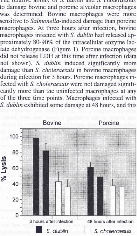
Salmonelln-induced macrophage damage
is
host-and serotype-dependent
The
relative
ability of
S.
dublin
and S. choleraesuis
to
damage
bovine
and
porcine alveolar
macrophageswas
determined.
Bovine
macrophages
were
more
sensitive
to
Salmonella-induced
damage thanporcine
macrophages.
At
three hours after infection,
bovine
macrophages
infected with
^S.dublin
had releasedap-proximately
80-907o
of the intracellular
enzyme
lac-tate dehydrogenase
(Figure
1).
Porcine
macrophagesdid not
releaseLDH
atthis time after infection
(datanot
shown).
S. dublin
induced
significantly
more
damage
than
^S.iholeraesuis
in bovine
macrophagesduring infection
for
3hours. Porcine
macrophagesin-fected
with
S. choleraesui,swere not
damagedsignifi-cantly more
than
the uninfected
macrophages
at
anyof
the three time points.
Macrophages
infected with
S.
dublin exhibited
somedamage
at48
hours,
andthis
Bovine
Porcine
3 hours after
infection
48 hours after infectionI
s.
orol, I
s, [image:3.595.47.282.309.712.2]"r,ot
roesursFigure
1.
Lysisof
alveolar macrophages by S. dublin nnd S.choleraesuis slrarns
Clinical features and
pathogenesis
221was
significantly
greater
than
theS.
choleraesuis
in-fected macrophages
(Figure
1).Salmonella induce macrophage
lysis
by
oncosis
The
ultrastructure
of
macrophages
after
infection
with
Salmonella
serotypes
was examined
by
trans-mission electron microscopy.
In uninfected
monolay-ers
of
bovine
mâcrophages,
the
majority
of
macro-phages appearedhealthy but there were
aminority
of
cells
exhibiting
typical
features
of
apoptosis
(con-densed
chromatin located very close
to
the
nuclear
membrane together
with
maintenance
of
the structure
of
organelles
and
membranes).
The
majority of
macrophages
incubated
with actinomycin D mannitol
for 5
hours exhibited
typical
features
of
apoptosis.Macrophage monolayers
which had
been infected
wrth
Salmonella
serotypes
contained
cells with
arange
of
morphological
changes.
Many
of
the
cells
appearednecrotic,
with
aloss
of
pseudopodia,
swol-len
organelles and
condensed
chromatin distributed
randomly through the nucleus.
These changes were
more
severe
at
18 hours
after infection and
at
this
time
there.
was
a
large
amount
of
debris
present,which presumably
was
aresult
of
macrophage
lysis.
These
ultrastructural
changes aremore typical of
ne-crosis
resulting
from
oncosis rather than
apoptosis.
There were more
relatively healthy cells in
monolay-ers infected
with
S. choleraesuis
compared
to
thoseinfected with
.1.dublin.
DNA from bovine
andporcine
macrophages
either
3or
18hpost
infection
with
either
S.dublin
or
S.chol-eraesuis
(with or without
the
addition
of gentamicin)
did
not show the characteristic
DNA
laddering
pat-tern characteristic
of
cells undergoing
apoptosis,
aswas
seenwith cells
treated
with actinomycin D
man-nitol.
Release
of
pro-inflammatory cytokine-like
activ-ity by infected porcine
macrophages
is
inde-pendent
of
Salmonel/a serotype
Uninfected macrophage monolayers released
little
IL-l
over the
time
course
of the
assay.Macrophages
infected
with
viable bacteria
released
large
amounts
of
IL-1
at
all
three
time points of
3,24
and48
hours.There was
no significant difference
between the
dif-ferent
serotypes.Uninfected macrophage monolayers released
little
IL-6
over the
time
course
of
the
assay.Macrophages
infected
with
viable bacteria
released
slightly
more
IL-6
than
the uninfected
macrophages at three hours
after infection
and
significantly more
IL-6
at
24
and
100
80
.2
o
à60
àe
40
48 hours after infection.
Macrophages
infected with
S.
choleraesals
released
slightly more
IL-6
at 3
and24 hours after infection than those infected
with
^9.dublin,
although
this difference
was
not significant.
Uninfected macrophage monolayers released
little
TNF-0, over the time
course
of
the
assay.Following
infection
with
all
three
serotypes, macrophages
re-leased
large amounts
of
TNF-o
at ali
three time
points. There were no
significant
differences
betweenthe
different
serotypes (p>0.1).
The recovery
of
Salmonella
from
infected
macro-phages
is
serotype-dependent
The number
of
bacteria
associatedwith
the
monolay-ers
was determined
atthe
sametime points
andfrom
the
same macrophage preparations
that
the
condi-tioned
macrophage supernatants were
collected.
The
number
of
bacteria
associated
with
monolayers
at
1hour after infection was
also measured
to
determine
whether differences between
the
serotype
could
beattributed to
thenumber
of
bacteria
initially
taken
up.At
t
hour after
infection,
there
wasno
significant
dif-ference
in
the
recovery of
S.dublin
and S.cholerae-szis.
At
three hours
after infection,
S.dublin
was
re-covered
in
higher
numbers
than
,S. choleraesuis.
Thereafter
both
serotypes persisted
within
porcine
macrophage
atcomparable
ratesup to
48hpost
infec-tion.
DISCUSSION
The aim of this
study
wasto
characterise the
interac-tion
of
S.
dublin
and S. choleraesuis
with
macro-phages
from
host animals
for
which the
serotypesshow
differing
host-specificty.
Salmonella-induced
macrophage
lysis
was dependent onboth
the serotype and.thehost.
However,
macrophage
lysis did
not
cor-relate
to
the virulence
of
each
serotype
for
pigs or
cattle.
S.dublin
induced more macrophage
lysis
than
S.
choleraesais
in both caltle
andpigs,
but is
associ:ated
with
severe
diseaseonly
in
cattle. Thus
macro-phage damage does
not
correlate
to Salmonella
sero-type-host specificity.
The
mechanisms
involved
in
mediating
Salmonella-induced macrophage
lysis
are unclear.
Salmonella
have been reported
to
induce lysis
of
celis
by
either
apoptosi5lO-l1 and
oncosisli.
We
found
no
correlation
between serotype-
and
host-dependent macrophage
damage
andthe mechanism
of
lysis. Primary
macro-phage
cultures
were
damageby
amechanism
resem-bling
oncosis
in all
serotype and host combinations.
Therefore, the
serotype- and host-dependent
macro-phage
lysis
appearsto
be related
to
the
susceptibility
of
the host
cell
andto
the
kinetics
of
oncosis
andnot
to
the involvement
of
different
mechanisms
of
cell
death.
Salmonella
serotypes canpersist
within
macrophages
in vitro. However,
studies
which
have
tried to
corre-late the
persistence
of
a Salmonella
serotype
to
its
virulence
in
aparticular
host
animal
areinconclusive,
as
failure
to
control
for
Salmonella-induced
macro-phage
lysis
ln
vitro
when
assessingintracelluiar
per-sistence
in
agentamicin protection
assayrenders
theresults
difficult to
interpret.
In
the
present
study
therefore,
the recovery
of
Salmonella
serotypes
dur-ing
macrophage
infection was re-evaluated.
It
wasnot possible
to
directly compare the relative ability
Salmonella serotypes
to survive in bovine or porcine
macrophages
becauseof
the higher susceptibility of
bovine
macrophages
to
be
damaged.
In
porcine
macrophages,
the
persistence
of
a Salmonella
sero-type did not correlate to its
virulence in
pigs. Thus
thepersistence
of
Salmonella
serotypes
in
macrophages
did not
correiate
to
serotype-host
specificity.
Macrophages
are important
in
the regulation
of
the
hosts
primary immune
responseto infection through
the
release
of
cytokines and other mediators
of
in-flammation.
This
response
may influence the
ability
of
the host to
controi infections, but
may
alsoexacer-bate
some
aspectsof
disease.
If
infection
of
macro-phages
with different Salmonellc
serotypes results
in
the
release
of
different types
or
amounts
of
inflam-matory
mediators,
this could affect the severity
andthe nature
of
the resulting
disease
and thus
Salmo-nella
serctypehost-specificity.
The
kinetics of
cytok-ine
release
from
bovine
macrophages
could
not
bequantified
becauseoftheir high susceptibility
for
Sal-monella-indtced damage.
Infection
of
porcine
macrophages
with
both
Salmonella serotypes
re-sulted
in
a rapid and
sustained release
of
IL-tr
andTNFo activity
and amore gradual increase
in
the
re-lease
of
IL-6
activity.
Therefore, the
ability of
differ-ent
Salmonella
serotypesto
induce the
releaseof
pro-inflammatory
cytokines
from
macrophages
in vitro
is
not influenced by
the serotype.
In
conclusion,
theoutcome of
theinteraction
between
Salmonella serotypes and
macrophages
ln
vitro
is
clearly
dependent on the
particular
derotype
andhost
animal involved.
Salmonella-induced
macrophage
lysis ln vitro
meansextreme
caution
is
required in
Suppl
I
-
1998macrophages
in
gentamicin protection
-experiments.The host specificity
of
S. dublinand
,S.choleraesuis
for
cattle
and
pigs respectively
cannot
be
explained
by any
1of
the
following
parameters:
Salmonella-in-duced macrophage damage,
bacterial uptake or
per-sistence
within
macrophages, mechanism
of
macro-phage damage
or
release
of pro-inflammatory
cytok-ines
by macrophages.
REFERENCES
1. Vladoianu I-R, HR Chang; J-C Pechère. Expression of host resistance to Salmonella typhi and Salmonella typhimurium: bacterial survival within macrophages of murine and human origin. Microbial Pathog 1990; 8: 83-90.
2.
Alpuche-Aranda CM, Berthiaume EP, Mock B, Swanson JA,Miller SI.
Spacious phagosome formationwithin
mouse macrophages correlateswith
Salmonella serotype patho-genicity and host susceptibility. Inf Immun 1995: 63:4456-62.
3. Ishibashi Y, Arai T.
A possible mechanism for host-specific pathogenesis of Salmonella serovars. Microbial Pathog 1996;21: 435-46.
4.
Schwan WR, Kopecko DJ. Serovar specific differences in Salmonella survival within macrophage cells. Mechanisms in the Pathogenesis of Enteric Diseases. Ed Paul etaI,Plentm
Press New York. Chapter 1997;46:277-8.
Clinical features and
pathogenesis
2235. Guilloteau
LA, Wallis TS, Gautier AV, Maclntyre S, platt DJ, Lax AJ. The Salmonella virulence plasmid enhances Salmo-nella-induced lysis of macrophages and influences inflamma-tory responses. Infect Immun 1996;64:3385-93.6. Zychlinsky
A, Prevost
MC, SansonettipJ. Shigeltaflenteri induces apoptosis in infected macrophages Nature 1992; 358: 167-8.7. Nakai
S, Mizuno K, Kaneta M, Hiraiy.
A simple, sensitive bioassay for the detection of interleukin- 1 using human mela-noma 4375 cell line. Biochem Biophys Res Commun 1988; 154:1189-96.8.
Van Snick J, Cayphas S, Vink A, Uyttenhove C, Coulie pG, Rubira MR, Simpson RJ. Purification and NH2+erminal amino acid sequenceof
a T:cell-derived lymphokine with growth factor activity for B-cell hybridomas. proc Natl Acad Sci USA 1986; 83: 96'79-83.9. Espevik
I
Nissen-Meyer J.A
highly sensitivecell line,
WEHI 164 clone 13, for measuring cytotoxic factor/hrmor necrosis factor from human monocytes. J Immunol Method 1986; 95: 99-105.10. Monack DM, Raupach B, Hromockyj AE, Falkow S. Salmo-nella,typhimurium inyasion induces apoptosis
in
infected macrophages. Proc Natl Acad Sci USA 1996; 93: 9833-8. 11. Chen LM, Kaniga K, Galân JE. Salmonella spp. are cytotoxic