http://mji.ui.ac.id
Acalypha indica
root extract increases post-hypoxic rat hippocampal
tissue culture cell viability via phospholipase A
2inhibition
Sophie Yolanda, Trinovita Andraini, Indra Kusuma
Department of Physiology, Faculty of Medicine, Universitas Indonesia, Jakarta, Indonesia
Abstrak
Latar belakang: Fosfolipase A2 (PLA2) terlibat dalam proses inlamasi dan kematian sel pada stroke, dan inhibisinya dapat mendorong terjadinya neuroregenerasi. Tujuan penelitian ini adalah mempelajari pengaruh pemberian ekstrak akar Acalypha indica Linn terhadap viabilitas relatif sel dan kadar enzim PLA2 pada kultur jaringan hipokampus pasca-hipoksia.
Metode: Studi eksperimental in vitro dilakukan pada 24 kultur primer jaringan sel saraf tikus Sprague Dawley dewasa yang dipajankan terhadap hipoksia dengan gas 5% O2 / 5% CO2 / N2 seimbang selama 24 jam. Pascahipoksia, ekstrak Acalypha indica Linn diberikan pada 3 kelompok perlakuan, masing-masing dengan dosis 10, 15, dan 20 mg/mL, sedangkan pada kelompok kontrol tidak diberikan apapun. Setiap kelompok terdiri atas 6 sampel. Setelah inkubasi selama 72 jam, viabilitas relatif sel diukur dengan 3-(4,5-dimethylthiazol-2-yl)-2,5-diphenyltetrazolium bromide (MTT), dan kadar enzim PLA2 diukur dengan menggunakan metode ELISA.
Hasil: Kadar enzim PLA2 kultur jaringan hipokampus tikus yang diberikan perlakuan ekstrak akar Acalypha indica Linn dalam dosis 10, 15, and 20 mg/mL menurun secara bermakna dibandingkan dengan kontrol (5,55 ng/mL, 6,85 ng/mL, 7,42 ng/mL vs. 7,96 ng/mL, p < 0,05).
Kesimpulan: Ekstrak akar Acalypha indica Linn meningkatkan viabilitas relatif sel dan menurunkan kadar enzim PLA2 pada kultur jaringan hipokampus tikus pasca-hipoksia dengan dosis optimal 10 mg/mL. (Med J Indones. 2013;22:136-40. doi:10.13181/mji.v22i3.581)
Abstract
Background: Phospholipase A2 (PLA2) is involved in inlammation and cell death following stroke, and inhibition of its
activity may promote neuroregeneration. This study aimed to observe the inluence of Acalypha indica Linn root extract towards relative cell viability and PLA2 enzyme level in post-hypoxic hippocampal tissue culture.
Methods: Experimental in vitro study using 24 primary neuronal cell cultures obtained from Sprague Dawley rat exposed to hypoxia with 5% O2 / 5% CO2 / N2 balanced gas for 24 hours. Post-hypoxia, Acalypha indica Linn root
extract was added at doses of 10, 15, and 20 mg/mL to three treatment groups. No treatment was given to the control group. Each group consists of six samples. After 72 hours of incubation, relative cell viability was measured using 3-(4,5-dimethylthiazol-2-yl)-2,5-diphenyltetrazolium bromide (MTT) examination, and phospholipase A2 enzyme level
was determined using ELISA.
Results: PLA2 enzyme level of rat hippocampal tissue culture treated with Acalypha indica Linn root extract at 10, 15,
and 20 mg/mL were signiicantly lower than that of control (5.55 ng/mL, 6.85 ng/mL, and 7.42 ng/mL vs. 7.96 ng/mL, p < 0.05).
Conclusion:Acalypha indica Linn root extract increases the relative cell viability and decreases the PLA2 enzyme
level of post-hypoxic mouse hippocampal tissue with the optimal dose of the extract at 10 mg/mL. (Med J Indones. 2013;22:136-40 doi:10.13181/mji.v22i3.581)
Keywords: Acalypha indica Linn, cell viability, hypoxia, neurogenesis, phospholipase A2
Correspondence email to: [email protected]
Stroke is the leading cause of morbidity and a common
cause of mortality worldwide.1 Thrombolytic is
currently the primary therapy for ischemic stroke2 due
to its effect in improving neurological function; the
only proven therapeutic approach that has this effect.3
Unfortunately, this therapy has a 3-hour therapeutic
window, which is very brief,2 and increases the risk of
hemorrhagic transformation.4 Thus, neurorestorative
therapy with longer therapeutic window that can improve neurological function to restore and optimize
the brain function is necessary.3,4 Neurorestorative
therapy enhances the development of new nerve cells (neurogenesis) in brain tissue following ischemic
stroke.4
One mechanism of stroke is the activation of
phospholipase A2 (PLA2) enzyme, which will produce
arachidonic acid (AA),5,6 leading to inlammation
and eventually cell death.7 There are at least 22
different PLA2 enzymes that have been identiied
in various mammalian tissues, broadly classiied into three families: calcium-dependent cytosolic
(cPLA2), secretory (sPLA2), and calcium independent (iPLA2).8 Cytosolic PLA2 (cPLA2) and secretory PLA2 (sPLA2) are involved in neuroinlammation and
neurodegeneration.8,9 Inlammation has been proven
to inhibit basal and post-hypoxic neurogenesis.10 In
cerebral hypoxia, there is an increase in cytosolic PLA2
http://mji.ui.ac.id
one of the site containing neural stem cells needed for
neurogenesis,3,12 is also very susceptible to hypoxia and
a prominent site of PLA2 expression.
13 Nowadays, all
PLA2 inhibitors that have been studied are non-speciic
so that the synthesis of a speciic inhibitor for cPLA2
is highly required.14 Studies have shown that
“knock-out” (cPLA2-/-) mice had smaller infarcts and fewer
neurological deicits compared to wild type following
cerebral ischemia.9 Drugs that target PLA
2 enzyme
have also shown neuroprotective effects.1,9,15
Acalypha indica Linn (akar kucing) is an indigenous
Indonesian plant that can easily be found in all regions of Indonesia. People have been empirically using the roots of the plant to cure nerve paralysis
caused by stroke.16 The plants have many active
chemical compounds; some that have been identiied are kaempferol (lavonoids), beta-sitosterol, HCN,
gamma-sitosterol, and acalyphin.17 Yolanda, et al18
have studied Acalypha indica Linn’s neurotherapeutic
effect on post-hypoxic nerve cells. Nirmal, et al19 have
proven that acalyphin and stigmasterol exert their
anti-inlammatory effects by forming PLA2 inhibitor
complexes. Decreasing the inlammatory response can increase the mobilization of endogenous adult stem cells population in the central nervous system, thereby increasing neurogenesis and neurological
function.16 This study aims to observe the effect of
Acalypha indica Linn root extract on PLA2 enzyme in post-hypoxic hippocampal tissue in vitro, and its
subsequent impact on cell proliferation.
METHODS
Study design and sample
An in vitro experimental study was conducted on
primary neuronal cell cultures, which were taken from
the hippocampus of adult Sprague Dawley rats aged
9-10 weeks weighing 150-200 grams. The experiment was conducted at the Oral Biology Laboratory, Faculty
of Dentistry Universitas Indonesia from September to
October 2011. The animals were sacriiced by cervical dislocation. The total number of samples was 24, divided equally into 4 groups (1 control group and 3 treatment groups) based on Federer formula. Ethical clearance was obtained from the Committee of the Medical Research Ethics of the Faculty of Medicine, Universitas Indonesia prior to the study.
Culture of nerve cells
Nerve cells were cultured in 10-cm petri dishes that have been coated with poly-L-lysine (Nacalai Tesque, Jakarta, Indonesia) for 1 hour. The nerve cells were then
seeded into 1x10⁶ cells/dish. Cultures were incubated
with 5% CO2 at 37°C. Dulbecco’s Modiied Eagle
Medium (DMEM) (GIBCO, Invitrogen) was used for culture medium and was replaced every 72 hours during the incubation period.
Cell harvest and plating
Nerve cells cultures were harvested on day 10 and identiied morphologically. The number of cells was counted using trypan blue and a hemocytometer. The nerve cells were then seeded into 24 well-plates at
5 x 10⁵ cells/well. The multiwell culture plates had
previously been coated with poly-L-lysine for 1 hour.
Exposure to hypoxia
Directly after plating, nerve cell were then exposed to
hypoxia by administering 5% O2 / 5% CO2 / N2 balanced
gas.20 The gas were administered for 5 minutes in a
vacuum container, then the vacuum container with the
hypoxic cells was incubated with 5% CO2 at 37°C for
24 hours.
Treatment with Acalypha indica Linn extract
Each treatment groups of nerve cells cultures were
given Acalypha indica Linn root extract at 10, 15,
and 20 mg/mL. The control group was given nothing.
Acalypha indica Linn root extract was obtained from
the Department of Pharmacy, Faculty of Medicine Universitas Indonesia. The plates were re-incubated
with 5% CO2 at 37°C for 90 hours.
Measurement of relative cell viability and proliferation
Relative cell viability was measured using 3-(4,5-dimethylthiazol-2-yl)-2,5-diphenyltetrazolium
bromide (MTT) assay (Sigma-Aldrich). MTT assay
was performed in duplicate. PLA2 enzyme level was
measured using Mouse Phospholipase A2, PLA2
ELISA Kit from EIAab (PT. Indogen Intertama, Jakarta,
Indonesia). PLA2 level examination was performed
in duplicate. The MTT examination was performed
on cells, while the PLA2 enzyme level examination
was done on the supernatant. MTT examination was performed to conirm the indings of the our previous
study.18
Data analysis
Mann-http://mji.ui.ac.id
Whitney test. Data processing was performed using Statistical Package for the Social Sciences (SPSS) version 11.5 software.
RESULTS
Results of relative cell viability were in accordance
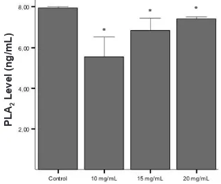
with our previous study.18 Relative mean PLA
2
enzyme level in the control group was 7.96 ± 0.060
ng/mL (n = 6). In groups treated with Acalypha indica
Linn extract at doses of 10, 15, and 20 mg/mL, mean
PLA2 enzyme level was 5.55 ± 0.970 ng/mL (n=6),
6.85 ± 0.590 ng/mL (n = 6), 7.42 ± 0.090 ng/mL (n = 6), respectively (p = 0.04 in all treatment groups vs control; Figure 1).
The lowest level of PLA2 enzyme was seen in group
treated with extract at dose of 10 mg/mL, and increased signiicantly in groups treated with extract at doses of 15 and 20 mg/mL (p = 0.016 and 0.04, respectively).
Signiicant difference was also found in the PLA2
enzyme level between groups treated with extract at doses of 15 and 20 mg/mL (p = 0.04; Figure 1).
DISCUSSION
MTT test results were in accordance with the author’s
previous study,18 indicating that Acalypha indica Linn
root extract has an effect on increasing the number of viable post-hypoxic nerve cells with an optimum dose at 10 mg/mL.
The mechanism of the increased cell proliferation is
thought to be due to the decrease in PLA2 enzyme
activity by acalyphin which will in turn decrease the inlammatory response and mobilize the endogenous stem
cells to proliferate.19,21 This mechanism is investigated
in this research relected by the measurement of PLA2
enzyme level. Our results indicated that the PLA2
enzyme level in all treatment groups were signiicantly
lower than control, with the lowest PLA2 enzyme level
found at the dose of 10 mg/mL. This result showed that
Acalypha indica Linn root extract decreased the PLA2
enzyme level in post-hypoxic nerve cell culture with the optimum dose at 10 mg/mL. This result also complies with the MTT assay result.
In this study, PLA2 data were obtained from the
supernatant of the nerve cells culture, therefore the
detected PLA2 enzymes were non-speciic and most
likely be the PLA2 enzyme secreted by cells (sPLA2).
Other than sPLA2, cPLA2 is also regarded as the
inlammatory precursor in cerebral hypoxia.8,11,14
Further research based on this inding is needed to
examine the level of cPLA2 enzyme in post-hypoxic
hippocampal tissue using a more speciic marker.
Figure 1. Comparison of PLA2 enzyme level among control and treatment groups given Aca-lypha indica Linn root extract with consecutive doses of 10, 15, and 20 mg/mL. Data was given in means ± SD from 6 different samples.
* showed signiicant difference compared to countrol group
PLA
2
Level
(ng/mL)
*
http://mji.ui.ac.id
Structurally, Nirmal, et al19 have shown that acalyphin
exerts its anti-inlammatory effects by forming an inhibitory complex in Asp-49, Lys-69, and Gly 30 from the
active site of sPLA2 Russell viper, and with Ca
2+ ion from
the active site of sPLA2 bovine pancreas. The mechanism
of this inhibitory complex formation has not been investigated in the central nervous system, thus further research is needed to assess whether or not in the central
nervous system Acalypha indica Linn root extract would
generate a similar sPLA2 inhibitory complex mechanism
similar to that in Russell’s viper and bovine pancreas.
In this study, the doses used were 10, 15, and 20 mg/
mL based on experiences from previous studies.16 It was
discovered that the optimum dose of Acalypha indica Linn
root extract to increase relative cell viability and decrease
PLA2 enzyme level was 10 mg/mL. This result suggests
that a lower dose than 10 mg/mL might give the same or better result, hence further study to determine the lowest
optimum dose of Acalypha indica Linn. root extract for
neurorestorative therapy post-hypoxia is needed.
In addition to reducing the inlammatory response,
Acalypha indica Linn’s root extracts may also increase
cell viability by increasing neurotrophic factors such as BDNF, VEGF, and bFGF; which are required for the
survival of neuroblast.3 Increased neurotrophic factors
as a mechanism of increased relative cell viability
caused by Acalypha indica Linn root extract has not
been studied, hence further research is needed to assess
whether Acalypha indica Linn root extract can enhance
post-hypoxic cell viability through enhancement of neurotrophic factors by examining the levels of growth factors (e.g. BDNF).
The limitation of this research lies in the morphologic identiication of the neural cells. This method cannot fully guarantee 100% identiication of neural cells with certainty because other cells also such as glial cells and ibroblasts also reside in the hippocampus, which may be identiied through this method. Thus, further research is needed with more precise identiication of the neural cells using speciic markers or antibodies such as neuron-speciic enolase type 2.
From this research we can conclude that Acalypha
indica Linn root extract at doses of 10, 15, and 20 mg/
mL with the optimum dose of 10 mg/mL can increase relative cell viability of rat hippocampal cells
post-hypoxia, in vitro.
Acknowledgments
The research was funded by HibahRiset Unggulan
Universitas Indonesia (RUUI) 2011 contract number
1318/H2.R12/PPM.00.01 Sumber Pendanaan/2011.
The authors would like to thank the expert consultant of this research, dr. Nurhadi Ibrahim, PhD for all his help and support. We would also like to thank the staff of the Oral Biology Laboratory, Faculty of Dentistry Universitas Indonesia for their work during the research; and Department of Physiology, Faculty of Medicine Universitas Indonesia for their
support.
REFERENCES
1. Wang Y, Ma T, Li M, Sun X, Wang Y, Gu S. Regulated hypoxia/reperfusion-dependent modulation of ERK1/2, cPLA2, and Bcl-2/Bax: a potential mechanism of neuroprotective effect of penehycidine hydrochloride. Int J Neurosci. 2011;121(8):442-9.
2. Becker JU, editors. Stroke, ischemic. 2008. Available from: http://www.emedicine.com/EMERG/topic558.htm. 3. Zhang RL, Zhang ZG, Chopp M. Neurogenesis in the
adult ischemic brain: generation, migration, survival, and restorative therapy. Neuroscientist. 2005;11(5): 408-16.
4. Ding G, Jiang Q, Li L. Magnetic resonance imaging investigation of axonal remodeling and angiogenesis after embolic stroke in sildenail-treated rats. J Cereb Blood Flow Metab. 2008;28(8):1440-8.
5. Titsworth WL, Liu NK, Xu XM. Role of secretory phospholipase A2 in CNS inlammation: implications in traumatic spinal cord injury. CNS Neurol Disord Drug Targets. 2008;7(3):254-69.
6. Wang Q, Tang XN, Yenari MA. The inlammatory response in stroke. J Neuroimmunol. 2007;184(1-2):53-68.
7. Culmsee C, Krieglstein J. Ischaemic brain damage after stroke: new insights into eficient therapeutic startegies. EMBO Reports. 2007;8(2):129-33.
8. Adibhatla RM, Hatcher JF. Phospholipase A(2), reactive oxygen species, and lipid peroxidation in CNS pathologies.. BMB Rep. 2008;41(8):560-567.
9. Gabryel B, Bielecka A, Stolecka A, Bernacki J, Langfort J. Cytosolic phospholipase A2 inhibition is involved in
the protective effect of nortiptyline in primary astrocyte cultures exposed to combined oxygen-glucose deprivation. Pharmacol Rep. 2010;62(5):814-26.
10. Ekdahl CT, Claasen JH, Bonde S, Kokaia Z, Lindvall O. Inlammation is detrimental for neurogenesis in adult brain. Proc Natl Acad Sci USA. 2003;100(23):13632-7. 11. Vexler ZS, Tang XN, Yenari M. Inlammation in adult and
neonatal stroke. Clin Neurosci Res. 2006;6(5):293-313. 12. Yagita Y, Kitagawa K, Ohtsuki T, et al. Neurogenesis by
progenitor cells in the ischemic adult rat hippocampus. Stroke. 2001;32(8):1890-6.
13. Arai K, Ikegaya Y, Nakatani Y, Kudo I, Nishiyama N, Matsuki N. Phospholipase A2 mediates ischemic injury
in the hippocampus: a regional difference of neuronal vulnerability. Eur J Neurosci. 2001;13(12):2319-23. 14. Farooqui AA, Ong WY, Horrocks LA. Inhibitors of brain
http://mji.ui.ac.id A2 involvement in neurodegeneration: differential testing
of prosurvival and anti-inlammatory effects of enzyme inhibition. PLoS ONE 2012;7(6):e39257.
16. Purwaningsih EH, Ibrahim N, Zain H. The nerve protection and in vivo therapeutic effect of Acalypha indica extract in frogs. Med J Indones. 2010;19:96-102.
17. Indian Medicinal Plants Growers’ Consortium. Acalypha indica L [Internet]. 2007 [cited 2009 Jan 12]. Available from: impgc.com.
18. Yolanda S, Bachtiar EW, Ibrahim N. Increased cell viability and proliferation in post-hypoxic hippocampal tissue
culture treated with Acalypha indica root extract. Med J Indones. 2011;20(2):94-9.
19. Nirmal N, Praba GO, Velmurugan D. Modeling studies on phospholipase A2-inhibitor complexes. Indian J Biochem Biophys. 2008;45(4):256-62.
20. Gozal E, Sachleben Jr LR, Rane MJ, Vega C, Gozal D. Mild sustained and intermittent hypoxia induce apoptosis in PC-12 cells via different mechanisms. Am J Physiol Cell Physiol. 2005;288(3):535-42.