176
Typhoid Fever and SalmoneLlosis Med J IndonesVMB.3
Factors influences on the survival and
death
of macrophages infected
with
Salmonella typhi
Kenji Hirose, Li-Cheng Zhao,Y;han Abdul Quayum, Masaki Miyake, Yoshiaki Kawamura, Takayuki Ezaki.
Abstrak
Dalam penelitian ini biakan jaringan makrofag manusia THP-I diùfeksi dengan Salmonella typhi dan S. typhimurium. Apa yang terjadi dengan makrofag tersebut setelah infelcsi oleh kedua patogen tersebut ternyata berbeda. Makrofag yang diinfeksi ol.eh S. typhi
tetap hidup lebih dari 24 jam, sedangknn 80Vo makrofag yang diinfeksi olelr S. typhimurium mati dalam waktu 16 jam. Karakter
apop-tosis makrofag yang diinfeksi oleh kedua patogen tersebut juga berbeda. Proses apoptosis pada makrofag yang diirfeksi oLeh S.
ty-phimurium terjadi lebih awal daripada infeksi oLeh S. typhi. Untuk mengetahui mengapa makrofag yang diùfeksi oLeh S. typhi tlapat bertahan lama, dilakukan analisis protein yang disekresi oleh S. typhi dan produksi TNF-r yang dihasilkan oLeh makrofag yan.g sudah
diinfeki. Galur S. typhi dengan kapsul V dan galur yang Vt antigennya delesi tentyata mensekresi protein dengan profiI berbeda. Tingkat transkripsi rnRNA TNF-a. dari kedua kelonryok makrofag inipun berbeda.
Abstract
Salmonella typhi and S. typhimurium were infected 1o human macrophage cell line THP-I. Fate of macrophage aJter infectiott of these two pathogetric species was different. Most of macrophages infected wltlr S. typhi were survived even after 24h4, howeve4, 80Vo of macrophages irdected wltlr S. typhimuritm were dead after 16 hour Apoptosis of macrophnges infected with these two pathogens were
alsodffirent.Atearlystageof infectionwithS.typhimuriuq macrophageswereinctpoptoticprocessbutS.typhiinfectedmacrophages remained at normal stage. To find the reason wfty S. typhi infected macrophages can survive for a long time, we analyzed secreted pro-teins of S. typhi and TNF-u production of infected macrophages. Vi capsulated S. typTti strains and V deleted S. typhi strains had com-pletely dffirent secreted proteitx profiLes. Iævel of tanscribed aRNA of TNF-r was also remarkably dffirent between macrophages infected with the two pathogens.
INTRODUCTION
SaLmonello typhi is known as a facultative
intracellu-lar parasite and
it
can survive inside macrophages.But, the survival mechanism is still unclear, We
pre-viously
reportedthat Vi
capsular polysaccharide played an important role for S. typhi to survive inside macrophages, becauseVi
positive strainsdid
not stimulatehost
macrophages andas
a
result, the macrophage did not produce TNF-cr.In this study, we constructed rpos mutant of S. typhi
which was derived from S. typhi
Yi
positive strain(wild
type) and analyzed the survival under various stress. The death of THP-1 cells infected with S. typhiwild type, S. typhi rpoS mutant were also compared. S.
typhi
rpoS mutant could replicate inside THP-I cells at the similar level with S. typhi wild type strain. However, S. typhi rpoS mutant caused cell death lessD e pa rtment of M icro b io Lo gy,
Gifu University, School of Medicine, Gifu, 500 Japan.
extent to infected macrophage than .S. typhi
wildtype
straln.
MATERIALS
AND METHODSBacteria and cell line
SaLmonelLa typhi wild type (GIFU 10007), S.
typhiYi
negative mutant (GIFU 10388), S. typhi rpoS mutant (GIFU-PPP 330) and S. typhimurium (GIFUI2l4Z)
were used in this study. THP-1 cells, which derived from human monocytic leukemia, were usedfor
in-fection assay.Cell staining
Cell staining to distinguish
live
and dead cells wascarried out with Live/Dead Eukolight
Viability/cyto-toxicity
assay reagent providedfrom
MolecularSuppl
I -
1998Flowcytometric analysis
S. typhi and S. typhimurium were cultured in L-broth
for
indicatedtime. Bacteria
were
stained with Live/Dead Eukolight Viability/cytotoxicity assay re-agent and analyzed by flow cytometry (Bio Rad Co, Japan). Live bacteria were carefully gated by forward scatter and lateral scatterto
remove dead bacteria. .Live bacteria were analyzedby
lateral scatter andFITC intensity.
RT-PCR
Bacterial mRNA was extracted from S.
typhi
after culturedfor
indicated time. cDNA was synthesizedfrom
mRNA
by reverse transcriptase.
sigma32, sigma38 and sigmaT0 detected by RI-PCR usingspe-cific primers.
Phagocytosis assay
Bacteria and THP-1 cells were mixed in HBSS at the ratio of 50 bacteria
to
1 cell, and then cells were in-cubatedin
37"Cfor
t
hour. The cells were resus-pendedin
complete medium with gentamicin to re-move extracellular bacteria for 30 min.Staining
intracellular
bacteria\â
antigenof
S. typhi insideTHP-I
cells were de-tected by rabbit anti-Vi polyclonal antibody and goat FITC conjugated-anti-rabbit IgG. THP-1 cells were stainedwith
PE-conjugated anti-CD14 monoclonal antibody.RESUI.JTS
S. typhi replication inside activated macrophages
S. typhi Vi positive strain and Vi negative strain inter-nalized
into
resting THP-1 cells almostthe
same level (data not shown). S. typhi Vi positive strain sur-vived into restingTHP-I
cells, whereasVi
negative S. typhi could not survive (FigureI).
S. typhimurium internalizedto
a greater extent than ^S. typhi strains and survived inside resting THP-1 cells. However, in THP-1 cell which activated by IFN-y, S. typhi strains and S. typhimurium did not survivein
spiteof
the higher numbers of bacteria were internalized.S.
typhi
expressedVi
antigen insideTIIP-I
cellafter infection
We detected the
Vi
antigenof
intracellular S. typhi. After invasion to THP-I cells, S. typhi inside THP-Icells were stained with rabbit anti Vi polyclonal
anti-Vaccine and MoLecular Biology 177
400
350
300
>
2s0 tr{(J
zoo150
100
50
0
[image:2.595.305.541.88.337.2] [image:2.595.303.540.339.661.2]vi (+) Vi
(-)
S. typhimurium Figurel.
Replication o/S. typhi and S. typhimurium insiderest-ittg and activated THP-I cells. Data were represented as mean + SEM.
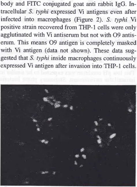
body and FITC conjugated goat anti rabbit IgG. In-tracellular S. typhi expressed
Vi
antigens even after infectedinto
macrophages (Figure2). S.
typhi W positive strain recovered from THP-1 cells were only agglutinated with Vi antiserum but not with 09 antis-erum. This means09
antigen is completely maskedwith
Vi
antigen (data not shown). These data sug-gested that S. typhi inside macrophages continuously expressed Vi antigen after invasion into TFIP-1 cells.Figure 2. S. typhi expressedViantigeninsideTHP-l ceLls.
178
Typhoid Fever and SalmonellosismRNA expression
of sigma factors
We analyzed the mRNA expression of sigma factor of
intracellular S. typhi. S. typhi were infected to
THp-l
cells, and intracellular S. typhi were recovered fromTHP-I
cells after indicated times (Figure 3). mRNAof Sigma32 was constitutively expressed in S. typhi.
mRNA of sigmaT0 was decreased after 20 hours. The mRNA
of
sigma3S was increased after infection. These results indicated that stationary phase genes under the controlof
sigma3S were expressed inside macrophages.oi 38
o70
Med J lndones
Analysis of S. typhi
viability by
FlowcytometryS. typhi Vi positive strain was cultured in L--broth for
4, 8 and 24 hours. The strain was stained with Live
/
dead cell eukolight viability assay reagent, and
bac-teria were analyzed by flowcytometer. After 8 hours
culture, S. typhi cells were separated into two
popu-lations shown in Figure 5, however, S. typhimurium
remained single population even after 24 hours. The upper population indicated brightly stained bacteria, and lower population indicated faintly stained
bacte-ria.
The bacteria
brightly
stainedin
green color means that these bacteria arestill
active. Anothergroup of bacteria stained in faint green means that
vi-ability were decreased. These results suggested that
S. typhi might change the metabolic phase and be-came stationary phase
in 24 hours when cultured
inordinary laboratory media.
S.
typhimurium
4hr
24hr
[image:3.595.48.283.220.415.2] [image:3.595.305.542.318.600.2] [image:3.595.45.283.422.724.2]LS1
Figure 5. Flowcytometric analysis o/S. typhi anri S. typhimu-rium cultured in L-broth for 4, B, and 24 hours. X axis
represents the intensity of lateral scatter and y ctxis
represents the intensity of FITC.
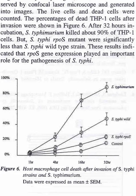
Host macrophage death after invasion of S. typhi rpoS mutant
We compared host cell death after S . typhi invasion
into
macrophagescell
line
THP-1 cells.
S.
ty-phimurium, S. typhi wild type and S. typhi rpoS
mu-04200420O42O
Time after phagocytosis (hr)
Figure 3. RT-PCR analysis of sigmafactorszlS. typhi inTHp-I cells.
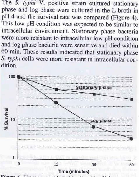
S. typhi suryival
in
low pH condition
The ^S. typhi
Yi
positive strain cultured stationary phase and log phase were cultured in theL broth in
pH 4 and the survival rate was compared (Figure 4).This low pH condition was expected to be similar to
intracellular environment. Stationary phase bacteria were more resistant to intracellular low pH condition and log phase bacteria were sensitive and died within 60 min. These results indicated that stationary phase
S. typhi cells were more resistant in intracellular
con-dition.
oo
oo
o
tr
100
0
Figure 4.
15
30ïme (minutes)
The survival o/S. typhi cubured in pH 4.
S.
typhi
q
Suppl
I
-
1998tant were infected to THP-1 cells and the cells were incubated for
I,4,
16 and 32 hours. After incubation,THP-1 cells were stained
with
Live/dead celleuk-olight viability assay reagent, then the cells were ob-served
by
confocal laser microscope and generatedinto
images,The
live
cells
and deadcells
werecounted. The percentages of dead THP-1 cells after
invasion were shown in Figure 6. After 32 hours
in-cubation, S. typhimuriumkllled abott90Vo of THP-1
cells. But, S. typhi rpoS mutant were significantly
less than S. typhi wild type strain. These results
indi-cated that rpoS gene expression played an important role for the pathogenesis of S. typhi.
S. typhinwiu
[image:4.595.42.280.133.475.2]lItr 4hr l6hr 32hr
Figure 6. Host macrophage cell death after invasion o/S. typhi strains and S. typhimurium.
Data were expressed as mean + SEM.
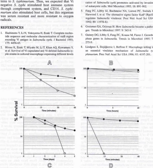
S. typhi survivals
in
various culture conditionsS. typhi wild type strain and r2oS mutant were com-pared on their survivals in various culture conditions.
In
Figure7A,
S. typhiwlld
type and rpoS mutantwere cultured
in
L-broth.After
culture transferring the cellsinto pH
4,
viable bacteria numbers were counted by the plating 15, 30, 60 min later, More than90 Vo of S. typhi 4poS mutant were dead after 60 min in pH
4
medium. This result indicated that S. typhirpoS mutant is sensitive to low pH conditions. In Fig-ure
78,
we compared intracellular replication insideTHP-1 cells. S. typhi were infected to
THP-I
cellsand incubated for further 4 or 20 hours in RPMI1640 with IÙVo FBS. After incubation, cells were lysed and
intracellular viable bacteria were counted.
After
4hour or 20 hours incubation, intracellular viable
bac-teria in THP-I cells were almost at the same level at each point. In Figure 7C, S. typhi wild type strain and
rpoS mutant were cultured in M9 medium for 5 days.
All
of S. typhi rpoS mutant were dead on day4
afterVaccine and MolecuLar
Biology
179inoculation, whereas 20Vo of S. typhi wild type strain
were
still
alive on day 5. This result means S. typhi rpoS mutant is sensitive to starvation. In Figure 7D,S. typhi rpoS mutant and wild type were cultured in
the medium containing 15 mM of hydrogen peroxide.
S. typhi rpoS mutant was rapidly died within 15 min.,
however, S. typhi wild type strain was resistant to 15
mM
hydrogen peroxide and 70Voof
bacteria werestill
viable after 60 min. incubation. From theseex-periment, S. typhi rpoS mutant was sensitive to low pH, starvation and hydrogen peroxide. This'means that ropes gene expression might be essential for S.
typhi to survive long time inside macrophages.
DISCUSSION
We previously reported that
Vi
capsularpolysaccha-ride played an important role for S. typhi to survive
inside macrophages. The macrophages infected with
S. typhi Vi mutant or S. typhimurium produced a large
amount
of
TNF-o, whereas,Vi
positive strains did not stimulate host macrophages to produce TNF-c,because Vi antigen covered the bacterial surface LPS
and
Vi
antigen prevent the LPSfrom
macrophagecD14.
In
this study, we constructed S. typhi rpoS mutantwhich derived from S. typhi
Yi
positive strain (wild type) and compared the survival of S. typht wild type,S. typhi
Vi
mutant, S. typhi rpoS mutant.S. typhi could not survive in the activated THP-1 cells
(Figure 1). The organisms, thus, internalizes into
rest-ing THP-1 cells to survive inside the cells, and intra-cellular S. typhi continuously expressed
Vi
antigeninside THP-1 cells (Figure
2).
Once entered intomacrophages, sigma38
of
S. ryphi is induced insideTHP-I
cells, and S. typhi might changes their meta-bolic phaseto
stationary phase, Stationary phase S. typhiis
resistantto low pH
(Figure4)
condition whichis
similar to intracellular conditions. S. typhi culturedin
L-broth for 24 hours separated into twogroups of bacteria (Figure 5), one population is still log phase bacteria, another population is changed into
stationary phase. RpoS mutant was less toxic to host
cell, and was sensitive to low pH, starvation and
hy-drogen peroxide. RpoS gene might be required for the pathogenesis
of
.S. typhi. Frcm these data, we con-cluded that Vi positive S. typhi shift metabolic phaseto
stationary phase after invasion into macrophagesto adapt to the intracellular environment, and
180
Typhoid Fever and Salmonellosis Med J Indonesof CD14 antigen. FEMS Microbiol Lett 1997; 147:259-65. Kogoma T, Yura T. Sensitization of Escherichia coli cells to
oxidative stress by deletion of the rpoH gene, which encodes
the heat shock sigma factor. J Bacteriol 1992; 174: 630-2.
Staendner LH, Rohde M, Timmis KN, Guzman CA.
Identifi-cation of Salmonella typhi promoiers activated by invasion of eukaryotic cells. Mol Microbiol 1995; 18: 891-902. Fang FC, Libby SI, Buchmeier NA, Loewn PC, Switala J,
Harwood J, et al. The alternative sigma factor KatF (RpoS) regulates Salmonella virulence. Proc Natl Acad Sci USA 1992;89: 11978-82.
Groisman EA, Ochman H. How Salmonella became a
patho-gen. Trends in Microbiol 1997:9:343-9.
Guiney DG, Libby S, Fang FC, Krause M, Fierer J. Growth-phase genes in Salmonella. Trends in Microbiol 1995:'l:
275-9.
Lindgren S, Stojiljkovic I, Heffron F. Macrophage killing is
an
essential virulence mechanismof
Sahnonellaty-phimurium. Proc Natl Acad Sci USA 1996; 93: 4197-201 .
We did not show another evidence that Vi positive S.
lyphi
sllain produced less amountof
secretorypro-teins into cultured media. Most of the secreted
pro-teins were flagellar antigens. However,
Vi
negativestrains produced almost equal level of secreted
pro-teins to S. typhimurium. Thus, we expected that Vi
negative
S. typhi
stimulated host immune systemthrough complement system, and CD14. S.
typhi-murium also stimulated host cells, but this organism
was serum resistant and more resistant
to
oxygenradicals.
REFERBNCES
l.
Hashimoto X Li N, Yokoyama H, Ezaki T. Complete nucleo-tide sequence and molecular characterization of vraB region encoding Vi antigen in Salmonella typhi. J Bacteriol 1993; 175 4456-65.2.
Hirose K, Ezaki T, Miyake M, Li T, Khan AQ, Kawamura Y,et al. Survival of Vi-capsulated and Vi-deleted Salmonella
ty-pÀi strains in cultured macrophage expressing different levels
[image:5.595.47.539.148.690.2]6.
Figure 7. S. typhi sumivals in various cuhure condilions. S. typhi wild type (-o-) and rpoS mutant (-o-) were cultured in Iôw pH
me-dium(pH 4) (A), cultured in M9 medium (C), and cultured in the medium containing 15 mM hydrogen peroxide (D). The sur-vival inside THP-I cells were compared benveen S. typhi wild type antl rpoS mutant (B).
3
4
5