Inluence of primaquine and ritonavir interaction on CYP3A4 mRNA
expression in HepG2 cell culture
Adam Iskandarmudasyah,1 Melva Louisa,2 Arleni,3 Sri W. A. Jusman,4 Franciscus D. Suyatna2
1 Master Program in Biomedical Sciences, Faculty of Medicine, Universitas Indonesia, Jakarta, Indonesia
2 Department of Pharmacology and Therapeutics, Faculty of Medicine, Universitas Indonesia, Jakarta, Indonesia 3Department of Biology, Faculty of Medicine, Universitas Indonesia, Jakarta, Indonesia
4Department of Biochemistry and Molecular Biology, Faculty of Medicine, Universitas Indonesia, Jakarta, Indonesia
Abstrak
Latar Belakang: Pengobatan antimalaria dan anti-HIV secara bersamaan merupakan tantangan baru dalam penanganan koinfeksi malaria dan HIV. Primakuin merupakan substrat sekaligus inhibitor bagi CYP3A4, sedangkan ritonavir merupakan substrat, inhibitor, sekaligus inducer bagi CYP3A4. Tujuan penelitian ini adalah untuk mengukur ekspresi mRNA CYP3A4 pada kultur sel HepG2 yang diinduksi oleh pemberian primakuin dan ritonavir secara bersamaan.
Metode: Pada penelitian pendahuluan, sel HepG2 diinkubasi dengan primakuin 30, 40, 50 uM; ritonavir 2, 10, 20 uM; DMSO <0,1 % sebagai kontrol negatif; atau rifampisin 20 uM sebagai kontrol positif. Adapun pada penelitian dengan perlakuan kombinasi obat, sel HepG2 diinkubasi dengan primakuin 40 uM+ritonavir 10 uM; DMSO <0,1 %; atau rifampisin 20 uM selama 72 jam. Sel dipanen menggunakan tripsin-EDTA dan RNA total diekstraksi menggunakan reagensia isolasi tripure. Setelah jumlah RNA total dikuantiikasi menggunakan alat spektrofotometer, ekspresi mRNA CYP3A4 diukur dengan real-time reverse transcription polymerase chain reaction (RT-PCR).
Hasil: Terjadi peningkatan ekspresi mRNA CYP3A4 (1,22 kali lipat terhadap kontrol) pada sel HepG2 yang diinkubasi dengan primakuin dan ritonavir secara bersamaan. Hal ini menunjukkan bahwa efek induksi oleh ritonavir lebih dominan daripada efek inhibisi oleh primakuin.
Kesimpulan: Pemberian primakuin dan ritonavir secara bersamaan meningkatkan ekspresi mRNA CYP3A4 in vitro. (Med J Indones 2012;21:3-7)
Abstract
Background: Concomitant treatment with antimalaria and antiretroviral drug is a new challenge in the management of malaria and HIV co-infection. Primaquine is a substrate and also an inhibitor of CYP3A4, while ritonavir is a substrate, an inhibitor, and also an inducer for CYP3A4. the objective of this study is to measure the CYP3A4 mRNA expression in HepG2 cell culture induced by primaquine and ritonavir co-treatment.
Methods: For the initial study HepG2 cells were treated with 30, 40, 50 uM of primaquine; 2, 10, 20 uM ritonavir; DMSO
≤0.1 % for negative control; or 20 uM rifampicin for positive control. While for the co-treatment study the cells were treated with 40 uM primaquine+10 uM ritonavir; DMSO ≤0.1 %; or 20 uM rifampicin for 72 hours. The cells were harvested using
trypsin–EDtA and total RNA was extracted using the tripure isolation reagent. After determining the quantity of RNA
spectrophotometrically, CYP3A4 mRNA expression was quantiied using real-time reverse transcription polymerase chain
reaction (Rt-PCR).
Results: the expression of CYP3A4 mRNA was up-regulated (1.22 fold over control) in HepG2 cells co-treated with primaquine and ritonavir. these data suggest that the induction effect of ritonavir was more dominant than the inhibitory effect of primaquine.
Conclusion: Concomitant administration of primaquine and ritonavir result in up-regulation of CYP3A4 mRNA expression in vitro. (Med J Indones 2012;21:3-7)
Keywords: CYP450 induction, CYP3A4, drug interaction, primaquine, ritonavir
Correspondence email to: [email protected]
Cytochrome P450 system (CYP) are the main enzyme system in the liver responsible for drug metabolism of
nearly 70-80% of clinically used drugs.2 Many drug
interactions are a result of the inhibition or induction of these CYP enzymes.3 CYP450 have many different isoform, including CYP1A1, CYP1A2, CYP2C19, CYP2D6, and CYP3A4.2 However, CYP3A4 is responsible for approximately 60% of
CYP450-mediated metabolism of drugs in therapeutic use today.4 thus, it is important to assess CYP3A4 in identifying drug interactions.
Primaquine (PRQ) is the only drug available for preventing relapse in malaria caused by dormant liver stage/hypnozoites form of Plasmodium vivax or P. ovale.5,6 this drug is a substrate and also an inhibitor of CYP3A4.7,8 In the other hand, ritonavir (RtV) is a protease inhibitor (PI) that frequently used as a booster to increase plasma concentration of other PI in the highly active antiretroviral therapy (HAARt) regimen for HIV patients.9 Ritonavir is a substrate, inhibitor, and also an inducer for CYP3A4.10 Accordingly, it is a curiousity whether ritonavir would act as an inducer or inhibitor of CYP3A4 when given concomitantly with another drug.
Louisa et al. reported that concomitant administration of
primaquine and ritonavir lead to signiicant decrease on
plasma concentration of each drugs in rats in comparison to the single administration of either primaquine or ritonavir only.11,12 to explain whether the decreased plasma concentration of both drugs was caused by the induction effect of primaquine or by the autoinduction effect of ritonavir, we measure the expression of CYP3A4 mRNA in HepG2 cell culture incubated with primaquine, ritonavir, and primaquine+ritonavir.
METHODS
this in-vitro experimental study was conducted from August 2010 to April 2011. Cell culture was conducted at Makmal terpadu Imunoendokrinologi, Faculty of Medicine. Universitas Indonesia. RNA isolation was performed at the Laboratory of Biochemistry and Molecular Biology, Faculty of Medicine. Universitas Indonesia. the purity and concentration of total RNA was measured at the Laboratory of Pharmacology and therapeutic Department, Faculty of Medicine. Universitas Indonesia. Real-time Rt PCR was performed at Eijkman-EOCRU Laboratory.
Chemicals
Primaquine, ritonavir, rifampicin (RIF), and dimethylsulfoxide (DMSO) were purchased from Sigma- Aldrich Ltd (Singapore). Dulbecco minimal essential medium (DMEM) and foetal bovine serum (FBS) were purchased from Gibco Ltd (Singapore). tripure isolation reagent was purchased from Roche Diagnostic (Singapore). Nuclease free water was purchased from Promega Corporation (Singapore).
Primers for β-actin and CYP3A4 were purchased from
1st BASE Ltd (Singapore). iScript one-step Rt-PCR kit with SYBR Green were purchased from Bio-Rad Laboratories Inc (Singapore). All other chemicals used were of the highest purity commercially available.
Cell culture and drug treatment
HepG2 cells were purchased from BPPt (Serpong, Indonesia). the cells were cultured in DMEM
supplemented with 10% FBS and maintained at 37oC
in a humidiied incubator of 5% CO2. Cells were
subcultured at conluence onto 12-well plates (seeding
density 2.0 x10⁵ cells/mL medium). Hepatocyte culture were maintained in culture medium for one days before incubation with the test-drug. In the second day, the medium were aspirated and replaced with fresh medium containing the test-drug. Drugs were dissolved for treatment in DMSO. For the initial study, cells were treated with one of the following: 30 uM PRQ, 40 uM PRQ, 50 uM PRQ, 2 uM RtV, 10 uM RtV, 20 uM RtV,
DMSO ≤0.1 % for negative control, or 20 uM RIF for positive control. While for the co-treatment study, cells
were treated with one of the following: 40 uM PRQ+10
uM RTV, DMSO ≤0.1 % for negative control, or 20 uM
RIF for positive control. In all cases the concentration
of DMSO was less than 0.1%. All treatments were done in duplicates. The cells were harvested at 72 hours after
drug treatment.
RNA isolation
Cells were harvested using trypsin–EDtA and total RNA was extracted using the tripure isolation reagent according to the manufacturer’s instructions.
The RNA was quantiied spectrophotometrically by
measuring absorbance at 260 nm. the purity of the
inal preparations was determined by calculating the absorbance ratio at 260 and 280 nm.
Real-time RT PCR
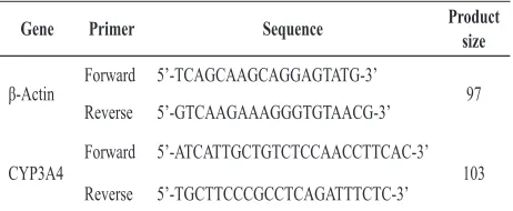
the sequences of the primers used for real-time PCR are shown in table 1. the samples were prepared by mixing 30 mM of each primer, 12.5 uL SYBR Green Rt PCR reaction mix, 0.5 uL iScript reverse transcriptase, 100 ng template RNA, and 9.5 uL RNAse free water. the samples were then incubated in real-time thermal detection system with the following condition: 10 minutes at 50oC for cDNA synthesis, 5 minutes at 95oC for iScript reverse transcriptase inactivation, and PCR cycling and detection (40 cycles) which was 10 seconds at 95oC for denaturation stage and 30 seconds at 58oC for annealing and extension stage. threshold values (Ct) were calculated automatically by the software. the Ct data was processed according to the method
described by Pfal.
table 1.β-actin and CYP3A4 primers
A: adenine, C: cytosine, G: guanine, t: timin
Gene Primer Sequence Product size
Forward 5’-tCAGCAAGCAGGAGtAtG-3’
97
Reverse 5’-GtCAAGAAAGGGtGtAACG-3’
CYP3A4
0
Figure 1. Concentration-response relationship of PRQ treatment on the expression of CYP3A4 mRNA in HepG2 cells.
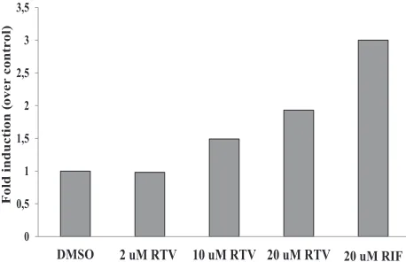
Figure 2. Concentration-response relationship of RTV treatment on the expression of CYP3A4 mRNA in HepG2 cells.
Figure 3. Effect of primaquine and ritonavir co-treatment on the expression of CYP3A4 mRNA in HepG2 cells. RESULTS
HepG2 cells were not changed morphologically during the drug treatment period under an inverted phase contrast microscope. Figure 1 shows normalized CYP3A4 mRNA expression in various dose of primaquine. Exposure to 30, 40, and 50 uM primaquine did not induce CYP3A4 mRNA expression, while exposure to 20 uM rifampicin as positive control increased CYP3A4 mRNA expression by 3-fold in comparison to DMSO treated cell as negative control.
the normalized CYP3A4 mRNA expression in various doses of ritonavir is demonstrated in Figure 2. Exposure to 2 uM ritonavir did not induce CYP3A4 mRNA expression. However, a slight increase (1.49 and 1.93 fold over control) of CYP3A4 mRNA expression was detected when ritonavir concentration was increased to 10 and 20 uM.
the expression of CYP3A4 mRNA was slightly increasing (1.22 fold over control) in HepG2 cells co-treated with primaquine and ritonavir. these data suggest that the induction effect of RtV was more dominant than the inhibitory effect of PRQ.
DISCUSSION
To our knowledge, this is the irst study reporting
the interaction between anti-malarial and anti-HIV drug at the mRNA level. From the initial study, it is
conirmed that even with the highest concentration
(50 uM), primaquine did not increase the CYP3A4 mRNA expression. the highest concentration of primaquine used in this study is much higher than the maximum concentration in blood (Cmax) following single administration of primaquine of a normal dose in human for radical cure or prevention of relapsing vivax or ovale Malaria.13 Nevertheless, 30 uM of primaquine was used by Backlund et al. to assess the induction effect of primaquine on CYP1A1 and 40 uM of primaquine was used by Kanebratt et al. to assess HepaRG cells as an in vitro model for evaluation of CYP450 induction in human.14,15 Consequently, it is unlikely that the induction of CYP3A4 mRNA expression by primaquine will happen under the in vivo circumstances. the results are in agreement with other studies that suggest primaquine as a substrate and also an inhibitor, but not an inducer of CYP3A4.7,8
From the co-treatment study, it is demonstrated that the net result of concomitant administration of primaquine and ritonavir is a slight increase of CYP3A4 mRNA expression, where ritonavir behave as an inducer. the behavior of ritonavir in this study is in contrast with other study reported by Ikezoe et al. Co-administration of 10 uM ritonavir and 10-3 uM docetaxel in DU145 cells resulted in no increase of CYP3A4 mRNA expression compared to DMSO treated control group, although docetaxel alone increased CYP3A4 mRNA expression by 2.5-fold.17
the slight increase of CYP3A4 mRNA expression by ritonavir in this study can be explained by receptor-mediated induction mechanism. CYP3A induction commonly occurs via the binding of the inducer to pregnane X receptor (PXR) and/or constitutive androstane receptor (CAR). Once activated, these receptors form heterodimers with other factors, retinoid X receptor (RXR for both PXR and CAR) and then bind to the target xenobiotic response elements (XRE) located in both the proximal and distal P450 gene promoters, resulting in the elevation of transcription of the respective CYP gene.18
In the other hand, Ikezoe suggest that ritonavir may block docetaxel-induced expression of CYP3A4 by affecting co-regulators such as silencing mediator for retinoid and thyroid receptor and steroid receptor coactivator-1 that mediated basal and xenobiotic-induced transcriptional activity of CYP3A4.17
the increase of CYP3A4 mRNA expression reported in this study could lead to the increase of CYP3A4 enzyme that could decrease both plasma concentration of primaquine and ritonavir. the result from this study
gives us a signal that both the eficacy of ritonavir in
anti-HIV regimen and primaquine as an anti-malarial agent could be reduced if both drugs were administered concomitantly in human.
From the elimination half-life (t1/2) point of view, ritonavir has relatively short t1/2 which is3-5 hours.9,13 this fact possibly will cause the effect of ritonavir induction on primaquine would not last long. However, we should consider that the duration of induction of a drug does not solely depend on the t1/2, but also depend on the time needed for CYP enzyme degradation. It is stated that t1/2 of CYP450 ranged between 1-6 days.3 therefore, ritonavir may only induce the CYP3A4 for 3-5 hours, but the induction effect would probably last for several days.
In addition, as a monotherapy or as a booster for other antiretroviral, ritonavir is given twice daily (every 12 hours).13 For that reason, the induction effect of ritonavir would last a full day.
The elimination half-life of primaquine is 3.7-9.6
hours.13 If primaquine is given concomitantly with
ritonavir, the t1/2 of primaquine will be shortened, so the duration of primaquine’s therapeutic effect would also be shortened.
there are several limitations from this study. According to Hewitt et al, it was found that HepG2 cells have very low CAR, PXR, CYP3A4 expression, but relatively
high CYP3A7 expression. This proile is in keeping with a fetal and not adult expression proile of hepatocytes.
this lack or low abundance of PXR and CAR makes them unsuitable for induction assays involving these pathways.18
this probably explain why 10 uM ritonavir in this study only gives slight increase (1.49 fold over control) of CYP3A4 mRNA expression but the same dose of ritonavir in the study using primary hepatocyte culture could produce 2.2 to 21 fold over control of CYP3A4 mRNA expression.16 However, the mechanism controlling CYP3A4 expression in HepG2 cells are identical to those of human hepatocytes.19
Another limitation from this study is that we did not measure the CYP3A4 enzyme activity. Although there is a good relationship between mRNA levels
and enzyme activities for most CYPs, the deinitive
endpoint is the activity of the enzyme rather than its expression because the overall change in enzyme activity will ultimately affect the clearance of the drug itself or of a co-administered drug.18
In conclusion, concomitant administration of primaquine and ritonavir result in a slight increase of CYP3A4 mRNA expression in vitro, where ritonavir
act as an inducer. Nevertheless, the signiicance of this inding should be further clariied by performing
CYP3A4 enzyme activity measurement.
Acknowledgments
the authors would like to express gratitude to DRPM UI for funding this research by giving Postgraduate Research Grant UI 2010, to our colleague taniawati Supali, PhD and Decy Subekti, PhD for the opportunity to perform real-time Rt-PCR in Eijkman-EOCRU Laboratory, and to Mrs. trimiatun for excellent technical assistance in culturing HepG2 cell in Makmal terpadu Imuno-Endokrinologi FKUI.
REFERENCES
Xu Y, Zhou Y, Hayashi M, Shou M, Skiles GL. Simulation 1.
of clinical drug-drug interactions from hepatocyte CYP3A4 induction data and its potential utility in trial designs. Drug
Metab Dispos. 2011;39(7):1139-48.
Ingelman-Sundberg M, Rodriguez-Antona C. 2.
Pharmacogenetics of drug-metabolizing enzymes: implications for a safer and more effective drug therapy.
Phil Trans R Soc B. 2005;360:1563-70.
Leucuta SE, Vlase L. Pharmacokinetics and metabolic drug 3.
Raucy JL. Regulation of CYP3A4 expression in human 4.
hepatocytes by pharmaceuticals and natural products. Drug Metab Dispos. 2003;31(5):533-9.
Baird JK. A rare glimpse at the eficacy of primaquine. Am
5.
J Trop Med Hyg. 2007;76(2):201-2.
Hill DR, Baird JK, Parise ME, Lewis LS, Ryan Et, Magill 6.
AJ. Primaquine: report from CDC expert meeting on malaria
chemoprophylaxis I. Am J Trop Med Hyg.
2006;75(3):402-15.
Li XQ, Bjorkman A, Andersson tB, Gustafsson LL,
7.
Masimirembwa CM. Identiication of human cytochrome
P450s that metabolise anti-parasitic drugs and predictions of in vivo drug hepatic clearance from in vitro data. Eur J Clin Pharmacol. 2003;59:429-42.
Baune B, Furlan V, taburet AM, Farinotti R. Effect of
8.
selected antimalarial drugs and inhibitors of cytochrome P450 3A4 on halofantrine metabolism by human liver
microsomes. Drug Metab Dispos. 1999;27:565-8. Warnke D. Antiretroviral drugs. J Clin Pharmacol.
9.
2007;47:1570-9.
Venkatakrishnan K, von-Moltke LL, Greenblatt DJ. Human 10.
drug metabolism and the cytochromes P450: application and relevance of in vitro models. J Clin Pharmacol.
2001;41:1149-79.
Louisa M, Soetikno V, Nafrialdi, Setiabudy R, Suyatna FD. 11.
Primaquine decreased plasma concentration of ritonavir: single- and multiple-dose study in Spraque Dawley rats. Med J Indones. 2011;20:190-4.
Louisa M, Soetikno V, Nafrialdi, Setiabudy R, Suyatna FD. 12.
Ritonavir decreases plasma concentration of primaquine:
single- and multiple-dose study in the rats. 2012. (Unpublished).
Drugs.com [Internet]. Source of drug information online 13.
[cited 2010 Jul 31]. Available from: http://www.drugs.com Backlund M, Ingelman-Sundberg M. Different structural 14.
requirements of the ligand binding domain of the
aryl hydrocarbon receptor for high- and low-afinity
ligand binding and receptor activation. Mol Pharmacol. 2004;65:416-25.
Kanebratt KP, Andersson tB. HepaRG cells as an in vitro 15.
model for evaluation of cytochrome P450 induction in
humans. Drug Metab Dispos. 2008;36:137-45.
Luo G, Cunningham M, Kim S, Burn t, Lin J, Sinz M, 16.
et al. CYP3A4 induction by drugs: correlation between a pregnane x receptor reporter gene assay and CYP3A4 expression in human hepatocytes. Drug Metab Dispos.
2002;30:795-804
Ikezoe t, Hisatake Y, takeuchi t, Ohtsuki Y, Yang Y, Said
17.
JW, et al. HIV-1 protease inhibitor, ritonavir: a potent
inhibitor of CYP3A4, enhanced the anticancer effects of docetaxel in androgen-independent prostate cancer cells in
vitro and in vivo. Cancer Res. 2004;64:7426-31.
Hewitt NJ, Lecluyse EL, Ferguson SS. Induction of
18.
hepatic cytochrome P450 enzymes: methods, mechanisms, recommendations, and in vitro–in vivo correlations.
Xenobiotica. 2007;37(10-11):1196-224.
Modarai M, Silva E, Suter A, Heinrich M, Kortenkamp A. 19.
Safety of herbal medicinal products: Echinacea and selected alkylamides do not induce CYP3A4 mRNA expression.