DOI: 10.12928/TELKOMNIKA.v14i4.3714 1493
Exudate and Blood Vessel Feature Extraction in
Diabetic Retinopathy Patients using Morphology
Operation
Siswo Wardoyo*, Anggoro Suryo Pramudyo, Erika Diana Rizanti, Imamul Muttakin
Department of Electrical Engineering, Universitas Sultan Ageng Tirtayasa,
Jl. Jenderal Sudirman Km 3 Cilegon 42435, telp/fax: +62-254-376712 (ext 20)/ +62-254-395440 *Corresponding autjor, e-mail: [email protected]
Abstract
Diabetic Retinopathy is one of the retina complications caused by diabetic disease with observable symptoms such as emergence of exudate and new blood vessels. The tool used to screen it is a fundus camera. However, analyzing the fundus image should be done by doctor who is an expert and will require a lot of time. Therefore, automatic feature detection can assist doctor in processing the retinal image in analyzing diabetic retinopathy disease. The proposed method has been tested on the morphological operations of the fundus image from Cicendo Eye Hospital, Bandung. The calculation results on feature extraction exudate area has a range of 0 pixels for normal retinal image, 17-21213 pixel for retinal image NPDR, and 125-12299 retinal image pixel for PDR. The calculation results on the extraction area of blood vessels has a range of 13319-46681 pixel to the normal retina, the retinal image 7435-49938 pixel for NPDR, and 13.81-53.802 retinal image pixel for PDR.
Keywords: Diabetic Retinopathy, Exudate, Blood Vessel, Morfology Operation, Area Centroid
Copyright © 2016 Universitas Ahmad Dahlan. All rights reserved.
1. Introduction
One of the chronic diseases among the growing number of today's society is a diabetic with a high number of people in the world. World Health Organization (WHO) reported that the number of diabetic cases in the world was about 171 million people in 2000, and Indonesia was fourth by the number of people with diabetes is 8.5 million inhabitants in 2000. This number is expected to reach 21.3 million by 2030 [1].
Diabetic retinopathy is one of the complications diabetes that is the leading cause of blindness in adults. Research in America, Australia, Europe and Asia reported that the numbers of diabetic retinopathy patients will increase from 100.8 million in 2010 to 154.9 million in 2030 with 30% of them are threatened with blindness [2]. The DiabCare Asia 2008 Study involved 1,785 diabetes patients in 18 primary and secondary health centers in Indonesia and reported that 42% of people with diabetes had retinopathy complications, and 6.4% was proliferative diabetic retinopathy [3].
The main problem is the delay in treatment of diabetic retinopathy diagnosis because most of the patients in the early stages are not impaired vision [4]. Screening program for diabetic retinopathy need computer assistance to analyze fundus images obtained from the fundus camera. Detection system requires a computational model to transform pixel retinal image into a retinal feature of diabetic retinopathy indicated by exudate and blood vessels that appear using morphological operations. Thus, with early detection of diabetic retinopathy, it can prompt the healing action quickly.
2. Diabetic Retinopathy Detection
hemorrhage, blood vessels, macula, and exudates through manual inspection of fundus images. A computer aided diagnosis system can significantly reduce the burden on the ophthalmologists and may alleviate the inter and intra observer variability. The review in [7] discussed the available methods of various retinal feature extractions and automated analysis.
Image recognition for the screening of diabetic retinopathy was explored in [8]. The presence of exudates within the macular region is a main hallmark of diabetic macular edema and allows its detection with a high sensitivity. Therefore, detection of exudates is an important diagnostic task. Exudates are found using their high grey level variation, and their contours are determined by means of morphological reconstruction techniques. A new algorithm for detection of exudates was presented and discussed in [9].
In another study [10], a technique based on morphological image processing and fuzzy logic to detect hard exudates from DR retinal images was proposed. At the initial stage, the exudates were identified using mathematical morphology that includes elimination of the optic disc. The fuzzy output for all the pixels in every exudate was calculated for a given input set corresponding to red, green and blue channels of a pixel in an exudate. This fuzzy output was computed for hard exudates according to the proportion of the area of the hard exudates. Similarly, report [11] discussed a hybrid fuzzy image-processing system for situation assessment of diabetic retinopathy to support the early detection of diabetic retinopathy in a primary-care environment.
In publication [12], classifiers such as the Gaussian Mixture model (GMM), k-nearest neighbor (kNN), support vector machine (SVM), and AdaBoost were analyzed for classifying retinopathy lesions from nonlesions. An algorithm to detect the presence of exudates automatically was proposed [13]. Research in [14] presented a method for automated identification of exudate pathologies in retinopathy images based on computational intelligence techniques. The color retinal images are segmented using fuzzy c-means clustering following some preprocessing steps. A genetic-based algorithm is used to rank the features and identify the subset that gives the best classification results. The selected feature vectors are then classified using a multilayer neural network classifier.
Segmentation method without initialization process was proposed in [15]. The segmentation was conducted by using the maximum value selection results of convolution 8 directions. Publication [16] showed a method for vascular pattern ehnacement and segmentation. An automated system which uses wavelets to enhance the vascular pattern was proposed and then subsequently applied a piecewise threshold probing and adaptive thresholding for vessel localization and segmentation respectively. In this article [17], a method to improve the quality of input retinal image was presented and considered as a preprocessing step in automated diagnosis of diabetic retinopathy. The preprocessing consists of background estimation and noise removal from retinal image by applying coarse and fine segmentation.
3. Feature Extraction Method
Flowchart of the research process shown in Figure 1. In this research, the input image is an image of the retina of diabetic retinopathy patients obtained by using a fundus camera or Zeiss visucam non medriatic pro cam 4726 with a capture 5.0 MP sensor produces image files in the format of the Joint Photographic Group (JPG) with a resolution of 2448 x 3696 pixels from Eye Hospital Bandung Cicendo that have been validated. Images were obtained 75 pieces, divided into three classes, i.e. 25 pieces of normal eyes, 25 eyes Non Proliferative Diabetic Retinopathy (NPDR), and 25 eyes Proliferative diabetic retinopathy (PDR).
Figure 1. Flowchart of the research process
A feature extraction method used in this research is the morphological operations. Morphological operations as a method for extracting image components are useful in image representation and description of the area of exudate and blood vessels. In exudate feature extraction phase, the input image data that has been converted into a gray scale image, then carried on morphological operations which applied morphological closing operation. It serves to increase the area of exudate and remove blood vessels. The next process is the column wise neighborhood. In this process, the image is reset into columns thus forming a matrix while using the function "colfilt" in matlab which is useful for marking exudate area.
The results of the process are then proceeding through thresholding. The threshold value applied is 0.9, which if the image is converted into a binary image, can be written:
If x <0.9 then x = 0, otherwise x = 1
The next stage is a morphological opening operation. It applies dilation followed by an erosion process, which aims to fill a hole or gap of exudate. Optic disc contains the highest pixel values in the image of retina; therefore, removing the optical disk is by searching for the highest pixel values of a gray scale image. The size of the optic disc defined at 576 x 720 color images have a size of optical disk defined by 80 pixels greatest, so it will be created mask to remove the area of the optic disc. Then the optic disc is removed together with the border. Resulting data is carried back with morphological operations namely erosion, which aims to remove noise that is not exudate. Thus, the feature extraction of exudate is available.
For feature extraction of blood vessels, the input image data which has been converted into a gray scale image is then complemented or inverted. In this process, the previous retinal image has a black on white background, so that objects which form blood vessels would be more visible. Next process is known as the Contrast Limited Adaptive Histogram Equalization (CLAHE). In this process the complemented retinal image is averaged image histogram to improve the contrast of the image in order to make the hidden feature can be seen more clearly. After the contrast of the retinal image is flattened, then the median filter process is carried out. This process aims to eliminate unnecessary noise in the extraction of blood vessels. The next stage is a morphological opening operation; the implementation of erosion followed by a dilation operation aims to protect small blood vessels while dilation aims to increase the details of large blood vessels and then removed. Subsequently, boundary extraction conducted by subtracting the result of the median filter with the results of morphological dilation, which aims to separate the blood vessels out of background. The results of the blood vessels are proceeded into thresholding, with threshold value of 0.1. If the image is converted into a binary image, it can be written:
with median filter. It will provide the final result of blood vessels extraction.
After obtaining the results of exudates extraction and blood vessels, value of area which is extensive or exudates is calculated. Centroid is useful to determine the position of objects. This value is obtained from the following equation:
∑ ∑ , (1)
, ∑ ∑ . , ; ∑ ∑ . , (2)
Where: f(i.j)=1 if (i,j) is an object pixel.
Feature extraction process employs Fast Fourier Transform (FTT) basic principle, decomposition calculation of Discret Fourier Transform (DFT) from length N into a series of smaller DFT respectively. For simplicity purpose, it is assumed that the samples N in row X (n) is the result of powers of 2; otherwise it is necessary to add 0. So, being the number nearest results of the powers. The next process is data normalization. Only magnitude parts of Fourier transform result are taken into account, omitting the imaginary parts. The maximum magnitude value is used as divisor factor for the rest value of magnitudes, so that the maximum value of each characteristic pattern of exudates is worth 1.
4. Results and Analysis
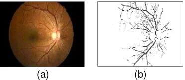
Exudates feature extraction is done by using morphological operations. At the final stage, generated candidate of exudates is carried in the operation process of erosion. It aims to remove noise. Results are shown in Figure 2.
(a) (b) Figure 2. (a) Original image; (b) Exudate extraction result
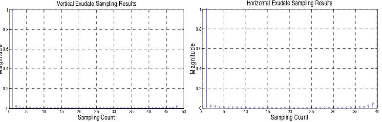
After obtaining the results, the centroid area can be calculated and the results obtained from the exudates. It can be described as shape of vertical sampling in every 15 pixels of 720 pixels to obtain 48 samplings in a vertical; while horizontal sampling process in every 14 pixels of 576 pixels. So it obtained 39 numbers of horizontal sampling of exudates using a fast Fourier transform. Figure 3 illustrates sampling results.
Figure 3. Sampling result in vertical and horizontal of Exudates
0 5 10 15 20 25 30 35 40 45 50 0
0.2 0.4 0.6 0.8 1
Vertical Exudate Sampling Results
Sampling Count
Ma
g
n
it
u
d
e
0 5 10 15 20 25 30 35 40
0 0.2 0.4 0.6 0.8 1
Horizontal Exudate Sampling Results
Sampling Count
M
agn
it
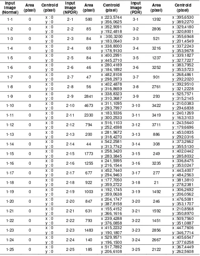
It can be seen from Table 1 that the characteristic of exudates can be extracted using morphological operation method. In normal eyes, there are no exudates, so the results of the calculation will produce a characteristic with a value 0 in the area. In the eyes of patients with diabetic retinopathy disease in NPDR class, there will be more exudates with characteristic area that is producing around 17-21.213 pixels, and in PDR class of the resulting characteristic range is 125-12.299 pixels. The result of centroid value indicates the position of exudates in the retinal image.
Table 1. The results of feature extraction area and centroid of exudate
Input
to remove unnecessary noise. Results are shown in Figure 4.
(a) (b)
Figure 4. (a) Original image; (b) Extraction result of blood vessels
After obtaining the results, the centroid area can be calculated and the results obtained from the blood vessels. Then, it can be translated into form with the vertical sampling process in every 15 pixels of 720 pixels to obtain 48 sampling in a vertical; and horizontal sampling process in every 14 pixels of 576 pixels to obtain 39 horizontal sampling of blood vessels by using a fast Fourier transform. Figure 5 illustrates sampling results.
Figure 5. Sampling result in vertical and horizontal of Blood Vessels
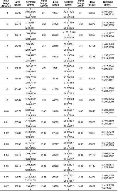
It can be seen from Table 2 that the characteristic of exudates can be extracted by using morphological operation. The calculation results may indicate that the more severe diabetic retinopathy disease, the more characteristics value are count in blood vessels areas. Especially in PDR class, the range characteristic is 13.319-46.681pixels on a normal class, 7.435-49.938 pixels on NPDR class, and 13.891-53.802 pixels on PDR class. Because in PDR class new blood vessels emerge, it causes many areas that counted. The resulting of centroid value indicates the position of the blood vessels in retinal images. The result of sampling in vertically and horizontally by using the FFT of the blood vessels shows similarity between the results of extracted image pattern of each class. Therefore, it will be difficult to distinguish normal eyes image, NPDR, and PDR.
0 5 10 15 20 25 30 35 40 45 50 0
0.2 0.4 0.6 0.8 1
Blood Vessels Vertical Sampling Results
Sampling Count
M
a
gn
it
ud
e
0 5 10 15 20 25 30 35 40
0 0.2 0.4 0.6 0.8 1
Blood Vessels Horizontal Sampling Results
Sampling Count
M
agn
it
u
1-18 23674 :406,1873 y :233,8725
2-18 12344 :403,0478 y
Morphological operation has been used as feature extraction method. It can be used to extract the characteristic of diabetic retinopathy disease that is exudates and blood vessels. The results obtained from the extraction of feature area exudates has a range 0 pixel for normal eyes, 17-21.213 pixels for retinal image with NPDR class, and 125-12.299 pixels for retinal image with PDR class. Centroid calculation result obtained in the exudates of feature extraction has a range of x=0; y=0 for normal eyes, x=150.9715-568.9565; y=167.0133-445.8784 pixels for retinal image with NPDR class, and x=187.1098-535.2328 pixels; y=176.0333-468.7908 pixels for retinal image with PDR class.
The results obtained from the extraction of feature area blood vessel has a range 13.319 - 46.681 pixel for normal eyes, 7.435 - 49.938 pixel for retinal image with NPDR class, and 13.891 - 53.802 pixel for retinal image with PDR class. Centroid calculation result obtained in the blood vessel of feature extraction has a range of x = 295,5133 - 489,0853 pixel ; y = 222,4509 - 365,1226 pixel for normal eyes, x = 309,8893 - 454,2538 pixel ; y = 167,118 - 317,1532 pixel for retinal image with NPDR class, and x = 302,4443 - 443,2236 pixel ; y = 202,8827 - 315,6003 pixel; y=176.0333-468.7908 pixel for retinal image with PDR class.
References
[1] Wild S, Roglic G, Green A, Sicree R, King H. Global Prevalence Of Diabetes: esstimates for the year 2000 and projection for 2030. 2004; 27: 53-1047.
[2] Wong TY, Yau J, Rogers S, Kawasaki R, Lamoureux EL, Kowalski J. Global prevalence of diabetic
retinopathy: Pooled data from the United States, Australia Europe and Asia. Prosiding The
Association for Research in Vision and Opthalmology Annual Meeting. 2011.
[3] Soewondo P, Soegondo S, Suastika K, Pranoto A, Soeatmadji DW, Tjokroprawiro A. The DiabCare Asia 2008 Study - Out - Comes on Control and Complications of Type 2 Diabetic Patients in Indonesia. Med J Indonesia. 2010; 19(4): 43-235.
[5] June Chu, Yusuf Ali. Diabetic Retinopathy: A Review. Drug Development Research. 2008; 69: 1-14.
[6] Jennifer L, Wilkinson-Berka, Antonia G Miller. Update on the Treatment of Diabetic Retinopathy. The
Scientific World Journal. 2008; 8: 98-120.
[7] Muthu Rama Krishnan Mookiah, U Rajendra Acharya, Chua Kuang Chua, Choo Min Lim, EYK Ng,
Augustinus Laude. Computer-aided diagnosis of diabetic retinopathy: A review. Computers in Biology
and Medicine. 2013; 43(12): 2136-2155.
[8] R Pires, HF Jelinek, J Wainer, S Goldenstein, E Valle, A Rocha. Assessing the Need for Referral in
Automatic Diabetic Retinopathy Detection. IEEE Transactions on Biomedical Engineering. 2013;
60(12): 3391-3398.
[9] T Walter, JC Klein, P Massin, A Erginay. A contribution of image processing to the diagnosis of
diabetic retinopathy-detection of exudates in color fundus images of the human retina. IEEE
Transactions on Medical Imaging. 2002; 21(10): 1236-1243.
[10] NG Ranamuka, RGN Meegama. Detection of hard exudates from diabetic retinopathy images using
fuzzy logic. In IET Image Processing. 2013; 7(2): 121-130.
[11] G Zahlmann, et al. Hybrid fuzzy image processing for situation assessment [diabetic retinopathy].
IEEE Engineering in Medicine and Biology Magazine. 2000; 19(1): 76-83.
[12] S Roychowdhury, D Koozekanani, K Parhi. DREAM: Diabetic Retinopathy Analysis Using Machine
Learning.IEEE Journal of Biomedical and Health Informatics. 2014; 18(5): 1717-1728.
[13] SW Franklin, SE Rajan. Diagnosis of diabetic retinopathy by employing image processing technique
to detect exudates in retinal images.IET Image Processing. 2014; 8(10): 601-609.
[14] A Osareh, B Shadgar, R Markham. A Computational-Intelligence-Based Approach for Detection of
Exudates in Diabetic Retinopathy Images. IEEE Transactions on Information Technology in
Biomedicine. 2009; 13(4): 535-545.
[15] A Muntasa, IA Sirajudin, MK Sophan. Matrix Mask Overlapping and Convolution Eight Directions for
Blood Vessel Segmentation on Fundus Retinal Image. TELKOMNIKA Telecommunication Computing
Electronics and Control. 2014; 12(3): 631-638.
[16] MU Akram, I Jamal, A Tariq. Blood Vessel Enhancement and Segmentation for Screening of Diabetic
Retinopathy. TELKOMNIKA Telecommunication Computing Electronics and Control. 2012; 10(2):
327-334.
[17] I Jamal, MU Akram, A Tariq. Retinal Image Preprocessing: Background and Noise Segmentation.