Electroretinogram analysis of relative spectral sensitivity in
genetically identified dichromatic macaques
Akitoshi Hanazawa*, Akichika Mikami†, Puti Sulistyo Angelika‡, Osamu Takenaka§, Shunji Goto¶, Akishi Onishi‖, Satoshi Koike**, Tetsuo Yamamori‡‡, Keichiro Kato†, Aya
Kondo§, Bambang Suryobroto‡, Achmad Farajallah‡, and Hidehiko Komatsu*,††
+ Author Affiliations
1. *Laboratory of Neural Control, National Institute for Physiological Sciences, Myodaijicho, Okazaki 444-8585, Japan; Departments of †Behavioral and Brain Sciences and §Cellular and Molecular Biology, and ¶Center for Applied Primatology and Human Evolutionary Modeling Research, Primate Research Institute, Kanrin, Inuyama, Aichi 484-8506, Japan; ‡Department of Biology, Bogor Agricultural University, Jalan Pajajaran, 16143 Bogor, Indonesia; ‖Department of Biophysics, Graduate School of Science, Kyoto University, Kyoto 606-8502, Japan; **Department of Microbiology and Immunology, Tokyo Metropolitan Institute for Neuroscience, Fuchu, Tokyo 183-8526, Japan; and ‡‡Laboratory for Speciation Mechanisms I, National Institute for Basic Biology, 38 Myodaijicho, Okazaki 444-8585, Japan
1. Communicated by Robert H. Wurtz, National Institutes of Health, Bethesda, MD (received for review January 15, 2001)
Abstract
The retinas of macaque monkeys usually contain three types of photopigment, providing them with trichromatic color vision homologous to that of humans. However, we recently used molecular genetic analysis to identify several macaques with a dichromatic genotype. The affected X chromosome of these animals contains a hybrid gene of long-wavelength-sensitive (L) and middle-wavelength-sensitive (M) photopigments instead of separate genes encoding L and M photopigments. The product of the hybrid gene exhibits a spectral sensitivity close to that of M photopigment; consequently, male monkeys carrying the hybrid gene are genetic protanopes, effectively lacking L photopigment. In the present study, we assessed retinal expression of L photopigment in monkeys carrying the hybrid gene. The relative sensitivities to middle-wavelength (green) and long-wavelength (red) light were measured by electroretinogram flicker photometry. We found the sensitivity to red light to be extremely low in protanopic male monkeys compared with monkeys with the normal genotype. In female heterozygotes, sensitivity to red light was intermediate between the genetic protanopes and normal monkeys. Decreased sensitivity to long wavelengths was thus consistent with genetic loss of L photopigment.
chromosome, and loss of one or the other— caused by unequal chromosome recombination— results in dichromatic color vision (1). Dichromatism in humans has been studied for the purposes of making clinical diagnoses and achieving a better understanding of the mechanisms responsible for trichromatic color vision; the roles of lost and retained photopigments become more apparent in dichromats. Macaque monkeys have trichromatic color vision homologous to that of humans (2) and have served as subjects in a variety of physiological and psychophysical studies of color vision. Therefore, the dichromatic macaque may be a useful animal model of dichromatism with which to conduct clinical studies and investigate the mechanisms underlying color vision.
In many species of New World monkeys, both dichromatic and trichromatic animals are mixed within a species because of the alleles whose products exhibit different spectral sensitivities in a single cone photopigment locus on the X chromosome (3–8). On the other hand, dichromatic macaques were not recognized until recently (9, 10). However, our molecular genetic analysis showed the existence of a dichromatic genotype of the crab-eating macaque (11). By using PCR to specify genotype, we found male protanopes and female heterozygotes spread among some troops in Pangandaran National Park, Indonesia. The genome of male protanopes contains a single hybrid gene—consisting of the 5′ part of the L photopigment and
the 3′ part of the M photopigment (R4G5, exons 1–4 come from the former, exons 5 and 6 from the latter; see ref. 11)—instead of separate L and M photopigment genes. The absorbance spectrum of R4G5 photopigment, which was characterized by photobleaching analysis, is very close to that of M photopigment. Consequently, males carrying only the R4G5 gene on their X chromosome should exhibit the retinal sensitivity of a protanopic dichromat, effectively lacking L photopigment.
In the present study, we examined the retinal chromatic sensitivity of monkeys carrying the R4G5 hybrid gene to confirm the correspondence between the protanopic genotype and the phenotype. We used electroretinogram (ERG) flicker photometry to measure the relative sensitivities of these animals to long- and middle-wavelength light. ERG flicker photometry has been used to measure the spectral sensitivity of macaque monkeys (10) and also has proved to be quite useful to show correlation between the genotype and phenotype in humans (12–15) and New World monkeys (4, 16). We chose this noninvasive approach from among a number of alternatives to minimize the likelihood of injury to the animals, as these carrier monkeys are rare and invaluable. Measurements were made in three genetic groups (normal males and females, heterozygous females, and protanopic males), and our findings show that sensitivity to long wavelengths corresponds to predicted levels of L photopigment.
Methods
The genotype of the crab-eating macaque (Macaca fascicularis) was determined by using PCR to selectively detect normal L and M photopigment genes and the R4G5 hybrid gene in samples of genomic DNA isolated from peripheral blood. To accomplish this, we prepared two
5′ PCR primers, each of which is specific for a region within exon 4 of the L and M photopigment genes (Ex4R: GTCCTCATGGTCACCTGCTGCATCATTCCACTGGCT and
and Ex5G: GGTGTAGGGTCCCCAGCAGAAGCAGAACGCCAGGAA). PCR was done in a 25-μl reaction mixture containing 0.4 μM of 5′ and 3′ primers, 0.2 mM dNTP, ≈10–100 ng of template genomic DNA, and 0.5 units of ExTaq DNA polymerase (Takara Shuzo, Kyoto) in a buffer recommended by supplier. The amplification condition was 94°C for 10 min and 40 cycles each of 94°C for 0.5 min, 60°C for 0.5 min, 72°C for 1.5 min followed by 72°C for 10 min. The genomic DNA was analyzed by using four possible combinations of the primers. Successful amplification using the Ex4R-Ex5R, Ex4G-Ex5G, and Ex4R-Ex5G combinations was indicative of the presence of the normal L gene, the normal M gene, and the R4G5 hybrid gene, respectively.
Four male and three female monkeys were used in our experiment: two males (M30 and M63) and a female (F67) carried the normal L and M photopigment genes, but not the R4G5 hybrid gene (normal); two males (M5 and M39) carried only the R4G5 hybrid gene (protanopic); and two females (F49 and F58) carried all three genes (heterozygous). With the approval of the Ministry of Forestry, all of the monkeys were caught in Pangandaran National Park, Indonesia and are being kept at Bogor Agricultural University (Bogor, Indonesia), which is where all of our experiments were conducted. All procedures related to animal care and experimentation were in accordance with the National Institutes of Health Guide for the Care and Use of Laboratory Animals (1996).
Figure 1
Schematic representation of ERG flicker photometry. Counter-phase flicker of green and red lights was presented as stimuli (combined stimulus). One of the lights was referred to as the fixed stimulus, the other the varied stimulus. The luminance of the fixed stimulus was constant at 20 cd/m2 during the on-period and at 0 cd/m2 during the off-period. The luminance of the varied stimulus during the on-period was changed across measurements. When both lights have the same effective intensity, retinal responses will be minimal. We refer to the luminance inducing the minimal response as the equation point.
The luminances of the stimuli were measured by using a photometer (J17, Tektronix) with a luminance head (J1083, Tektronix), while their spectral intensity distributions were measured by using a spectrophotometer (PR-650, Photo Research, Chatsworth, CA). Retinal responses to the stimuli were recorded by using a contact-lens electrode (Z7816, Kyoto Contact-Lens, Kyoto). Stimulus flicker as well as amplification and measurement of the retinal response was achieved by using a portable ERG system (PE-3000, TOMEY modified by MAYO, Nagoya, Japan). The resultant retinal signal was passed through a band-pass filter (1.6–100 Hz) and averaged, and its amplitude was measured.
Before making ERG measurements, the subject was anesthetized with an intramuscularly injected mixture of medetomidine hydrochloride (0.15 mg/kg) and ketamine hydrochloride (4 mg/kg), and the pupil of one eye was dilated by topical application of tropicamide (0.5%) and phenylephrine hydrochloride (0.5%). Before installation of the contact lens electrode, the cornea was anesthetized by topical application of oxybuprocaine hydrochloride (0.4%). The animals were positioned for recording by using a holder that supported the head with pads.
Results
View larger version:
In this page In a new window
Download as PowerPoint Slide
Figure 2
Results of ERG flicker photometry from three normal monkeys (two males and one female). Each panel plots the amplitudes of the retinal responses against the luminance of the varied stimulus. The symbols depict the averages of three measurements (○) as well as the individual measurements (+). (Upper) Green was the varied stimulus, and red the fixed stimulus. (Lower) Red was the varied stimulus, and green the fixed stimulus.
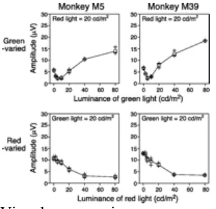
Fig. 3 shows the results from two genetically protanopic dichromats (M5 and M39). In each of these monkeys, the equation point was 5 cd/m2 when green was the varied stimulus (Fig. 3 Upper), making the R/G sensitivity ratio 0.25:1. By contrast, when red was the varied stimulus, the retinal response monotonically declined as the luminance of the red light increased (Fig. 3 Lower), and no clear equation point could be identified within the range of luminances examined. Nevertheless, retinal response was fairly small when the luminance of the red light was maximal (80 cd/m2). If we assume that 80 cd/m2 is the equation point for the red-varied measurements, the R/G sensitivity ratio becomes 0.25:1, the same as that for the green-varied measurements. Both measurements showed the relative sensitivity to red light to be only one-fourth that of normal monkeys, which is consistent with a genotype that does not encode L photopigment.
In this page In a new window
Download as PowerPoint Slide
Figure 3
Results of ERG flicker photometry from two protanopic males. Conventions are the same as in Fig. 2.
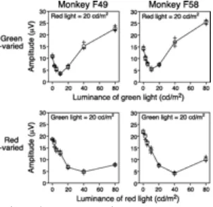
Fig. 4 shows the results from two heterozygous females (F49 and F58), carrying both the hybrid and normal genes. In this case, the equation point was 10 cd/m2 for the green-varied measurements and 40 cd/m2 for the red-varied measurements, which corresponds to an R/G sensitivity ratio of about 0.5:1. The relative sensitivity to the red light was thus about half that of normal monkeys and twice that of the protanopic monkeys, which is consistent with the notion that female protan heterozygotes have fewer retinal L-cone photoreceptors than the normal trichromats but more than the protanopes (see Discussion).
View larger version:
In this page In a new window
Download as PowerPoint Slide
Figure 4
Discussion
We conducted ERG flicker photometry with macaque monkeys identified by molecular genetic analysis as carrying the protanopic genotype (11). Protanopic males exhibited severely diminished retinal sensitivity to long wavelengths, as compared with normal monkeys. Heterozygous females also exhibited diminished sensitivity to long wavelengths, but to a lesser extent. Molecular genetic analysis predicts the complete absence of L cones in protanopic males and smaller numbers of L cones in heterozygous females. This event would be expected to lead to severe and moderate decreases in retinal sensitivity to long wavelengths, respectively. Thus, the present results are consistent with those predicted by the genotypes of the monkeys and provide electrophysiological evidence for the existence of dichromatism in macaque monkeys.
ERG flicker photometry is well established as a useful method for measuring retinal spectral sensitivity (13, 17). The data obtained reflect the relative quantities and absorbance spectra of the photopigments (18), and by using a 30-Hz flicker rate, the contribution made by short-wavelength-sensitive cones to the ERG response can be made negligible (19). Consequently, this method has been widely used to estimate L/M cone ratios in both monkeys and humans (10, 18, 20–22) and to detect differences in spectral sensitivity between human trichromats and dichromats (12–15). This method also has been successfully applied to detection of dichromatic and trichromatic New World monkeys (4, 16). These studies recorded full spectral sensitivity curve by using ERG flicker photometry to estimate the relative contribution of different types of cones. In the present study, we obtained relative sensitivity to red and green lights instead of full spectral sensitivity. Although full spectral measurement is more informative than only a single equation of red and green lights, we avoided long-lasting experiments to minimize the risk of trouble in the physical condition of the monkeys, which were very important genetic resources. In the present study, our priority was to indicate that dichromatic genotypes had much lower sensitivity to long-wavelength light compared with normal monkeys to confirm the correspondence between dichromatic genotype and phenotype. Our method was simple but accurate enough to distinguish normal, heterozygous, and protanopic genotypes. R/G sensitivity ratios estimated from green-varied and red-varied measurements were consistent in all monkeys tested. Differences in R/G sensitivity ratios were quite small within each genetic group, but were clearly distinguishable between groups.
In human females, the reduced sensitivity to long wavelengths observed in protan heterozygotes is known as Schmidt's sign (23, 24). The photopigments of these individuals are thought to derive from one normal and one defective X chromosome as a result of random X chromosome inactivation (25), which would lead to a decrease in the number of L photopigments and a decline in the L/M cone ratio. Our result of heterozygous females exhibiting moderately decreased sensitivity to long wavelengths is consistent with this scheme.
of sensitivities to red in normal, heterozygous, and protanopic animals to be about 1:0.5:0.25, whereas it was found to be 1:0.48:0.16 (26), 1:0.47:0.21 (27), or 1:0.51:0.30 (28) with the psychophysical approach.
Our study has provided phenotypical evidence of the dichromatism in macaque monkeys, but further psychophysical and anatomical studies are necessary to understand the details of color vision in these monkeys. We think these monkeys will become a good animal model of human protanopia, which may advance studies on color vision in a variety of fields. We are now preserving and breeding monkeys carrying the hybrid gene and consider them to be a precious genetic resource. Physiological and further genetic analyses also are within the scope of future studies. Moreover, behavioral observations of protanopic monkeys in their natural habitat may provide important insights into the evolution of trichromatic color vision.
Previous SectionNext Section
Acknowledgments
We thank Dr. M. Horiguchi and Dr. M. Kondo for technical advice in ERG measurement, and M. Togawa and N. Takahashi for technical assistance. This work is supported by
Grant-in-Aid for Scientific Research (A)11691189 from the Ministry of Education, and ―Research for the
Future‖ Program JSPS-RFTF96L00202 from the Japan Society for the Promotion of Science.
Previous SectionNext Section
Footnotes
↵†† To whom reprint requests should be addressed. E-mail: [email protected]. Abbreviations:
ERG,
electroretinogram; L,
long-wavelength-sensitive; M,
middle-wavelength-sensitive; R/G,
red to green
Copyright © 2001, The National Academy of Sciences
References
1. ↵
1. Nathans J
(1999) Neuron 24:299–312, pmid:10571225. 2. ↵
(1974) Vision Res 14:53–67, pmid:4204837. 3. ↵
1. Mollon J D, 2. Bowmaker J K, 3. Jacobs G H
(1984) Proc R Soc London Ser B 222:373–399, pmid:6149558. 4. ↵
1. Jacobs G H, 2. Neitz J
(1987) Proc Natl Acad Sci USA 84:2545–2549, pmid:3470811. 5.
1. Jacobs G H, 2. Neitz J
(1987) Vision Res 27:1263–1268, pmid:3424673. 6.
1. Jacobs G H, 2. Neitz J, 3. Crognale M
(1987) Vision Res 27:2089–2100, pmid:3128920. 7.
1. Tovee M J, 2. Bowmaker J K, 3. Mollon J D
(1992) Vision Res 32:867–878, pmid:1604855. 8. ↵
1. Jacobs G H, 2. Neitz J, 3. Neitz M
2. Deegan J F, 2nd
(1997) Visual Neurosci 14:921–928, pmid:9364728. 11.↵
(1999) Nature (London) 402:139–140, pmid:10647004. 12.↵
(1998) Vision Res 38:3391–3396, pmid:9893854. 15.↵
1. Kremers J, 2. Usui T, 3. Scholl H P, 4. Sharpe L T
(1999) Invest Ophthalmol Visual Sci 40:920–930, pmid:10102289. 16.↵
1. Jacobs G H, 2. Neitz M, 3. Deegan J F, 4. Neitz J
2. Jacobs G H
(1985) J Physiol (London) 363:135–150, pmid:4020697. 20.↵
1. Brainard D H, 2. Calderone J B, 3. Nugent A K, 4. Jacobs G H
(1999) Invest Ophthalmol Visual Sci 40:2840–2847, pmid:10549644. 21.
(1934) Klin Monatsblatter Augenheilkunde 92:456–467. 24.↵
1. Schmidt I
(1955) Am J Optometry 32:404–408, pmid:13238560. 25.↵
(1961) Nature (London) 190:372–373, pmid:13764598. 26.↵
1. Crone R A
(1959) Am J Ophthalmol 48:231–238, pmid:13670291. 27.↵
1. Adam A
(1969) Proc Tel-Hashomer Hospital (Tel-Aviv) 8:2–6. 28.↵
1. Majima A