Annular pancreas: the failure of side-to-side gastrojejunostomy
anastomosis and the success of repair with Roux-en-Y
gastrojejunostomy
Keywords:annular pancreas, infant, Roux-en-Y gastrojejunostomy, side-to-side gastrojejunostomy
pISSN: 0853-1773 • eISSN: 2252-8083 • http://dx.doi.org/10.13181/mji.v24i2.1207 • Med J Indones. 2015;24:115-9 • Received 17 Feb 2015 • Accepted 17 Jun 2015
Correspondence author: Isaac A. Deswanto, isaacdeswanto@yahoo.com
Copyright @ 2015 Authors. This is an open access article distributed under the terms of the Creative Commons Attribution-NonCommercial-ShareAlike 4.0 International License (http://creativecommons.org/licenses/by-nc-sa/4.0/), which permits unrestricted non-commercial use, distribution, and reproduction in any medium, provided the original author and source are properly cited.
Isaac A. Deswanto, Janta F. Barus
Department of Surgery, Tarakan General Hospital, Tarakan, North Kalimantan, Indonesia
C a s e Re p o r t
ABSTRAK
Pankreas annular adalah kelainan kongenital yang khas dengan terbentuknya cincin pankreas yang mengelilingi duodenum secara utuh atau sebagian. Kami melaporkan sebuah kasus pankreas annular pada pasien anak berusia 15 hari yang datang ke rumah sakit dengan keluhan muntah berulang dengan penurunan berat badan yang bermakna. Foto rontgen abdomen menunjukkan pembesaran lambung dengan pasase gas yang terkumpul pada duodenum proksimal. Laparotomi terbuka dilakukan dan ditemukan adanya pankreas annular yang mengelilingi duodenum proksimal yang menyebabkan hambatan pasase melewati duodenum. Pasien ini dilakukan
side-to-side gastrojejunostomy dan stoma dibuat melewati obstruksi, dan didapatkan pasasenya memuaskan. Empat hari setelah operasi, pasien kembali muntah-muntah dengan abdomen yang membesar. Foto abdomen menggunakan kontras menunjukkan tidak adanya pasase kontras melalui stoma yang dibuat. Terjadi striktur atau re-stenosis pada stoma yang pertama dibuat. Hasil yang memuaskan tampak dengan teknik Roux-en-Y gastrojejunostomy bypass anastomosis.
Pasien diperbolehkan pulang setelah 24 hari perawatan di rumah sakit tanpa sekuele yang tidak diinginkan.
ABSTRACT
Annular pancreas is a rare congenital anomaly characterized by a partial or complete encirclement of ectopic pancreas tissue around the duodenum. We report a case of annular pancreas in a 15 day-old infant admitted to the hospital with complaints of profuse and recurrent vomiting and loss of body weight. Non-contrast abdominal X-ray showed a dilated stomach with bubbles formation around the upper abdomen. An obstruction was noted and open laparotomy was performed. Upon laparotomy, pancreatic ring encircled the proximal duodenum causing an obstruction. Side-to-side gastrojejunostomy was performed and passage through the bypass was satisfactory. Four days after the operation, vomiting and bulging abdomen
ensued. Contrast abdominal X-ray demonstrated filling
Annular pancreas is one of the rarest causes of acute duodenal obstruction in both children and adults. It is characterized by the presence of a ring of pancreatic tissue that completely or partially surrounds the duodenum. Approximately one to three out of 20,000 babies were born with this congenital disorder with higher predisposition towards male.1
Defects during the embryologic development of the foregut are responsible in the development of annular pancreas. The formation of pancreas begins during the 5th week of gestation. Primitive
foregut develops into one dorsal and two ventral buds, in which during the 7th week of gestation
ventral buds will rotate with gut as the axis to unite with dorsal bud after rotating behind the duodenum. During this embryologic process, it was postulated that annular pancreas was formed by the abnormal union of the edge of ventral bud with duodenum resulting in fibrous tissue formation around the duodenum.2-4
Annular pancreas causes complete or incomplete duodenal obstruction where this may lead to recurrent profuse vomiting and poor feeding. This will manifest as malnutrition and failure to thrive if there is a delay in hospital admission.5,6 Abdominal
distension is commonly found and may prompt immediate nasogastric decompression. Diagnosis of annular pancreas is usually confirmed upon laparotomy, but radiographic images with the help of contrast may aid in displaying evidences of the presence of duodenal obstruction. Immediate management of annular pancreas in children should include fluid resuscitation to prevent hypovolemic shock, correction of any electrolyte abnormalities, gastric decompression and surgical management to relieve the obstruction. Surgical options include duodenoduodenostomy, duodenojejunostomy, gastrojejunostomy or very rarely pancreatic resection.7
CASE REPORT
A 15 days old infant was admitted into Tarakan General Hospital (Rumah Sakit Umum Daerah Tarakan, North Kalimantan) with chief complaint of persistent vomiting. The first episode of vomiting occurs one hour after breastfeeding. The vomit was projectile and not bile-stained. From the date of delivery until hospital admission
patient had been vomiting repetitively, look more exhausted and lost a lot of body weight.
The baby was delivered full-term through normal birth delivery and assisted by midwife with the birth weight of 4,000 grams. He immediately cried after delivery and passed meconium on small amount within 24 hours after delivery. Breastfeeding was initiated as soon as possible.
During physical examination, the baby looked severely ill and dehydrated. The infant’s heart rate was 160 x/minute, respiratory rate 48 x/minute, and temperature 36.6°C. The baby’s weight upon
admission was 2,800 grams and his body length was 68 cm. The upper abdomen was moderately distended, no mass was palpable and peristalsis was prominently seen in the upper abdomen. There was no abnormal peristalsis heard upon auscultation.
Laboratory examination showed no significant abnormalities. However, abdominal x-ray photo revealed distended stomach with bubbles of gas in the right upper quadrant of abdomen. Distended abdomen and bubbles of gas raised suspicion of the presence of obstruction although the level of obstruction still could not be determined as of this moment. Barium meal was not given at this point, because the diagnosis of obstruction was already appropriately established to start surgical exploration without further investigation.
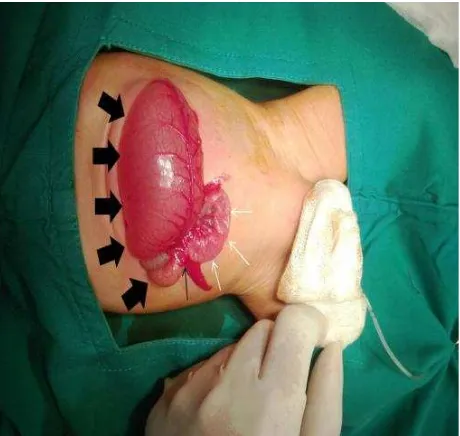
Operation was carried out after both the state of dehydration and malnutrition had been resolved. After general anesthesia was performed, supraumbilical transverse incision was made. Upon opening the peritoneum, a dilated stomach approximately as large as eight centimeter was exposed containing fluid and no passage of fluid was apparent. The second part of the duodenum was apparently constricted by a ring of pancreatic tissue that completely surrounded the duodenum and was continued with the head of pancreas. Upon identifying the obstruction, it was decided to bypass the obstruction by performing side-to-side gastrojejunostomy. Upon the completion of side-to-side gastrojejunostomy, passage through the by-pass was tested and seemed to be patently working well. After that, abdominal cavity was adequately irrigated with normal saline and surgical wound was closed.
Postoperatively the nasogastric tube was inserted and remained in place for the next three days. The patient was programed to start breastfeeding again three days after the operation in which during that period of time he received parenteral nutrition under the close supervision of pediatrician. After three days, nasogastric tube was taken off because of production of vomit through
Figure 2. Intraoperative: dilation of the stomach and proxi-mal duodenum is clearly visualized (black arrowheads) due to a complete annular band of pancreatic tissue that con-stricted the second part of the duodenum (black arrow). Distal to site of obstruction, the distal or third part of was collapsed (white arrows)
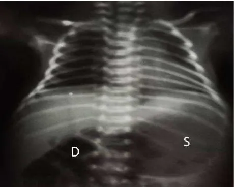
Figure 3. Barium Study taken after side-to-side
gastrojeju-nostomy. Barium contrast only fills the dilated stomach (S)
and duodenum proximal to the obstruction (white arrow) nasogastric tube was minimal. Unfortunately, the patient started to vomit repetitively on the next day after the nasogastric tube was taken off. The vomit was again projectile and bilious; therefore initiatives were taken to re-examine the patient. Barium meal examination was performed. It was clear that the barium contrast only going to fill the stomach and proximal part of duodenum. No contrast was clearly seen entering the by-pass that has been made during the operation.
Consequently, another laparotomy was
DISCUSSION
Annular pancreas is a congenital abnormality that is characterized by the formation of pancreatic tissue that partially or fully encircles the duodenum. Annular pancreas, with its rare incidence, may be symptomatic in the early stage of life or can go unnoticed until adulthood depending on the degree of obstruction.8,9
Symptoms of annular pancreas in adults are commonly associated with complications of peptic ulcer, pancreatitis, duodenal, and biliary obstruction.9,10 In infants, on the other hand,
clinical manifestations revolve around duodenal obstruction in which leads to abdominal distention and profuse vomiting that leads to failure to thrive if left unresolved. Annular pancreas sometimes associates with other congenital abnormalities such as Down syndrome, esotracheal fistula, anal imperforation, Hirschsprung disease11 and
Smith-Lemli-Opitz syndrome.12
The diagnosis of annular pancreas can be suspected prenatally with the findings of double-bubble signs accompanied with hyperechogenic band around the duodenum during ultrasound examination.13 After birth, simple radiograph
of the abdomen may signify the presence of duodenal obstruction. These signs, however, are also commonly observed in other conditions such as duodenal web, duodenal atresia, and midgut volvulus.6 Despite its role as pathognomonic
finding, double bubble signs may not be present in all cases of duodenal obstruction14 as with
the present case. A barium enema can also be performed to further confirm the presence as well the level of obstruction. Nevertheless, in 40% of annular pancreas cases, the diagnosis was made by visual confirmation during laparotomy.14 Other
imaging modalities such as CT-scan, endoscopic retrograde cholangiopancreatography (ERCP) and magnetic resonance cholangiopancreatography (MRCP) may provide rapid anatomic depiction of pancreatic ring encircling the duodenum in a number of adult patients.3,15
The relief of duodenal obstruction surgically is the main goal in the management of patients with annular pancreas. The relief of obstruction is preferably achieved by creating anastomosis that will bypass the obstruction.
The procedures that may be used include gastrojejunostomy, duodenojejunostomy or duodenoduodenostomy.4,14,15 The choice of which
procedure to be performed has been an argument among different authors. Fu, et al1 stated that each
procedure is proper for certain type of patient. In younger patients, duodenoduodenostomy or duodenojejunostomy is preferred because the need of vagotomy may be minimized. Up until today, however, there has not been enough data to conclude which procedure provides better outcome and it requires further research. Attempts to resect or divide the annulus can be extremely difficult to achieve as the pancreatic tissue can lie intramurally with no dissection plane.14 Furthermore separation of annulus
from the duodenum has been associated with many complications, including duodenal leakage, pancreatitis and pancreatic fistula.1,4,14,15
In our patient, side-to-side gastrojejunostomy was initially performed. This procedure, however, was complicated with incidental anastomotic strictures or re-stenosis. Therefore, this necessitates the repair of by-pass with Roux-en-Y gastrojejunostomy. In a study conducted by Ezomike, et al16 repair of anastomosis is
as high as 17.4% (out of 23 patients) which is due to anastomotic leakage and strictures. They also stated that repair of anastomosis or reoperation is related to higher mortality.16 In
our case, nevertheless, result from post-repair has been satisfactory and uneventful until the date of the most recent follow-up. This is probably attributable to the time of presentation to the hospital and meticulous pre-operative resuscitation with the appropriate parenteral nutrition.16 A number of studies also report high
mortality rate associated with prolonged gastric stasis and sepsis16,17 which is in contrast to our
patient.
Adequate pre-operative resuscitation without delaying definitive surgical by-pass is important to achieve better prognosis for the patient with lower mortality. The patency of the anastomosis must be also monitored carefully before patient is discharged. The presence of anastomotic stricture prompts immediate repair of anastomosis.
Conflicts of interest
The authors affirm no conflict of interest in this study.
Acknowledgment
Many thanks is given to Tarakan General Hospital in Tarakan, North Kalimantan and the close relatives of the patient in giving their consent in regard to the publication of this article.
REFERENCES
1. Fu PF, Yu JR, Liu XS, Shen QY, Zheng SS. Symptomatic adult annular pancreas: report of two cases and a review of the literature. Hepatobiliary Pancreat Dis Int. 2005;4(3):468 - 71.
2. White J, Wilson J. USMLE Step 1 Anatomy Lecture Notes. Kaplan Medical. 2011: 247-8 p.
3. Türkvatan A, Erden A, Türkoğlu MA, Yener O.
Congenital variants and anomalies of the pancreas and pancreatic duct: imaging by magnetic resonance cholangiopancreaticography and multidetector computed tomography. Korean J Radiol. 2013;14(6):905-13. 4. Mahdi B, Selim S, Hassen T, Mongi MM, Fadhel CM, Fathi
C, et al. A rare cause of proximal intestinal obstruction in adults - annular pancreas: a case report. Pan Afr Med J. 2011;10:56.
5. Mulugeta PG, Hilmes MA. Annular pancreas in a toddler. Pediatr Radiol. 2010;40(Suppl1):S106.
6. Pansini M, Magerkurth O, Haecker FM, Sesia SB. Annular pancreas associated with duodenal obstruction. BMJ Case Reports. 2012;10:1-2.
7. Bouassria A EH, Elbouhaddouti H, Mouaqit O, Benjelloun E B, Ousadden A, Mazaz K, et al. Annular pancreas producing duodenal obstruction: A case report. Open J Gastroenterol. 2013;3(3):202-4.
8. Zeineb M, Sadri BA, Nizar M, Hassen H, Nafaa A, Taher K. Annular pancreas intra operatively discovered: a case report. Clin Pract. 2011;1(4):e82.
9. Patra DP, Basu A, Chanduka A, Roy A. Annular pancreas: a rare cause of duodenal obstruction in adults. Indian J Surg. 2011;73(2):163-5.
10. Tas A, Köklü S, Kocak E, Akbal E, Ergul B. An unusual cause of acute pancreatitis: annular pancreas and papillary opening of the cystic duct. Gut Liver. 2012;6(3):403-4. 11. Kandpal H, Bhatia V, Garg P, Sharma R. Annular
pancreas in an adult patient: diagnosis with endoscopic ultrasonography and magnetic resonance cholangiopancreatography. Singapore Med J. 2009;50(1):e29-31.
12. Demirdöven M, Yazgan H, Korkmaz M, Gebeşçe A, Tonbul
A. Smith-lemli-opitz syndrome: a case with annular pancreas. Case Rep Pediatr. 2014;2014:623926. 13. Dankovcik R, Jirasek JE, Kucera E, Feyereisl J,
Radonak J, Dudas M. Prenatal diagnosis of annular pancreas: reliability of the double bubble sign with periduodenal hyperechogenic band. Fetal Diagn Ther. 2008;24(4):483-90.
14. Whittingham-Jones PM, Riaz AA, Clayton G, Thompson HH. Annular pancreas - a rare cause of gastric obstruction in an 82-year-old patient. Ann R Coll Surg Engl. 2005;87(1):W13-5.
15. Alahmadi R, Almuhammadi S. Annular pancreas: a cause of gastric outlet obstruction in a 20-year-old patient. Am J Case Rep. 2014;15:437-40.
16. Ezomike UO, Ekenze SO, Amah CC. Outcomes of surgical management of intestinal atresias. Niger J Clin Prac. 2014;17(4):479-83.