Scholars Research Library
J. Nat. Prod. Plant Resour., 2014, 4 (5):39-48 (http://scholarsresearchlibrary.com/archive.html)
ISSN : 2231 – 3184 CODEN (USA): JNPPB7
Identification of compounds from extract methanol of ketepeng
leaves (
Cassia alata
)
Purwantiningsih Sugita
1*, Budi Arifin
1, Selvia Rachmawati
2and Wahyu Yuly Annas
21
Departement of Chemistry, Bogor Agricultural University, Bogor, West Java, Indonesia
2
Department of Chemistry Graduate student, Bogor Agricultural University, Bogor, West Java, Indonesia
_____________________________________________________________________________________________ obtained. From B fraction was obtained one of phenolic compounds, methyl p-hydroxybenzoate, has been isolated
for the first time from the leaves of this species. The structure of the compound has been established by 1H and 13C
NMR spectroscopic methods. Meanwhile from the H fraction was obtained 21 fractions. One of them, the H9
fraction then was analyzed further by using LC-MSand 1H NMR. The chromatogram and 1H NMR spectrum of this
fraction still indicated mixture of compound, but a dominant peak at retention time (tR) 6.7 min was observed.
Further analysis to this peak, particularly to its mass spectrum, indicated that the peak is predicted as a flavonoid adduct, that is 4’-hydroxy-6-C-xylofuranosyl flavanone.
Keywords: Cassia alata, 1H NMR, 13C NMR, LC-MS, methyl p-hydroxy benzoate, flavonoid adduct 4’-
hydroxy-6-C-xylofuranosyl flavanone
_____________________________________________________________________________________________
INTRODUCTION
Cassia is a main genus of Fabaceae family consisting of around 600 species. Someof them are spread out widely,
particularly in tropical countries and they are abundant in India. C. alata, C. tora, C. siamea, and C. fistula are some species researched frequently [1]. C. alata is a herb which is well known in Indonesia as ketepeng. C. alata
generally used as traditional medicine mainly in tropical area such as Malaysia, Brazil, and Indonesia. This plant is believed to be able to cure constipation, hernia, syphilis, diabetes [2], malaria and influenza [3], bronchitis, asthma [4], as anti-obesity [5], strong laxative, and insects repellant [6]. Its leaves methanolic extract was proved to has anti-fungi and antibacterial activity [2]. Chloroform fraction of leaves methanolic extract was proved to have antimalarial activity [7]. Besides, leaves methanolic extract also has anti-angiogenic and cytotoxic activity in breast cancer cell lines [8]. In Indonesia, one of cleaning tissue product made from this plant claimed to be able to prevent and treat leucorrhoea (fluor albus) [9].
leaves ethanolic extract from Nigeria contains flavonol glycosides, 5,7-dihydroxy-2-(4-hidroxyphenyl)-3-[(2S,3R,4S,5S,6R)-3,4,5-trihydroxy-6-(hidroxymethyl) oxan-2-il] oxy-chromene-4-one [11]. The seed ethanolic extract contains alkaloid compound, cannabinoid dronabinol ((S)-3-buthylamino-9-methyl-10,10a-dihydro-7H -benzo[c] chromene-1-ol) [13]. A flavonoid compound, kaempferol also found in C. alata leaves methanolic extract from Jamaica [14].
A typical research to C. alata leaves has not been much conducted yet in Indonesia, mainly related to chemical components information of its leaves. Growing site difference will influence the type and amount of secondary metabolites existing in this plant. Therefore, this research was conducted to characterize chemical components in C.
alata leaves growinginIndonesia. Isolation and characterization were conducted to leaves methanolic extract. This
result is desired to be able to enlarge information database of secondary metabolites existing in C. alata whichcan be used to explore this plant potency further next time.
MATERIALS AND METHODS
Materials and Equipment
Materials used were ketepeng leaves (obtained from Cikasungka village, Solear, Tangerang, Indonesia), methanol, ethyl acetate (EtOAc), chloroform-ammonia, concentrated sulphuric acid, Mayer, Dragendorf, and Wagner reagent, diethyl ether, concentrated acetic acid, Mg, HCl, FeCl3 1% reagent, n-hexane, acetone, chloroform (CHCl3),
dichloromethane (DMC), silica gel Merck 60G, silica gel Merck 60 PF254, silica gel 60 (0.2–0.5 mm), and TLC on
silica gel GF254. The equipment used were liquid vacuum chromatography (LVC) kit, column for gravitational
column chromatography (CC), liquid chromatography-mass spectrometer (LC-MS) with electrospray ionization (ESI-MS) mode Mariner Biospectrometry, proton nuclear magnetic resonance (1H NMR) spectrometer Agilent 500 MHz (1H), the 1H NMR spectra were recorded on a JEOL ECA 500 (500 MHz) instrument in CDCl3 with TMS as
an internal standard (chemical shifts in δ, ppm), and 13C NMR spectra were recorded on a JEOL ECA 500 (125 MHz) instrument.
Extraction and Isolation of C. alata leaves
C. alata leaves powder (1 kg) was macerated by methanol (4 L) in room temperature for 24 hours. Macerate was
separated, then the residue was macerated again with the same type and amount of solvent. This step was repeated for 3 times. All of macerate were collected and concentrated. A dark green crude methanolic extract obtained and its recovery was counted with water content as correction factor. Then, phytochemical test was conducted to that extract. The chemical components process separation of C. alata leaves methanolic extract was initiated by separating chlorophyll and tannin. This separation was conducted by diluting crude methanolic extract (150 g) with 1 L of methanol: water (1:1), shaking it, and letting it for a night. The precipitated chlorophyll was separated and weighed, then free-chlorophyll methanolic extract was extracted by EtOAc and was let for a while until 2 layers formed. The bottom layer was predicted as water extract containing tannin, while the other one was predicted as EtOAc layer containing chemical components without chlorophyll and tannin. The EtOAc extract obtained was concentrated by rotary-evaporator, and then fractionated by LVC with increasing polarity of n-hexane-EtOAc eluent system and 8 fractions were obtained (A-H fractions). B fraction (68 mg) was fractionated further by radial chromatography (RC) with the former eluent system in LVC and 8 fractions were obtained (fraction-B1-8). B5
fraction (9.40 mg) then was fractionated further by preparative-thin layer chromatography (TLC) with n- hexane-CHCl3-EtOAc (7:1:2) eluent system and 4 fractions were obtained (fraction-B51-54). The B53 fraction then was
analyzed further by using 1H and 13C NMR. H fraction (4 g) was then fractionated by gravitational column chromatography by n-hexane-EtOAc gradient elution system and 21 fractions were obtained. The H9 fraction was
further purified by liquid partition with n-hexane-methanol (1:1). Methanol-soluble component was then analyzed under TLC to ensure that was a single component and analyzed further by 1H NMR and LC-MS spectrometer.
RESULTS AND DISCUSSION
______________________________________________________________________________
Phytochemical Constituents in Crude Methanol Extract
Phytochemical test result of its extract showed the existence of phenolics, flavonoids, steroids/triterpenoids, saponins, and alkaloids. Flavonoids and steroids/triterpenoids have ever been reported by Soetjipto et al. [15], Veerachari and Bopaiah [16]. Phenolics and saponins were not found by Veerachari dan Bopaiah [16], while alkaloid was not found by Soetjipto et al. [15]. In Bangladesh, 2,3,7-tri-O-methylellagic acid which belongs to phenolic group was successfully isolated from C. alata methanolic leaves extract by Alam et al. [12].
Kaempferol-3-O-gentio-bioside, a flavonoid compound, which detected in C. alata methanolic leaves extract in Indonesia was already reported by Moriyama et al. [17]. Singh et al. [18] isolated flavonoid compounds, kaempferol and rhein, from hydromethanolic leaves extract in India. An alkaloid, cannabinoid dronabinol, was successfully isolated from ethanolic seed extract in Nigeria [13]. This difference of secondary metabolites constituents is caused by age difference and place where the plant grows.
Methyl p-hydroxybenzoate
Result of B fraction (68 mg) which was fractionated further by radial chromatography (RC) with the former eluent system in LVC obtained 8 fractions (fraction-B1-8). B5 fraction (9.40 mg) then was fractionated further by
preparative-thin layer chromatography (TLC) with n-hexane-CHCl3-EtOAc (7:1:2) eluent system were obtained 4
fractions (fraction-B51-54). The B53 fraction then was analyzed further by using 1H and 13C NMR and . Fraction-B53
was predicted as methyl p-hydroxy benzoate or p-methoxybenzoate acid.
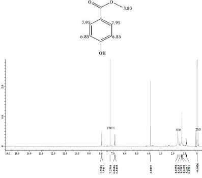
Analysis on 1H NMR spectrum (Figure 1) shows proton chemical shifts (δ) at 3.90; 6.85, and 7.95 ppm. A singlet signal (3H) at 3.90 ppm is identified as proton chemical shift of methoxy group. Two doublet signals (2H) at 6.85 and 7.95 ppm with same coupling constant value (J = 8.43) are identified as proton chemical shift of para
disubstituted-benzene aromatic ring. One signal in upfield area (6.85 ppm), might be caused by electron donation from substituent in ortho position. That substituent with electron donating property may be a methoxy (–OCH3) or
hydroxy (–OH) group. Another signal at 7.95 ppm (in downfield area) indicates the existence of electron attracting substituent in ortho position. Substituents predicted having this property are –CO2H, –CO2R, and –COH. Generally,
proton chemical shift of aromatic without substituents is around 7.4 ppm.
Figure 1. The 1H NMR spectrum (CDCl
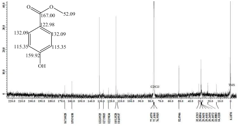
The 13C NMR spectrum (Figure 2) shows signals at 52.09; 115.35; 122.98; 132.09; 159.92; and 167.00 ppm. Signals at 52.09 ppm and 167.00 ppm are identified as carbon signal from methoxy group and carbonyl group of carboxyclic acid or ester. Two sp2 carbon signalsat 132.09 and 115.35 ppm shows twice as higher intensity indicating that each of them represents 2 equivalent C atom (homotopic), while sp2 carbon signals at122.98 and 159.92 ppm with lower intensity are predicted as quaternary C. Signal which is more downfield (159.92 ppm) is predicted because of stronger electron attracting substituent attached to quaternary C. The resulted 1H and 13C NMR data spectrum from this study and the calculated 1H and 13C NMR chemical shift [19] are compared to strengthen the compound prediction of B53 fraction.
Figure 2. The 13C NMR spectrum of fraction-B
53
Table 1 shows comparison between chemical shifts of B53 to calculation of methyl p-hydroxybenzoate and
p-methoxybenzoate acid based on theory [19] and Spectral Data Base (SDBS) literature [20,21], respectively. SDBS is an organic compounds databases spectrum built by Japan to whole world. The result showed the 1H and 13C NMR chemical shifts of B53 have suitable values with calculated chemical shift values of methyl p-hydroxybenzoate,
either according to theory or SDBS literature. Base on this, therefore, B53 was deducted as methyl
p-hydroxybenzoate.
Table 1 The1H and 13C NMR chemical shifts of methyl p-hydroxybenzoate and p-methoxybenzoate acid as compared to theoretical
calculation and SDBS Fabaceae family [22], but this existence has ever been reported from the other family. This compound was isolated from Nerium oleander leaves which come from Shambat [23], Stocksia brahuica in Pakistan [24], from Oxalis
______________________________________________________________________________
Identification of H Fractions
Fractionated by gravitational column chromatography for H fraction (4 g) was obtained 21 fractions. The H9 fraction
was further purified by liquid partition with n-hexane-methanol (1:1). Methanol-soluble component was then analyzed under TLC and analyzed further by 1H NMR and LC-MS spectrometer.
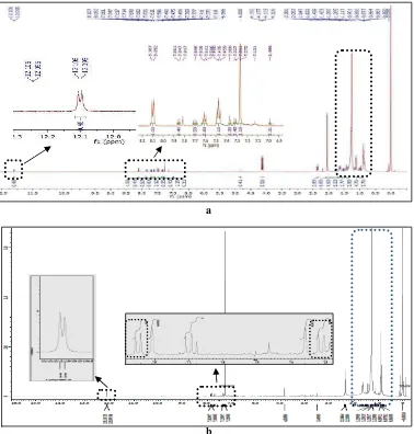
The H9 fraction 1H NMR spectrum shows that H9 still not pure. (Figure 3a). It is indicated with the existence of
complex enough signal peaks in aliphatic range (δH 0.7–1.7 ppm) and signal peak at δH 12.1 ppm (indicates –OH
carboxylic acid functional group) splitting into 2 peaks (Figure 3b). Further analysis with LC-MS strengthen the former hypotheses since the chromatogram (Figure 4) shows 4 peaks with retention time (tR) respectively 3.0, 3.7,
4.7, and 6.7 minute with the area 325.30, 302.264, 264.99, and 2822.93 respectively.
H9 peak with tR 6.7 minute (then signed as H94) has the highest abundance in H fraction. Therefore, secondary
metabolite structure characterization was focused to compound in that fraction. Fragmentation pattern obtained is relatively simple. (Figure 5). Due to that fact, compound inside was predicted to contain relative stable functional group and was not fragmented overall. This may be caused by low energy used in ionization process. Tsimogiannis
et al. (2007) [27] said that if the collision energy in ionization process is low, spectrum obtained from [M+H]·+
cannot be fragmented overall. This kind of fragmentation pattern was predicted to be produced by compound with aromatic cyclic structure stabilized by conjugated system. This compound does not contain aliphatic hydrocarbon chain since fragment having [M+H 14]·+ or multiples is not observed. Based on those characterizations, this compound was predicted to have flavonoid basic skeleton. The 1H NMR spectrum also shows signals in aromatic range (δH 6.5–8.0 ppm), that are at δH 7.2 ppm (1H, d) and 7.8 ppm (1H, dd) with coupling constant respectively 9.1
and 7.8 Hz (3Jortho= 7–10 Hz) (Figure 3b). This pattern in accordance with benzene structure unit substituted in para
Figure 3 The H9 fraction 1H NMR spectrum before partition (a) and after partition by n-hexane-methanol (b)
39.0 271.2 503.4 735.6 967.8 1200.0
position. One signal which is more unshielded (δH 7.8 ppm) than the other (δH 7.2 ppm) indicated that 2 substituents
in para position have different electron-withdrawing power.
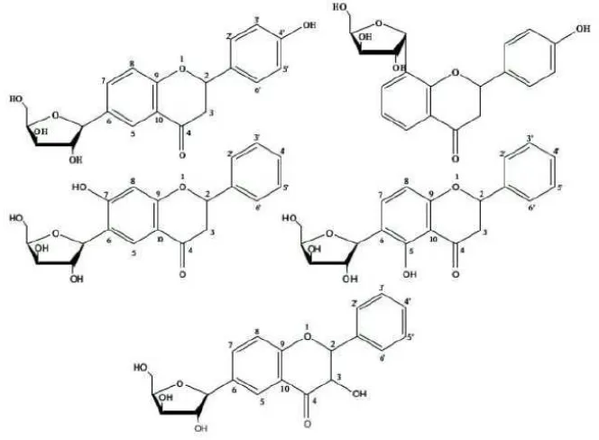
Flavonoid has C6-C3-C6 basic skeleton. Based on C ring structure, flavonoid is distinguished to 5 big groups. Those
are flavonol, flavone, dihydroflavonol or flavanonol, flavanone, and flavanol. Flavonoid compounds isolation and identification frequently are hard to do since structure and polarity similarity [27]. Flavonoid is generally found as flavonoid O-glycoside form in nature that one or more of hydroxyl groups from aglycone are bound to sugar through hemiacetal bond which labile to acid. Yet, glycosylation may also happens through C-C bond resisting to acid, forms C-glycoside flavonoid. The C-glycoside flavonoid generally found are mono-C-glycoside, di-C-glycoside, and C-glycosyl-O-glycoside flavonoid. Sugar group usually found in nature is glucose. Galactose, rhamnose, xylose, and arabinose are not common. Similarly, manose, fructose, glucuronic acid, and galacturonic acid are also rare. That sugar group is substituted at position 3, 5, and 7 in flavonoid O-glycoside, while in flavonoid C-glycoside is only found at position 6 and 8 [28].
Based on C-C bond resistibility of sugar group with flavonoid aglycone compared to O-C bond of O-glycoside flavonoid, this H94 compound was predicted belong to C-glycoside flavonoid. The bonded sugar probably was a
pentose. A pentose was predicted bonded to flavonoid basic skeleton based on some combinations made which were suitable with base peak appearing at m/z = 372 (Figure 6), with assumption that the base peak showed molecular ion
0 2.8 5.6 8.4 11.2 14.0
Figure 4 H9 fraction-methanol soluble LC chromatogram with methanol-water 95:5 as eluent and flow rate 1
mL/minute
1 2 3
______________________________________________________________________________
[M+H]·+ from H94 fraction. This pentose sugar existence was predicted gave weak signals in 1
H NMR spectrum at δH 3.4–4.3 ppm (Figure 7).
Based on structure prediction made before, the H94 compound having m/z= 371 most probably belong to flavanone
derivative compound hydroxylated and glycosylated by a pentose sugar. A hydroxyl group was predicted at position 4’ of B ring based on 1H NMR spectrum analysis showing para-disubstituted existence. While a pentose sugar group was predicted to be bound at position 6 or 8 since naturally sugar groups usually exist at those positions in C -glycoside flavonoid (Figure 8). Pentose sugar groups which have ever been identified to be combined with flavonoid are xilose and arabinose [28].
Fragmentation pattern of this compound was proposed as in Figure 9, yet none of m/z values of these fragments was observed in mass spectrum (Figure 5). It may due to the low energy used in ionization process. When a weak ionization method like ESI and APCI are used to analyze flavonoid compounds, generally fragmentation is not
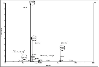
observed. This problem can be overcome by using CID integrated with MS-MS [29]. This compound was predicted to have molecular weight, 372 g/mol with [M+H]·+ at m/z= 371. The [M+H+1]·+ fragment also appears at m/z= 372.
The molecular ion existence at this m/z value is supported by probability of pseudomolecular ion formation. This ion is formed as adduction result between molecular ion or its fragments and cation existing in the solvents or with the solvent mediated by that cation. In ESI-MS, complex formation from analite with alkali metal cations commonly is observed since Na+ or K+ cation exists in the solvent or glassware used [30]. Those cations generally are extracted from glassware during storage and are easier formed in flavonoid substituted at position 3, like flavonol 3-O -glycoside and isoflavone [28]. The presumed compound, H94 has ketone functional group with free electron pair as a
ligand which will complex metal cation, Na+ or K+. Besides, Na+ ion can form 5 or 6 coordination with ligand so that one Na+ ion can coordinate with free electron pair from that ketone functional group and with the other molecules like solvent [31]. Based on this fact, it was predicted that molecular ion appearing at m/z= 371 formed an adduct as [0,4B++Na+ 5H2O]
·+
and [M+Na]·+ so peaks having low abundance appear respectively at m/z= 259 and m/z= 393 with abundance less than 10% and around 39%. Besides, this molecular ion can also form [M+Na+H2O+CH3OH]·+ and [2M+Na]·+ adduct so peaks having low abundance also appear respectively at m/z=
444 and m/z= 765 with abundance less than 10% and around 18% (Figure 10).
The m/z values obtained and probability of those adducts formation strongly supported the former hypotheses that H9 compound having retention time (tR) equal with 6.7 minute, or simply the H94 is predicted belong to C-glycoside
______________________________________________________________________________
CONCLUSION
The phytochemical result of C. alata leaves methanolic extract from Cikasungka village, Solear, Tangerang, Indonesia was identified to contain phenolics, flavonoids, steroids/triterpenoids, saponins, and alkaloids. Two components characterized successfully were belong to phenolic compounds, methyl p-hydroxybenzoate, and flavonoid adduct which presumed as pelargonidin-3-(feruloyl)diglucoside-5-(malonyl)glucoside, respectively.
Acknowledgements
The authors wish to express appreciation to the Directorate General of Higher Education, the Ministry of Education and Culture for funding through 2013 BOPTN research program granted to Dr. Gustini Syahbirin, MS.
REFERENCES
[1]Phongpaichit S, Pujenjob N, Rukachaisirikul V, Ongsakul M. J Sci Technol, 2004, 26(5): 741-748. [2]Makinde AA, Igoli JO, Ama LTA, Shaibu SJ, Garba A. African J Biotechnol, 2007. 6(13):1509-1510. [3]Kusmardi, Kumala S, Triana EE. Makara Kesehatan, 2007. 11(2):50-53.
[4]Savithramma N, Sulochana C, Rao KN. J Ethnopharmacol, 2007. 113:54-61. [5]Hernandez CLC, Leonido FMG. J Med Plants Res, 2011. 5(3):452-455.
[6]Odunbaku OA, Ilusanya OAF. Adv Environ Biol, 2011. 5(8):2162-2165. ISSN 1995-0756
[7]Kayembe JS, Taba KM, Ntumba K, Tshionga MTC, Kazadi TK. J Med Plant Res, 2010. 4(11):991-994. DOI: 10.5897/JMPR09.145
[8]Levy A, Lewis A. West Indian Med J, 2011. 60(6):608-614. [9]Yacob T, Endriani R. J Natur Indones. 2010. 13(1):63-66.
[11]Saito ST, Silva G, Santos RX, Gosmann G, Pungartnik C, Brendel M. Int J Mol Sci, 2012. 13:2846-2862. DOI:10.3390/ijms13032846
[12]Alam A, Mamedov VA, Gubaidullin AT, Kalita D, Tsuboi S. Nat Med, 2003. 57(2):73. [13]Okwu DE, Nnamdi FU. Der Chemica Sinica, 2011. 2(2): 247-254 . ISSN: 0976-8505
[14]Levy AS, Carley SK. TropPharmaceut Res. 2012. 11(2):201-207. http://dx.doi.org/10.4314/tjpr.v11i2.5 [15]Soetjipto H, Kristijanto AI, Asmarowati RS. Biota. 2007, 12, 78
[16]Veerachari U, Bopaiah AK. J. Chem Pharm Res. 2012, 3, 574
[17]Moriyama H, Toru I, Masahiro N, Yoshimi M. Fitoterapia, 2003, 74 425 doi:10.1016/S0367-326X(03)00058-3.
[18]Singh B, Nadkarni JR, Vishwakarma RA, Bharate SB, Nivsarkar M, Anandjiwala S. JEthnopharmacol. 2012, 141, 469 doi: 10.1016/j.jep.2012.03.012
[19]Silverstein RM, Webster FX, Kiemle DJ. In Spectrometic identification of organic compounds D Brennan (ed.) (New Jersey (US): Wiley) 2005, p. 222
[20][SDBS] Spectral Database for Organic Compounds. Anisic acid, (http://www.chemspider.com/ Datasource Details)
[21][SDBS] Spectral Database for Organic Compounds. Methyl Paraben, (http://www.chemspider.com/Data sourceDetails)
[22]Wink M. South African Botany. 2013, 51, 1026 [23]Almahy AH, Khalid EH. Int J of Trop Med. 2006, 1, 58
[24]Ali Z, Viqar UA, Muhammad Z, Rasool BT..Phytochemistry. 1998, 8, 1271 [25]Bais HP, Ramarao V, orge MV. Plant Physiology and Biochemistry. 2003, 41, 345 [26]Cheng Mj, Tsai IL, Chen IS. J Chin Chem Soc. 2003. 50, 1241
[27]Tsimogiannis D, Samiotaki M, Panayotou G, Oreopoulou V. Molecules, 2007. 12:593-606. ISSN 1420-3049 [28]Cuykens F, Claeys M. J Mass Spectrom,2004. 39:1-15. DOI: 10.1002/jms.585 www.interscience. wiley.com [29]Andersen ØM, Markham KR. Flavonoids. Boca Raton (US): CRC Pr; 2006.