Cyclo (Tyrosyl-Prolyl) Produced by
sp.:
Bioactivity and Molecular Structure Elucidation
Streptomyces
ROFIQ SUNARYANTO , BAMBANG MARWOTO , LIESBETINI HARTOTO ,
ZAINAL ALIM MAS'UD , TUN TEDJA IRAWADI
1,2* 2 1
3 AND 3
1
2 3
Department of Agro-Industrial Technology, Institut Pertanian Bogor, Darmaga Campus, Bogor 16680, Indonesia; Center of Biotechnology, Badan Pengkajian dan Penerapan Teknologi,
Kawasan Puspiptek Serpong, Tangerang, Banten 15314, Indonesia;
Department of Chemistry, Institut Pertanian Bogor, Darmaga Campus, Bogor 16680, Indonesia
Streptomyces Streptomyces
Escherichia coli Pseudomonas aeruginosa Staphylococcus aureus Bacillus subtilis
Streptomyces Streptomyces
Escherichia coli Pseudomonas aeruginosa Staphylococcus
aureus Bacillus subtilis
Streptomyces
Determination of bioactivity by minimum inhibitory concentration (MIC) methods and molecular structure identification of antibiotic produced by sp. have been carried out. The antibiotic was produced by liquid culture using sp. isolate. Purification of antibiotic was carried out by silica gel column chromatography and preparative HPLC. Molecular structure identification was carried out using ESI-MS, H NMR, C NMR, and C DEPT NMR. Pure antibiotic showed inhibition activity to Gram-negative and Gram-positive bacteria. MIC to ATCC 25922,
ATCC 27853 , ATCC 25923, and ATCC 66923 were 27.0,
68.7, 80.2, and 73.7 µg mL , respectively. Identification using ESI-MS showed that the molecular weight of this antibiotic was 260 g mol , and molecular formula was C H N O . Elucidation of molecular structure using H NMR, C NMR, and C DEPT NMR showed that antibiotic was cyclo(tyrosyl-prolyl).
Key words: antibiotic, cyclo(tyrosyl-prolyl), inhibitory concentration, bioactivity
Telah dilakukan penentuan bioaktivitas antibiotik menggunakan metode konsentrasi hambatan minimum dan identifikasi struktur molekul antibiotik yang dihasilkan oleh sp. Antibiotik diproduksi dengan menggunakan isolat sp. dengan kaldu fermentasi. Pemurnian antibiotik dilakukan menggunakan kromatografi kolom silika gel dan HPLC preparatif. Identifikasi struktur molekul dilakukan menggunakan ESI-MS, H NMR, C NMR, dan C DEPT NMR. Antibiotik hasil purifikasi menunjukkan daya hambat terhadap bakteri Gram-negatif dan Gram-positif. Konsentrasi
hambatan minimum terhadap ATCC 25922, ATCC 27853,
ATCC 25923, dan ATCC 66923 masing-masing ialah 27.0, 68.7, 80.2, dan 73.7 µg mL . Identifikasi menggunakan ESI-MS menunjukkan bobot molekul antibiotik sebesar 260 g mol , dengan rumus molekul C H N O . Hasil elusidasi struktur molekul menggunakan H NMR, C NMR, dan C DEPT NMR menunjukkan antibiotik yang dihasilkan oleh sp. adalah cyclo (tyrosyl-prolyl).
Kata kunci:antibiotik, siklo (tirosil-prolil), konsentrasi hambatan, bioaktivitas
1 13 13
Increasing microbial resistance due to the use of antibiotics and the emergence of new pathogenic microbes has inspired the search of new antibiotics from microbes. This situation encourages the growing importance of business to get cheap antibiotic materials continuously available in large quantities and has all the elements needed for the manufacture of antibiotics.
Approximately 800 classes of peptide antibiotics have been discovered and studied by several researchers. Most of these peptide antibiotics have been included in clinical trials and has been used in several clinical drugs such as actinomycin, gramicidine, bacitracin, polymyxyn, tyrocidine, and many other peptide antibiotics (Dubin . 2005).
Developments in science and inventions peptide antibiotics initiated new drug developments, especially for the treatment of infections (Kesting . 2010). The discovery of peptide antibiotics produces
broad-et al
et al
spectrum antimicrobial activity with the potential to make peptides as anti-cancer drugs (Lu and Chen 2010) and anti-virus (Lee 2011) or parasite infections (Manfredini 2010). One member of the class of the simplest peptide antibiotics with low molecular weight was cyclo dipeptide group. Although cyclo dipeptide antibiotic have a low molecular weight and simple structure, this antibiotic is known have antimicrobial activity with a broad spectrum (Rhee 2004). For example antibiotics cyclo (leu-pro) has a minimum inhibition concentration (MIC) 32 µg mL
against 99-38, and the MIC
µg mL against K-01-511. Cyclo(phe-pro)
has a minimum inhibition concentration µg mL
against K-99-38. Combination of
cyclo(leu-pro) with cyclo(phe-cyclo(leu-pro) was able to increase anti microbial activity, where the MIC against
99-38 K-1 was 1 µg mL and MIC against
K-01-511 was 0.5 µg mL (Rhee 2004). The objective of this research was to obtain information on bioactivity of an antibiotic produced by
sp. and elucidate molecular structure of antibiotic. et al.
*Corresponding author, Phone/Fax: +62-21-7560208, Email: [email protected]
ISSN 1978-3477, eISSN 2087-8575 Vol 5, No 2, June 2011, p 81-87
MATERIALS AND METHODS
Microorganism
Liquid Culture and Extraction of Active Substance
. sp. was isolated
from sediment of marine site in Banten West Java (Sunaryanto . 2010). The isolate was deposited in strain collection of the Biotechnology of Microbial Culture Collection (Bio-MCC) BPPT Puspiptek Serpong, Tangerang.
. An established slant of isolate was inoculated into a 250 mL flask containing 100 mL of vegetative medium (YEME medium) consisting of: bacto peptone 5 g L , yeast extract 3 g L , malt extract 3 g L , glucose 3 g L , demineral water 25 mL, and sea water 75 mL. The pH value of the medium was adjusted to 7.6 before sterilization. The flask was incubated at 30 °C for 2 days in an incubator-shaker. Fifty milliliters of the culture was transferred to 1000 mL of the fermentation medium. Fermentation medium consisted of bacto peptone 15 g L , yeast extract 3 g L , Fe (III) citrate hydrate 0.3 g L , demineralized water 250 mL, and sea water 750 mL (Nedialkova and Mariana 2005). The pH value of the medium was adjusted to 7.6 before sterilization. The fermentation was carried out at 30 °C for 5 days in incubator-shaker.
For extraction of active substance, the culture broth was centrifuged at 14 000 x g for 15 min. Biomass was dried and weighed than extracted twice using methanol 500 mL. Methanol extract was concentrated by evaporation under vacuum to the least possible volume, after dehydration with anhydrous Na SO and weighed. The broth supernatants were extracted using ethyl acetate. Supernatant and the organic solvent were mixed thoroughly by shaking them in 2 L capacity separating funnel and allowed to stand for 30 min. Two layers were separated; the aqueous layer and the organic layer, which contained the solvent and the antimicrobial agent. The organic layer was concentrated by evaporation under vacuum to the least possible volume, after dehydration with anhydrous Na SO The aqueous layer was re-extracted and the organic layer added to the above organic layer. The organic layer was concentrated by repeated cycle of evaporation under vacuum.
The dry extract of the supernatant was purified using silica gel column chromatography. Dry extract was injected onto the column and then eluted stepwise with chloroform-methanol solvent system as follows: first the column was eluted with 100% chloroform (fraction 1). Then repeated with reducing the chloroform by 10% in each fraction while the methanol was increased by 10% in each fraction, until the percentage of methanol was 100%. Thirty fractions
Streptomyces
were collected (each of 20 mL) and then concentrated and dried for testing their antimicrobial activities. The active fractions obtained from chromatography column were further purified by preparative HPLC.
. Purification by preparative HPLC was conducted using a Waters 2695 HPLC, photodiode array detector (PAD), and column puresil 5 C18 4.6x150 mm. The sample volume was 100 L per injection under conditions of average pressure of 1267 psi, and the flow rate was 1 mL min where the mobile phase was 0-45% methanolwater and time period was 25 min.
. Molecular weight and formula were determined using ESI-MS (LCT Premier-XE waters), molecular structure elucidation of active compound were determined using
FTIR (Shimadzu 8300), H NMR, C NMR, and
DEPT C NMR (Buker AV-500 (500 Mhz)).
. Antimicrobial activity was monitored by the agar diffusion paper-disc (6 mm) method. Discs were dripped with methanol solution of extract, dried, and then placed over the agar surface plates freshly inoculated with
either ATCC 25922,
ATCC25923, ATCC 66923, and
ATCC27853 as test orga nisms. Suspensions of test organisms were adjusted to 10 CFU mL . Each experiment was run in duplicate, and the most potent isolates were noted for each test microorganism, based on the mean diameter of inhibition zones (Bonev . 2008). Rifampicin (500 ppm) was used as a control.
MIC determinations were performed using the agar-dilution methods according to methods of Andrews
(2001) and Bonev (2008). Active purified
compound was dissolved in methanol (6500 g mL ) and taken as standard stock. A series of two fold dilutions each solution were dripped on paper disc 6 mm, dried, then placed over on agar surface plates freshly inoculated with either ATCC 25922,
ATCC25923, ATCC 66923, or
ATCC27853. Minimum inhibitory concentration is defined as the lowest concentration of antimicrobial that will inhibit the visible growth of a micro-organism and determined toward a standard curve of clearing zone diameter and the concentration of active compound. The MIC of tetracycline and streptomycin as positive controls were also determined and each experiment was run in duplicate.
Analysis was performed using HPLC with an analytical Sunfire C18 column (4.6 x 250 mm, Shiseido Co. Ltd., Tokyo, Japan). Mobile
-1
Elucidation of Chemical Structure
Antimicrobial Activity Assay
Minimum Inhibitory Concentration (MIC)
HPLC Analysis.
μ
μ
E. coli Staphylococcus aureus
Bacillus subtilis
Pseudomones aeruginosa
-et al
.
et al.
E. coli S.
aureus B. subtilis P.
phase used methanol-water (0-100% linear gradient for 25 min and then isocratic elution with 100% methanol over 10 min), at a flow rate of 1 mL min , volume of injection 10 Lper injection, and detection was at a of 210 nm.
Liquid culture of isolate was carried out for 5 days by using yeast-peptone medium. From a 5 L volume of culture, 4.72 g of dry biomass was obtained and after extraction by methanol 2.72 g of extract was obtained. On the other hand, 0.33 g of ethyl acetate extract was obtained from supernatant. Antibacterial activity assay of the both extract against ATCC
25922, ATCC25923, ATCC
27853 ATCC 66923 showed that the extract
of supernatant was active, but no activity with the extract of biomass (Table 1).
Supernatant extract was further purified using column chromatography and preparative HPLC. Pure active fraction was collected and MIC was determined using four bacterial testes. The active fraction had a strong inhibition against positive and Gram-negative (Table 2). MIC of the compound to ATCC 25922 was 27.0 µg mL , while to
ATCC 27853 was 68.7 µg mL . When compared with tetracycline and streptomycin, the active fraction had the highest inhibition against ATCC 25922, but
lower inhibitions against ATCC 2785.
Active fraction also had a strong inhibition against
Gram-positive bacteria, MIC to ATCC 25923
was 80.2 µg mL while to ATCC 66923
was 73.7 µg mL . Compared with the tetracycline and streptomycin, this active fraction had higher
inhibition against ATCC 25923 and
ATCC 6692.
Molecular weight, formula and structure elucidation of active compound were determined using
RESULTS
S. aureus P. aeruginosa
, B. subtilis
E. coli
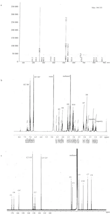
respectively (Fig 4). ESI-MS spectra were obtained on LCT Premier-XE waters. ESI-MS spectra showed that this active compound has molecular weight of 260.0 g mol . Data base from LCT Premier-XE Waters Program showed that this molecule had 14 carbon, 16 hydrogen, 2 nitrogen, and 3 oxygen. The most possible of molecular formula was C H N O The position each carbon, nitrogen, oxygen, and hydrogen atoms were confirmed by HNMR, C NMR, and FTIR. This chemical characteristics were indicated by ESI-MS at
261 (M+H) (Fig 1A).
High-resolution H NMR spectrum were obtained on a Bruker AV-500 (500 MHz) with tetramethylsilane (TMS) as internal standard in CDCL and give
1 13
Diameter of clear zone (mm)
Staphylococcus aureus
Extract of supernatant 10.39 24.43 9.64
Control (rifampicin 500 ppm) 21.27 44.57 10.08
Escherichia coli
ATCC 25922
-9.55
10.12
Table 1 Antibacterial activity of biomass and supernatant extract fromStreptomycessp.
Diameter of disc paper: 6 mm
Table 2 Minimum inhibitory concentration of active purified compound
Minimum inhibitory concentration µg mL-1 Sample
Active purified compound 27.0 80.2 73.7 68.7
Tetracycline (positive control) 64.0 256.0 128.0 12.5
Streptomycin (positive control) 50.0 130.0 120.0 61.0
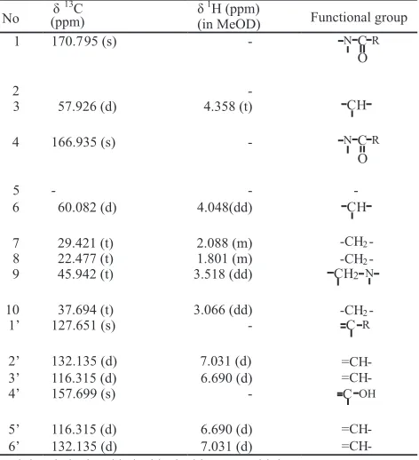
Table 3 Spectrum data of H NMR and C NMR of active compound
produced by sp.
1 13
Streptomyces
No δ
13
C δ1H (ppm)
(in MeOD) Functional group
1 170.795 (s)
-2
-3 57.926 (d) 4.358 (t)
4 166.935 (s)
-5 -
-6 60.082 (d) 4.048(dd)
7 29.421 (t) 2.088 (m)
t: triplet; d, duplet; dd, double doublet; m: multiplet
-a
b
c
Fig 1 Spectrum of bioactive compound produced byStreptomycessp.: a, ESI-MS m/z 261 (M+H) ; b, H NMR; c, C NMR.+ 1 13 350 000
300 000
250 000
200 000
150 000
100 000
50 000
100
0 200 300 400 500 600 700 800802.8 825.0 882.2900
670.4
679.0
583.0
541.2
544.2
543.2
521.2
Max: 346 333
523.2
522.2
410.0
374.0
303.0
284.0
262.0
228.4
191.2
methanol water
H3’ H5’
H2’ H6’
H8
H7 H10 H9
H6 H3
impurity impurity
(ppm) 0.5 1.0 1.5
2.5 2.0
3.5 3.0
4.5 4.0
5.5 5.0
6.5 6.0
7.5 7.0
8.0
0.21 0.21 2.09 0.46 2.55 0.62 1.00 0.24 1.04 1.38 1.47 1.27 1.00 0.24 0.27 1.46 2.10 0.64 1.16 0.41 0.72
C2’ C6’ C3’ C5’
methanol
C8 C7 C9
C10 C6
C3
C1’ C4’
C4 C1
170 160 150 140 130 120 110 100
(m/z)
Fig 3 Spectrum of DEPT C NMR active compound produced by13 sp. Streptomyces following data: δ : 4.358 (t, 1H), 4.048(1H, dd), 2.088
(2H, m), 1.801 (2H, m), 3.518 (2H, dd), 3.066 (2H, dd),
7.031 (2H, d), 6.690 (2H, d), and C spectrum :
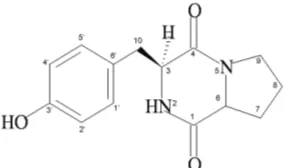
170.795 (s), 57.926 (d), 166.935 (s), 60.082 (d), 29.421 (t), 22.477 (t), 45.942 (t), 37.694 (t), 127.651 (s), 132.135 (d), 116.315 (d), 157.699 (s). The impurities of active compound was also showed at H NMR δ 1.7 -δ 0.9 (Fig 1B). Based on information of ESI-MS, H NMR, and C NMR (Fig 3) spectrums the molecular structure of active compound can be predicted as Fig 2. Spectrum data of H NMR and C NMR were presented on Table 3. Structure elucidation of active compound was also conducted using DEPT C NMR (Fig 3).
H
H H
13
1
1 13
1 13
13
Fig 2 Molecular structure prediction of active compound produced by sp.
Streptomyces
13
13
Two singlet carbons representing a ketone group
were evident in the C spectrum at 1)
and er
analysis of the C spectrum revealed two other non substituted carbons [ 127.651 (C1'), 157.699 (C4')], six methine carbons [ 57.926 (C3), 60.082 (C6), 132.135 (C2'), 116.315 (C3'), 116.215 (C5'), 132.135 (C6')], and four methylene carbons [ 29.42 (C7), 22.477 (C8), 45.942 (C9), 37.694 (C10)]. DEPT 45° spectrum on Fig 3 showed that there were 3 non-substituted carbon [ 127.651 (C1'), 157.699 (C4'), and 166.935 (C4)]. DEPT 135° and 90° showed that there were six methine carbons [ 57.926 (C3), 60.082 (C6), 132.135 (C2'), 116.315 (C3'), 116.215 (C5'), 132.135 (C6')] and four methylene carbons [ 29.42 (C7), 22.477 (C8), 45.942 (C9)]. Carbons at position 3' and 5' appeared more upfield than C2' and C6'. This was due to the shielding effect of the hydroxyl group at C4' toward its ortho coupled carbon (C3' and C5'). A similar phenomenon occurred on C1' (para coupled with C4') which shifted more upfield than C2' and C6'.
δ 170.795 (s) (C δ 166.935 (s) (C4) (Table 3, Fig 1C). Furth
δ δ
δ
δ
δ
δ
-1 -1
-1 -1
-1 -1 -1
-1 -1
-1
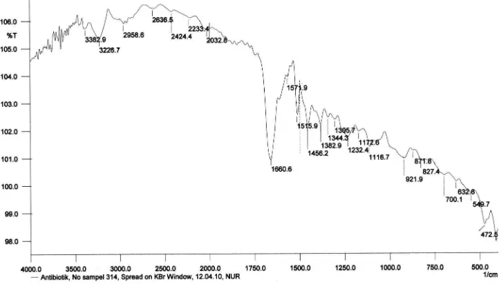
The infra red spectrum in a KBr pellet showed characteristic bands at 3383 cm (N-H), 3227 cm (O-H), 2959 cm (saturated C-(O-H), 1660 cm (C=O), 1515 cm (benzene ring), 1456 cm (methine), 1344 cm (methylene), 1232 cm (phenol), 1116 cm (C-O), 827 cm ( -disubstituted benzene ring) (Fig 4).p
DISCUSSION
The active compounds inhibited both Gram-positive and Gram-negative bacteria. According to Rhee (2004), most of antibiotic peptides especially cyclic dipeptide has antimicrobial properties with broad spectrum. Several cyclo peptides have
broad-DEPT 90
“Sample 4”3 1 /opt/topspin Mercian
[rel]
1.5
1.0
0.5
-0.0
160 140 120 100 80 60 40 (ppm)
C1 C4 C4’
C2 C6’
DEPT 135
DEPT 45
CH
CH
C3’
C5’
CH CH
CH CH
CH CH
C6 C3
C9
C10 C7 C8
“Sample 4”3 1 /opt/topspin Mercian
“Sample 4”4 1 /opt/topspin Mercian
“Sample 4”5 1 /opt/topspin Mercian
C1
Fig 4 Infra red spectrum of active compound produced byStreptomycessp.
spectrum activity and also able to act as an anti-viral and anti cancer. The same case were also presented by
(Lu and Chen 2010; Lee 2011). In addition to
having antimicrobial activity with a broad spectrum, our compound also had lower MIC compared with tetracycline suggesting higher activity than tetracyclin and streptomycin. The same result is shown by Rhee (2004).
Activity of cyclo(tyrosyl-prolyl) on the is lower compared with tetracycline. It is
suspected that the has a resistance to
cyclo(tyrosyl-prolyl). According to Macfarlane
(2000) and Reig . (2009), has a
resistance to type of peptide and aminoglycoside antibiotics. Cyclo(tyrosyl-prolyl) was peptide antibiotic and streptomycin was aminoglycoside
antibiotic. has highly adaptable nature,
including the ability to develop resistance. It can grow on a wide variety of substrates and alter its properties in response to changes in the environment (Lambert 2002). Thus, lower inhibition of cyclo(tyrosyl-prolyl) and streptomycin than that of tetracycline on
might be caused by resistance.
This active compound was included in group cyclo dipeptide, namely was cyclo(tyrosyl-prolyl). This active compound has the same profile such as H NMR, C NMR, Infra red spectrum, and molecular weight with cyclo(tyrosyl-prolyl) that found previously but
from different origin (Stierle 1988; Guo .
2007). Previously, this compound was produced by a host-specific phytotoxin for spotted knapweed (Stierle . 1988),
and produced by GcM5-1A isolated
from the pine wood nematode (PWN), and from
(Guo . 2007).
et al.
P. aeruginosa
P. aeruginosa
et al.
et al P. aeruginosa
P. aeruginosa
P. aeruginosa
et al. et al
Alternaria alternate and can be used as et al P. fluorescens
Bursaphelenchus xylophilus et al
1 13
ACKNOWLEDGMENTS
REFERENCES
1 13
13
We thank Hardaning Pranamuda, Anis Mahsunah and Evita Chrisnayani for their valuable advice and support our research, and Mercian Co. Japan for sample analyses using ESI-MS, H NMR, C NMR, and DEPT C NMR. First author was supported by Islamic Development Bank scholarship.
Andrews JM. 2001. Determination of minimum inhibitory concentration. J Antimicrob Chemother. 48(Suppl. 1):5-16. doi: 10.1093/jac/48. suppl_1.5.
Bonev B, James H, Judicael P. 2008. Principles of assessing bacterial susceptibility to antibiotics using the agar diffusion method. J Antimicrob Chemother. 61(6):12951301. doi: 10.1093/jac/dkn090.
Dubin AP, Mak, Dubin G, Rzychon M, Stec-Niemczyk J, Wladyka J, Maziarka K, Chmiel D. 2005. New generation of peptide antibiotics. Acta Biochim Pol. 52(3):633-638. doi:10.1093/jac/dkn090.
Guo Q, Daosen G, Zhao B, Xu J, Li R. 2007. Two cyclic dipeptides from Pseudomonas fluorescens GcM5-1A carried by the pine wood nematode and their toxicities to Japanese black pine suspension cells and seedlings in vitro. J Nematol. 39(3):243-247.
Kesting MR, Stoeckelhuber M, Holzle F, Mucke T, Neumann K, Woermann K, Jacobsen F, Steinstraesser L, Wolff KD, Loeffelbein DJ, Rohleder NH. 2010. Expression of antimicrobial peptides in cutaneous infections after skin surgery. Brit J Dermatol. 163(1):121-127. doi: 10.1111/j.1365-2133.2010.09781.x.
Lambert PA. 2002. Mechanisms of antibiotic resistance in J R Soc Med. 95(Suppl. 41):22-26.
Lee SB, Li B, Jin S, Daniel H. 2011. Expression and characterization of antimicrobial peptides retrocyclin-101 and protegrin to control viral and bacterial infections. Plant Biotechnol J. 9(1):100-115. doi: 10.1111/j.1467-7652.2010.00538.x.
Lu J and Chen Z. 2010. Isolation, characterization and anticancer activity of SK84, a novel glycine-rich antimicrobial peptide from . Peptides. 31(1):44-50. doi:10.1016/j.peptides. 2009.09.028.
Macfarlane EMA, Kwasnicka A, Hancock REW. 2000. Role of PhoP-PhoQ in resistance to antimicrobial cationic peptides and aminoglycosides. Microbiology. 146:2543-2554.
Pseudomonas aeruginosa.
Drosophila virilis
Manfredini F, Beani L, Taormina M, Vannini L. 2010. Parasitic infection protects wasp larvae against a bacterial challenge. Microb Infect. 12(10):727-735. doi:10.1016/j.micinf.2010.05.001.
Milne PJ, Oliver DW, Roos HM. 1992. Cyclodipeptides: Structure and conformation of cyclo(tyrosyl--prolyl) J Crystallog Spect Res. 22(6):643-649. doi: 10.1007/BF01160980.
Munekata M and Tamura G. 1981. Selective inhibition of SV40-transformed cell growth by diketopiperazines. Agric Biol Chem. 45: 2618-2628.
Nedialkova D and Mariana N. 2005. Screening the antimicrobial activity of actinomycetes strains isolated from Antartica. J Cult Collect. 4: 29-35.
Reig S, Hutha A, Kalbacherb H, Winfried, Kerna V. 2009. Resistance against antimicrobial peptides is independent of
.
Escherichia coli
AcrAB, MexAB and
NorA efflux pumps. Int J Antimicrob Agents 33(2):174-176. doi:10.1016/j.ijantimicag.2008.07.032.
Rhee KH. 2004. Cyclic dipeptides exhibit synergistic, broad spectrum antimicrobial effects and have anti-mutagenic properties. Int J Antimicrob Agent. 24(5):423-427. doi:10.1016/j.ijantimicag.2004. 05.005.
Sunaryanto R, Marwoto B, Irawadi TT, Mas'ud ZA, Hartoto L. 2010. Isolation and characterization of antimicrobial substance from marine sp. Microbiol Indones. 4(2):84-89. doi: 10.5454/mi.4.3.1.
Stierle A, Cardellina JH, Strobel GA. 1988. Maculosin, a host-specific phytotoxin for spotted knapweed from . Proc Nat Acad Sci. 85(21): 8008-8011.
Pseudomonas aeruginosa Staphylococcus aureus .
Streptomyces