F
t
I'ol 12, No I, Jan@ry, March2003
Paediatric .ataruct inplant swqerydtc:ne
2l
Paediatric cataract
implant
surgery outcome
Istiantoro
Abstrak
pef,etitian ini bettujuan nengewttasi hasil ddti bebqapa tek ik bedah kotardk rlan ihpldntdsi leasa intraokulel (UO) Pada aiak, .lt
Jaktrta E e Center. Jakafld, I4donesia. Penetitian ini netupakan studi retrospe+Jif pada 44 pendetita onok
(57
atd) ians nehjalaftibeddh katarak dah penosaagon LtO. Tiga na.an teknik lang dipakai ddalah: I - Ek:lrak'i katarak ekstrukapsulat dan pehuanga, UO ttengan kapsul posreriir tetap ihtak, yanE ditdkukan pada
2t
ata ftelontpokl).
2. Ekttruksi kotaruk eksttukopsular .laapnNangon LIO de;sM kapsuta/eksis pasEnor ecCC) dan oPtic ePture vang dilaklkti pada 21 nata (kelonpok 2) 3. Ekstrdksi
k'taruk ;k:mkapsutdt don penasangan LIO rldgan kopsutoteksis pasorior don
itreho
i antetior setla aptic coPt*e, tang dilafukaftpaia 2t hato (kelanpok 3). Seturuh pendentu nenjaldki oahasi tindak lanjur selana lebih tlai I tdhun. I{6i! penelitian nenunjukkon
bahwd keketuhan kapsut postenat eCO) terjddi pada 20 ndta Pada kelonPak
I
Senud dato henPravai akis risudl ranejenih podokelohpak 2, dan tetjadi pCO haara pada I nata pada kelonpok 3. Ka'itupulan : PCCC dengan dtau tdapa vitrelTodi antenot dan optic
capture adalah netoda ydnge:fettifuntukn cegah
ti
bulnta PcO Pana bayi atau atuk analL (Med.I Iddoaes 2003i 12:21'6)Abstract
This stud! ewt@ted the suryical attcohe ofvonous sttgi@l technique in Paediatrit .atdract inPld t srryery' at Jakana E e Centq, Jakarta. Indonesia Ihis
u6
a retraspecttue study of 57 eyes in 44 children *ho ha.l Prinary @taract inPlants s$gery' mrcesutgicol techniques tsed were : 1. btdcdpsular .otatuct dtroctian with intraocular lens ihplortation ||nh irtact poseiar capstle which ||6 peiomed an 2l eyes (group
t,
2. E tacapsrlat catqnct dtrcction wnh intraacalor le6 idPlantatioi ond Post riotcapsutathdis (PCCC) ahd aptic cdpturc which flas perJotued on 24 eles (8toup 2). 3. httucapsular cataruct %troctioh ||ith inio'
odlat te8 ituplantation, posznor .apsulorhsb and an@tior vitr@totur which v6 Peiomed an 24 eves (gtouP 3). All Patient: werc
lotlowed up hote than ohe tear. On resuLs shoeed that posteior capsule apaciry (PCO)
i6
deNelaped i4 20 ey6 with intact capsul5 ingroupt
AUeyeshadaclea/istal6isihsroup2.PCOddebpedonlyinaneeteinarcuP3Incorcl6ion, PCCC ud opti. caPtutewrh ot eithad antenor vitrectary arc elledive nethods ih PrcrentinA PCO in infmt a d childrer' (Metl J Ia.ton6 2003i 12: 2I'6)
Ketwlds: ponerbr coptule apaci|ication, Posknot caPsulothuis, oPtic capture, antdiot virectany
The
lotal
amountof
blindnessin
chjldren (bestcorected visual acuity less than 3/60) is 1.5 million in the world, and it is estimated that there will be 500
thousands
new
blindness.lTh€
incidenceof
blindness
in
childrenis
l5110,000in
developing countries, and the underlying cause of blindness arcmalnutrition, and infections
which
causea
highmortality Iate. Furthermore there is lack ofsupport
of
health services in blind children.
The visual outcone
of
r.rlaract rmplJnl surgery isexcellent
in
adults,but
in
the
past, implanting intraocDlar lens (IOL) in children has morc probtemsDeparhent of OpthalnolasJ, Focul, aI ]ttedicine, Unive$rt)
of blloDesid, JaLa a. Ihdonesia
with iis complications.'?'r However, the development
of
viscoelastic agents,IOL,
and the techniquesof
rmplanr in cataract.ur8ery in children have changed and \howed dramaric results.ot The high rncidmceof posterior capsule opacification (PCO) a.nd fibrosis led to rhe concept of primary posterior capsulorhexis (PCCC) and optic capture, followed by hsertion
of
optic IOL, wilh or without anterior vitrectomy at thetime of catlmct surgery to prevent PCO. Therefore,
there are three d€velopments
of
cataract implantsurgery
in
children:
1.
Extracapsular capsularcatamct extraction and
IOL
implantation (ECCEIOL) with intact capsule. 2. ECCE IOL, PCCC and
posterior optic capture.
l.
ECCE IOL, PCCC andposterior optic capture, and anterior vitrectomy. The purposc oflhis study is to evaluate lhe results
of
cataracl implant surgery
in
childrenwith
the
3METHODS
.This is a retrospective study of 57 eyes in 44 children
who got
pimary
catamct implants at Jakarta EyeCenter
ftom
1993
ro
1999.
Fifry-five
werecongenital, and
two
eyes were haumatic cataract. Total lens opacity wasfou
in 25 eyes, and the rest showed partial opacity. Themean
age at timeof
sugery was 5.7 yea$, that ranged lrom 4 months to
14 years. Th€ distribution
of
age group and thel
surgical techniques can be seen in Table L
Surgical technique
All
operations were perfiDrmed by a single surgeon. General anaesthesia was used in all cases. The sclemlor comeal tunnel incision was made with 2_5 or 3.0
mrn keratome. The aqueous humor was exchanged
with high viscosity sodium hyaluronate through the
tumel
incision. The anterior capsule was openedwith
a
technique called continuous curve linearcapsulotomy
(CCC)
using
a
bend
23
needle.Multilamellar hydrodisseclion were performed. Lens material was removrd using phaco hand piece or irrigation aspiration hand piece
of
phaco machinewith
aspirationmode.
The
posterior
capsule managemeni ardIOL
insertion were managed in 3different techniques.
1.
Afier
nuclear material removal, the posteriorcapsule
was
leavedintact,
and the
ovoidpolymethyl methacrylate
(PMMA)
IOL
was inserted in the bag. Vitrectomy was perfo.medif
there was posteriot capsule rupture with vitreouslost, and the IOL inserted to the ciliary sulcus.
Twenty-one
eyes were
performedin
this fashioned2.
After nuclear material r€moval, the visco-elasticmaterials
were
injected
into
the
anterior chamber, then post€rior capsule was opened atrhe cenlral area to orake a tsiangular flap using a
27-bend needle. The posterior capsular flap was grasped with Uhata forceps, and a 4.5 !o 5.5 mm diameter continuous curve linear capsulectomy (PCCC) was performed. Additional viscoelastic materials are injected as necessary through the posterior capsular tear to push the vikeous face
away as tear enlarging.
The
injectionof
theviscoelastic materials should be very carefully
done;
it
should not push rhe capsular tear, asit
may cause difliculties in grasping the diangrlartear. Too much viscoelastic material may extend
Metl J Indonq
the tear to the equator (tear escape). The ovoid
PMMA IOL
or
5.5nm
optic acrylic IOL was inserted in the bag, and then was pushed throughPCCC opening with
a
Sinskey hook (posterioroptic
capture).If
the
inregrityof
posterior capsule is lost (peripheral tear),it
is impossibleto do the posterior optic capture. In this study twelve eyes were pe.formed in this technique.
3.
Afier nuclear material removal and PCCC was cornpleted, an anterior vitectomy was performed through the incision and the ioigation cal)llula byparacenthesis. The ovoid PMMA IOL or 5.5 mm
optic acrylic
IOL
was inserted in the bag andthen pushed through PCCC opening
with
aSinskey hook (optic capture). Twenty-four cyes were performed in this technique (Tabte 1).
Table 1. The sugical techniques in age groups (yqr)
3
6
6-12
More 12Group I ECCE+IOL
Crcup 2 ECCE+TOL+
ECCE+IOL+
ll
t2
t5
l8 t2 57
ECCE = ExtFcapsular caMcr extmction, IOL = int6 ocutd tchs.
vr dr - "1rrio a.Fror.
ln all cases the residual visco-elastic substaDce was
removed
using
automaticirrigation
aspiration.During visco-elastic removal the IOL was h;ld wilh
the Sinskey hook to prevent optic our ofthe Dosrerior
optic capture because the positive vitreous pressure.
Tbe wound
\
as closed wirh inrenupted l0-0 nylonsutures and 0.01
%
carbachol and balance saltsolution (BSS R) was injected
into
the
anterio.chamber. The anterior chamber was check€d
if
there was viFeous strand and vitr€ctomy was performedif
necessary. At the end of surgeqT the eye was alressedwith subconjunctival injection of dexamethasone and
anlibiotic,
ar
eye patch and shietd. Dexameihasoneand antibiotic eyc drops was given four times a dqy. Postopemtive examination performed at day I , weeks
l,
2
and4,
tlen
every3
months_ Refraction. slitYal I 2, No I , JonuarJ - March 2n3
lamp, and indirect ophthalmoscope were performed
upon every visit. Special attention was paid
to
thefomation of posterior capsule opacification (PCO)
or flbrcsis. Second procedure was performed, either
Nd YAG Lasei capsulotomy or capsulectomy with
anterior vitrectomy
if
necessary, when evtdenceof
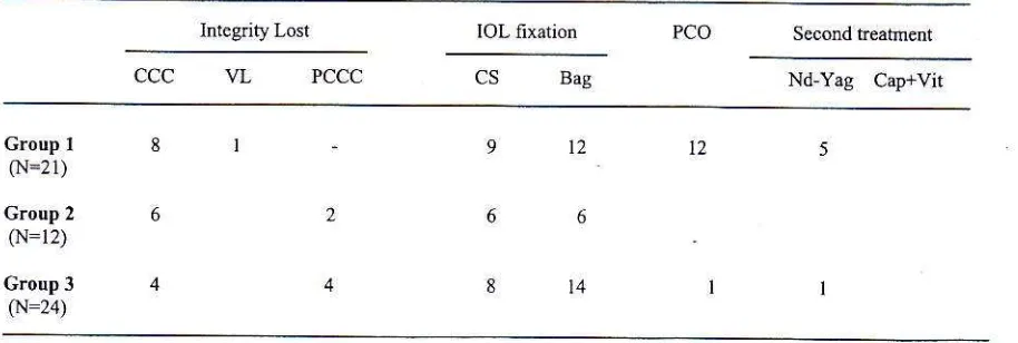
PCO or fibrosis reduce best corrected visual acuitv.RESULTS
The srrgical complications:
in-goup
I
(ECCE + IOL), integrity lost of CCC was found in 8 eyes, andposterior capsule rupture and vitreous lost
in
I
eye.Posterior capsule opacity was found
in
20
eyes,except
one eye that had
complication duringprocedure i.e. posterior capsule rupture and vitreous lost- The secondary treatment of Nd-Yag Laser was
performed
in
13 eyes, and posterior capsulectomyand anterior vitrectomy were performed in 5 eyes.
ln-group
2
(ECCE+IOL+PCCCTOPTIC CAPTURE),integrity lost
of
PCCC were folmdin 2
eyes, and anterior CCC in 6 eyes- Intraocular lens were implan&dat
ciliary sulcusof
2
eyes without optic caphne.Posterior capsule opacigT was not found in this group.
In-$oup
3
(ECCE+PCCC+Anterior Vitrectomy + IOL Optic Capture), fibrosis was found in 1 eye of aTable 2. Surgicat complications, post€.ior capsule opacity and secondary treaunenr
Paediotic catoract ituplaat surgery
olr:otue
234
monthsold
child. The
secondary tseatulant (capsul€ctomy and anterior vitrectomy) *"s performedThe surgical complications: integrity lost
of
CCCwas fomd in 4 eye, and PCCC in 4 eyes. The IOL
insertion at ciliary sulcus was done
in
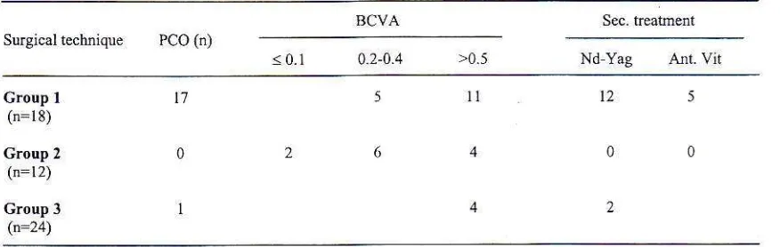
8 eyes, and without optic capture in 4 eyes (Table 2).Post opemtive visual acuity rang€d from
0-l
to 0.7. The visual outcomes of the patients in age group lesslhan
I
years were drfficult to measure. However, in15 eyes
(85
%o)ftom
group2
&
3,
visual axis remained clear al lasl visil. In age goupI
6 years.beslcorected
visual acuity (BCVA)
0.1
was achieved in 2 eyes, BCVA between 0.2 to 0.4 in 9 eyes, and BCVA more than 0.5in 9
eyes.In
agegroup more than 12 years, BCVA 0.2
to
0.4 was achieved in 2 eyes, and BCVA more than 0.5 in 2€yes.
In
group
l,
posterior capsule opacity hadsignificantly rcduced the visual outcome. Thercfor€, secondaq/ treatrnents of Nd-Yag Laser were done in
12 eyes, and capsulectomy with anterior vitrectomy
in
5 eyes. One eye had clear visual axis in -whichanterior vrtrecrom) had
lo
be
done as primarytreatment because posterior ruptue with vitreous lost
occmed (table 3). ln this series we found 3 patients
$bo developed myopi( shift dunng follo$ up
lnlegnty Lost IOL fixation PCO
ccc
PCCC B"g Nd-Yag CaFrVitc.oup 1
0\r=21)
Croup 2
cN=12) Croup 3
(N=24i
l2
l4
t2
CCC =
utdor
capsulorhexjs, VL = viteous Lost, PCCC = posierior capsulorhens, CS = cilliary $lcus, [image:3.596.44.501.504.658.2]24
Table 3. Coftiation ofsuAicll technique, PCO dd visual outcone
BCVA Pco
(i)
< 0.1 0.2-0.4
Group r
(n=18) Group 2
(n=12) Group 3
(n:24)
t7
ll
12PCO
:
posterior capsule opacity, BCVA:
besi conected visual dcuity, Ant-Vit = dtcrior vitrectomy,sec. (realmenr = secondary rrealment
DISCUSSION
Catamct
in
childrenis still
di{ficultto
manage-It
remains the cause
of
sig4ificant partial blindness.Cataract
ir
childrenis
differ€ntfrom
adults. A child's eye is not a miniature from an adult's.eye,and time
of
surgery and aphakic conection always have effects on visual development. Implanting anintraocular lens into an adult eye as an aphatic
corection
is
usually
extremely successful, Atpresent, both surgical technique to remove cataract
and the IOL
design
have
improved. Phacoemulsification has become the state of art andwidely used
in
many countries. The materials anddesign of
IoL
with high degree of biocompatibilify,soft
nexibiliry
allows smaller incisionand
lessinvasive surg€ry. However, such improvements are
not enough to be applicable for cataract in children.
Cataract surgery
in
chilalren has morc problems. Irnplanting IOL in newbom eye has many difTicultiesas
follows:
preope.ative, intraopemtive andpostoperative care. Preoperative difficulties: risk
of
general anaesthesia, associated conditions, late diagnosis and
lol-
power calculation. Intmoperativedifficulties: small eye,
low
scleml and
comealdgidity, solid vitreous tends to yield positive vitreous pressure, thin elastic lens capsule causes difliculty in capsulorhexis. Postoperative difficulties: exaggerated
uveal
responseto
inflammation, formationof
secondary
posterior opacification
or
fibrosis, secondary glaucoma, changein
axial
length with myopic shift and challeqc in amblyopic treaiment.Implanting
IOL into
a
child's
eye
for
aphakiqconection was pioneered by Binkhorst and Gobin,'
and Hiles.s
High
incidenceof
posterior capsuleopacity (PCO)
or
fibrosis was reported followingIOL
implantationin
the capsular bag with htact posterior capsule,9'ltPost
operative exaggerdteduveal responses/inflarnmations caused anterior and
ponerjor
capsule opaciryor
llbrosr..
posterior s)'nechiae, IOL optic capture, capsule contracture andIoL
decentmtion.In
this study 17 eyes($oup
l)
with intact posterior capsule developed PCO. Twelve eyes required Nd-Yag laser capsulotomies and five
eyes
required
capsulectomiesand
anterior vitrectomies. All capsulectomy and anterior vitectomy were performed in patients under6
years old with thick posterior capsule fibrosis. In this groupl,
only one e)e have a cl€ar visual axls aq a resuh ofprimary anterior vitrectomy dueto
posterior rupture andvitreous lost dudng surgery (Table
l).
Several studies reported
to
prevent PCO formationb1
openingthe
posrerior capsuleas
a
primary procedure. Gimbel 5 opened th€ postenor capsule by continuous cDrve linear capsulectomy (PCCC) andoptic capture. Posteior optic capture is a challenging procedure. Pedorming PCCC
is
difficult.It
needssurgeon
skill
and
experienceto
anticipate thediameter oi PCCC to ensure permanent optic
capture-The diameter of PCCC should be
I
rnm smaller thanoptic diameter
of
theIOL.
Zetterstromr2 rcportedclear visual
axis
by
implantingheparin-surface-modified IOLS after performing CCC of the anterior
and posterior capsules. Seveml studiese'rr-r5 also
reported prevention
of
PCOby
pedormirg bodr PCCC and anlcnor \ itrecromy. rcmovtng lhe t ttreousscaffold
for
lens epithelial migration- The capsular [image:4.596.44.482.111.250.2]ttol 12. No t..Ja
tury
March 2403diminish cenhal lens epithelial rnigration
or
cell movemenr o\er rhe lens uplrc. whichi.
not a suilablesubjn.rr(
lor
len. cell survi\al''
Besidc ro prerenr secondary PCO,oplic
capture enhancesIOL
andcentration. Consequently, deccntiircd PCCC could
cause
IOL
decentration. Vasavada" repoted that anterior vitrectomy along with PCCC is desirable asa
primary procedurein
children youngcr than 5years- Our experience showed that PCCC with optic capiure and anterior vilrectomy in younger children could prevent PCO
/
fibrosis. Only one eye of thc 4monrh. old rnfdnr dc\cloped po.rerior fibro{s orer
the antcrior ofthe vitreous body (Table 2, 3). It may
b€
causedby the
procedurcthal only
cut
ihc viscoelastic substance during anterior vitrectony. During the second surgical procedure the opticof
IOL was not captured because thc diamcter ofPCCCwas too small. Surgeon should keep in mind lhat the anterior vitrectomy sbould be performed intensively
in
the
entire anterior surfaceof
vitreous body, togethcr with as large as possible PCCC.The visual oulcome
of
paediatric calaract surgery created challenging problems.A
childwith
densecongenital cataract has to fight both amblyopia and speclacles correction
to
adjust eyegrowth
Post operative care inchdes repeared spectacle conectionsand iniensive care
to
promote visual development.Tl'e<e need
a
ream
$ork
ol
paedralricjan.ophthalmologist, optometrist
etc.
Intensive pos!operative care is necessary
to
achieve good visualacuity
in
the
future-Intra
occularlens
power calculation should anticipate the ocular growth. The eye of an adult is 40-5070 larger than that of an theinlanr. Ihe a)\ial lengrh
ol
a newbom child selc
rsl?
mm, while in adult's eyeit
is 23.5-mm.r7 Mostocular growth occurs during
the first
2
years,particularly
in
the first year. Myopjc shift due toa\ial
growth
during
childhoodhas
led
someinve511g61et.
Io
implanL undercorrecled lOL.rr o ln this study, best corrected visual acuity (BCVA) > 0.5 was achieved in2l
eyes (36.8 %); BCVA > 0-2 < 0.4in I I eyes (19.3 %) and BCVA s 0.1
h
2 eyes (1.5 %).In the rest I0 infant eyes (17.5 %), the visual acuity was not able to be measured. the same with the other
14 eyes (22.4
%)
that were amblyopia. However, thcse eyes had clear visual axis (surgical group 2&
3)
and
only
one
cye
(group3)
need anteriorr
itrectom)
a.
.econdary lieJlnrenr.
Inlelsi\r
postoperativ€ care from paediatric medical team was needed ro de\elop visual acurl) ro8elher
$ilh
rhecooperation rnd support from rheir parents
Paediotrir catdrdct inplant luryery
outcone
25In
conclusion, PCCCwirh optic
capture could prevenl PCO. Vitrectomy anterior along with PCCCand
optic
capiureis
more desimblein
youngerchildren,
and
anterior
vitrectomy should
be performed intensively (entire anterior) with as large as possible PCCC. Cataract implanl surgery should be done by a skilled experienccd surgeon to ensuregood visual outcome in the firture
RXI.'ERXNCES
l
wodd Heath Organization Prevendonof
Childhood Blindness. Genevar WHOi 1992.2
Menezo JL, Taboada JF, Feder E. Complications of int.aocul lenses in Child.cr. Trans Ophthalmol Soc UK1985: 104:5,16 52
3.
Hiles DA, Watson BA. Conplicarions ofimplant suryeryin childrcn. An lntd'Ocular implanl Soc J 1979; 5: 24,
32.
4.
Zette6t.omC,
KugelberSU,
LundgrcnB,
Syren-Nordqvisr S. Afler cahract fomatio. in newbom Ebbits ihplanled with intraocularlenses. J Cataract Reiiact Su.8
1996i 22r 85-8.
5.
Gimbel
HV.
Posierior continuous cNelinearcapsulorhexis and optic capture of intraoculd lens to prevenr secondary opacificarion
in
pediatric cataFct.urceD l adrddc. Rerrs,r surS taa_: 21: 052-s
6.
Vasavada A. Desai J. Prima.y Dosterior caDsulorhexiswilh snd wilhour anterior vihectomy
in
congerital calaEcts. . J Cataract Rcfract Srig 1997;21: 5-51.L
BinkioFt CD, Gobin MH. Treatment of congenital andjuvenile cata.act with inra@uiar lens (pseudophakoi) Br
J Ophthalnol 1970; 54:759-65.
8.
Hiles DA. Intraocllar leDs implantaiion in childr€n withmonocular cataractj 1974 1983. Ophthalmology 1984:
9l:1211-1.
9.
Buckley EG, Klombers LA, Seaber JH. Scalise-cordyA, Minster R. Management of of poste.ior capsnle duringpediatric
inlraoolar
lens
inplanration.Am
JOphihalmol 1993; ll5:722 8.
10. Hiles DA. Hered RW. Modm tens implmts in chil&€n with new age limitation. J CataEct Re&act Sug 1987;
ll:
493-7.I
L
Oliver M, Milstein A. Pollack A. Posrerior cbamber lensimplmtation in infants and juveniles_ Eui J Implant Refiact Sug 1990;2: 309-14.
12. Zettesftom C, Kugelberg U, Osc$son C.
Calarur
gery,n childrD wiLh r"pjulorhe\{ of brerio'
od
po,rer;",capsDles ud hepein-surface modified int
ieulr
lenss. J Catamci Refiact Sug 1994;20:599 601.ll.
Mackool RJ, Cbhatiawala H. Pcdiarric @raFct surgery and iDtraoculd lens implmt tion: a new technique forprevedling
or
exisling postop@nve secondarynenbtues. J Cat@ct Refinct Surg l99l i 17:62 6.
l4
Keecn Rv. lolg',F 4c. scon $i.
Compl'cadon;"n.'
sugery for congeniral
ad
inf&lile @tancl. Am J15. Koch
DD
Kohnen T- Retrospective @mpsison oftechniques to prcved seondr,ry csraBct forotion after post€rior chmbe! intnoculsr lqs inpluratior in infets dd childfen. J CataEcr Refiact SW 1997;23:567 -3.
Med J In.lones
Dahd B, Welsh NH, Salmelson BD. Posterior chmbd implmt
in
unilaleml congenit4led
developmolalcateets. Eur J inplul Refilct Surg 1990; 2:295-302. Gordon RA, Donzis PB. Refiactive developn.nt of tle
lrlmn
eye. Achof
Ophthalm@l 1985; l0l:785-9.t6.