Serum vascular endothelial growth factor as a predictor of clinical
outcomes in anterior circulation ischemic stroke
Keywords:angiogenesis, clinical outcomes, ischemic stroke, VEGF
pISSN: 0853-1773 • eISSN: 2252-8083 • http://dx.doi.org/10.13181/mji.v24i2.1196 • Med J Indones. 2015;24:109-14 • Received 31 Jan 2015 • Accepted 19 Jun 2015
Correspondence author: Vivien Puspitasari, [email protected]
Copyright @ 2015 Authors. This is an open access article distributed under the terms of the Creative Commons Attribution-NonCommercial-ShareAlike 4.0 International License (http://creativecommons.org/licenses/by-nc-sa/4.0/), which permits unrestricted non-commercial use, distribution, and reproduction in any medium, provided the original author and source are properly cited.
Vivien Puspitasari,1 Syarifuddin Wahid,2 Amiruddin Aliah,3 Budhianto Suhadi,4 Cahyono Kaelan,3
Suryani As’ad,5 Ilhamjaya Patellongi,6 Jan S. Purba,7 Eka Wahjoepramono8 1 Department of Neurology, Faculty of Medicine, Pelita Harapan University, Jakarta, Indonesia
2 Department of Anatomical Pathology, Faculty of Medicine, Hasanuddin University, Makasar, Indonesia
3 Department of Neurology, Faculty of Medicine, Hasanuddin University, Makasar, Indonesia
4 Department of Clinical Pathology, Faculty of Medicine, Pelita Harapan University, Jakarta, Indonesia
5 Department of Nutrition, Faculty of Medicine, Hasanuddin University, Makasar, Indonesia
6 Department of Physiology, Faculty of Medicine, Hasanuddin University, Makasar, Indonesia
7 Department of Neurology, Faculty of Medicine, Universitas Indonesia, Jakarta, Indonesia 8 Department of Neurosurgery, Faculty of Medicine, Pelita Harapan University, Jakarta, Indonesia
C l i n i c a l Re s e a rc h
ABSTRAK
Latar belakang: Respons inflamasi pada fase akut strok
iskemik akan memicu proses neuroplastisitas dan menentukan luaran klinis. Angiogenesis dan neurogenesis diinduksi oleh ekspresi vascular endothelial growth factor (VEGF) pada fase akut strok. Tujuan penelitian ini adalah untuk mengetahui hubungan antara kadar VEGF serum pada fase akut strok dengan luaran klinis.
Metode: Penelitian ini menggunakan desain kohort longitudinal terhadap 64 pasien strok iskemik sistem anterior dan baru pertama kali mengalami stroke, dibuktikan dengan difussion-weighted magnetic resonance imaging (DWI) kepala. Pemeriksaan kadar VEGF serum dilakukan pada 72 jam dan 7 hari pasca-strok dan dinilai luaran klinis pada 30 hari pasca-strok menggunakan skor National Institutes of Health Stroke Scales (NIHSS). Analisis data menggunakan uji T, uji Spearman, dan regresi logistik.
Hasil: Kadar VEGF jam ke-72 dan hari ke-7 yaitu 5,84 ± 0,736 ng/mL and 5,797 ± 0,96 ng/mL (p > 0,05). Kadar VEGF 72 jam pasca-strok yang tinggi dapat digunakan untuk memprediksi derajat klinis yang buruk pada 30 hari pasca-strok (OR = 6,5; 95% CI = 1,15-36,61; p = 0,034). Subjek yang mengalami peningkatan kadar VEGF pada 7 hari dibandingkan 72 jam pasca-strok menunjukkan derajat klinis hari ke-30 yang cenderung lebih ringan. (Skor NIHSS = 1,33 ± 1,22 vs 3 ± 3,78; p = 0,232).
Kesimpulan: Kadar VEGF pada fase akut strok iskemik menggambarkan derajat kerusakan otak, dinamika kenaikan kadar VEGF pasca-strok berkaitan dengan perbaikan luaran klinis.
ABSTRACT
Background: Inflammatory response in the acute phase of
ischemic stroke will trigger the process of neuroplasticity and determine the clinical outcomes. Angiogenesis and neurogenesis are induced by expression of vascular endothelial growth factor (VEGF) in the acute phase of stroke. The purpose of this study was to determine the association between VEGF serum level in acute phase of stroke with the clinical outcomes.
Methods: This longitudinal cohort study was conducted on
64 patients suffering from first-attack of anterior circulation
blockage as evidenced by cephalic diffusion-weighted magnetic resonance imaging (DWI). VEGF serum level was measured at 72 hours and 7 days after stroke and the clinical outcomes were assessed on day 30 post-stroke using the National Institutes of Health Stroke Scale (NIHSS).
Results: VEGF level at hour-72 and on day-7 were 5.84 ± 0.736 ng/mL and 5.797 ± 0.96 ng/mL, respectively (p > 0.05). High VEGF levels at hour-72 can be used to predict poor clinical outcome 30 days after stroke (OR = 6.5; 95% CI = 1.15-36.61; p = 0.034). Subjects who have increasing levels of VEGF on day-7 compared to hour-72 tend to have better clinical outcomes on day-30. (NIHSS score = 1.33 ± 1.22 vs 3 ± 3.78; p = 0.232).
Stroke is the third leading cause of death in developing countries among population above 60 years old, after heart disease and cancer. Approximately, more than 80% cases of stroke
is ischemic and the rest is hemorrhagic.1,2 In
Indonesia, stroke is the first leading cause of
death.3 It is not just an elderly disease, but also
affects young people. Stroke causes both physical and psychological burden for patients, families, and communities. The impact of this medical issue economically results in the decline of productivity and economic capacity of communities and
nations.1-4
Mechanism of brain injury after ischemic stroke is a complex process and is concomitant with inflammatory process that involves neuronal cells, glial cells, endothelial cells, extracellular
matrix, and peripheral leukocytes.5-7 Post-stroke
outcome is not solely determined by pro- and anti-inflammatory cytokines, but also by neurotrophic factors released in acute stroke as a result of neuronal injury. After cerebral ischemia, neuron plasticity is once again triggered on surviving cells to compensate the dead neurons and support the function of the damaged ones. The mechanisms underlying neuronal plasticity are dendrite reorganization, axonal sprouting and activation of endogenous pluripotent cells which are able to differentiate into neurons. Angiogenesis and arteriogenesis are the major processes of vascular plasticity. After an ischemic attack, angiogenesis is necessary to provide new capillary and increase blood flow to regenerate the suffering cells.8
Vascular endothelial growth factor (VEGF) is a vascular permeability factor, a major protein that induces angiogenesis in both normal and pathological conditions. VEGF binds to tyrosine kinase receptors, vascular endothelial growth factor receptor-1 (VEGFR-1) and VEGFR-2 / kinase insert domain receptor (KDR) / fetal liver kinase-1 (Flk-1), and mediates intracellular
signals for cell growth and cell survival.8 In the
brain, VEGF is produced primarily by neurons,
vascular cells, and astrocytes. Following
ischemic stroke, hypoxic condition induces VEGF expression followed by fibroblast growth factor (FGF), angiopoietins, transforming growth factor beta (TGFβ), platelet-derived growth factor
(PDGF) and tissue-plasminogen activator (tPA).9,10
Previous studies reported that the VEGF serum
level in acute phase of stroke (within 24 hours onset of symptoms) is associated with infarct volume and a better prognosis in three months. Plasma VEGF levels increase immediately after the onset of ischemic stroke in all types of strokes
up to 90 days.11
Although it seems to be constructive in long term, this response may contribute to deleterious edema formation during the acute phase of ischemic stroke. Several studies showed that VEGF contain an unfavorable effect towards outcome. High VEGF in acute ischemic stroke correlates with cerebral
micro-bleeding.12 Early post ischemic administration of
recombinant VEGF to ischemic rats significantly increased blood brain barrier leakage, hemorrhagic
transformation and ischemic lesions.13
This study aimed to explore the role of VEGF in the pathomechanism of acute ischemic stroke and its role in predicting clinical outcomes.
METHODS
This longitudinal cohort study was conducted from August to December 2014, in Siloam Hospital, Karawaci, West Java. Patients with acute onset (<72 hours) of anterior circulation ischemic stroke, were closely observed for one month after the initial stroke. Patients with history of previous stroke and thrombolytic therapy, decreased liver and kidney functions, congestive heart failure, acute myocardial infarction, and pneumonia were excluded from the study. Magnetic resonance imaging (MRI) was performed at admission to confirm the ischemic stroke and to assess cerebral
infarction volume. Formula ABC/2 (mm3) was
used to measure infarct volume (A:length;
B:width: C:number of slice).14
VEGF serum levels were measured with human
VEGF immunoassay Quantikine® (R&D Systems,
intensity of the color was measured at 450 nm wavelength.
Clinical outcomes were assessed at hour-72, day-7 and up to day-30 after stroke using National Institutes of Health Stroke Scales (NIHSS). The severity of stroke was evaluated with NIHSS and they were categorized as mild (0-3), moderate (4-15) and severe (16-42). Ethical clearance was obtained from Mochtar Riady Institute for Nanotechnology Ethics Committee Number 072/MRIN-EC/08/2014.
Data were analyzed with SPSS 20.0. T-test was used to compare NIHSS scores on day 30 in group with increased or decreased VEGF levels. Spearman correlation test was used to evaluate the correlation between VEGF levels with NIHSS score. Logistic regression analysis was used to predict 30 days outcome after stroke.
RESULTS
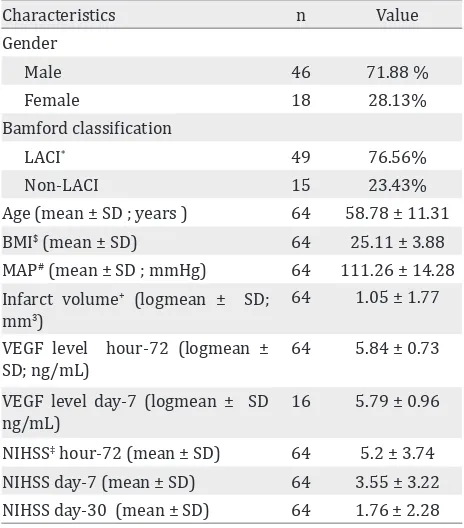
In total, 64 patients aged 37 - 89 year old (mean of 58.8 ± 11.3 years) met the inclusion criteria lacunar anterior circulation infarct (LACI). About 59.4 % of subjects had a moderate NIHSS score (4-15) on admission with an average score of 5.2 ± 3.7. On day 30, we found 89.5% of subjects had a mild NIHSS score (0-3). There were only 16 patients evaluated for VEGF serum level on day-7 after stroke(Table 1).
There was no significant correlation between VEGF level at hour-72 with NIHSS score on day-30 (r = 0.156; p = 0.217). VEGF levels on day-7
Table 1. Characteristic of patients, VEGF serum levels, and NIHSS score
* LACI: Lacunar anterior circulation infarct # MAP : Mean arterial pressure
$ BMI: Body mass index
+ Infarct volume measured using formula ABC/2 mm3
(A=length; B=width; C=number of slice) 14,20
‡ NIHSS : National Institute of Health Stroke Scale
n R p
Day-7 VEGF level vs NIHSS day-30
16 0.518 0.040
Table 2. Correlation between VEGF level and NIHSS score
day-7 can be categorized as “increased” and “decreased” (Figure 1). T-test was then used to see the difference in NIHSS score on day-7 and -30. In this category, the group with increasing VEGF serum level (n = 9) tended to have milder clinical severity than the group with decreased VEGF level (n = 7). NIHSS score in both groups were 1.33 ± 1.22 and 3.001 ± 3.78, respectively; p = 0.232) (Figure 2).
Figure 1. VEGF level at 72 hours compared to NIHSS score 30 days after stroke
Figure 2. NIHSS scores at day 30 after onset in group with increasing (n = 9) and decreasing (n = 7) VEGF level
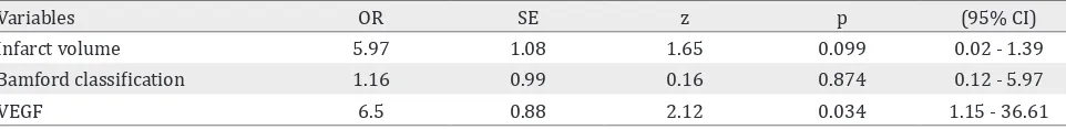
Variables OR SE z p (95% CI)
Infarct volume 5.97 1.08 1.65 0.099 0.02 - 1.39
Bamford classification 1.16 0.99 0.16 0.874 0.12 - 5.97
VEGF 6.5 0.88 2.12 0.034 1.15 - 36.61
Table 3. Logistic regression to predict 30 days outcome after stroke
and NIHSS score 30 days (r = 0.362; p = 0.005). There was also correlation between Bamford classification and NIHSS score 30 days after stroke. Multivariate analysis logistic regression showed that VEGF was the strongest clinical predictor on day 30 after stroke, compared to the infarct volume and Bamford classification with an OR = 6.5 and p = 0.034 (CI = 1.15-36.61) (Table 3).
DISCUSSION
VEGF showed a dramatic increase in serum of stroke patients with highest expression occurred on day 7, and it remained significantly elevated at 14 days after stroke, even after 3 months post-
stroke.11,15
Lee, et al16 reported a relationship between elevated
serum level of VEGF and infarct volume, and the highest level of VEGF was found in large vessel
disease (LVD) stroke.Moreover, he also found that
the high VEGF serum level in 24 hours post-stroke was related with more favorable prognosis in three months follow-up. This study seems to show different result, as the high VEGF serum taken in
acute phase of stroke was related with more severe prognosis (r = 0.264; p = 0.035). This could be
explained by result of Matsuo’s study.11 They found
that the VEGF serum level in acute ischemic stroke was higher than control, this elevation continued up to three months after onset. It is interesting that VEGF level varied in subtype stroke according to TOAST (Trial of Org 10172 in acute Stroke Treatment) classification of ischemic stroke type. There was a positive correlation between VEGF level and clinical presentation within three months after onset on cardioembolic infarct subtype and lacunar infarct. On the other hand, a negative correlation was found in atherothrombotic infarct subtype. In this study, VEGF serum levels taken within 72 hours post-stroke were positively correlated with early clinical presentation within 72 hours after onset (r = 0.264 and p = 0.035), and also have positive correlation with 30 days outcome (r = 0.15 and p = 0.217). Whereas VEGF serum level on day-7 after ischemic stroke had a positive correlation with 30 days outcome (r =
0.518 and p = 0.04). Referring to Matsuo study,11
After acute stroke, VEGF expression appears within six hours in response to hypoxia along
the ischemic area.17 In vivo study shows that
introducing intravenous Vascular Endothelial Growth Factor-A (VEGF-A) two days post-stroke would induce angiogenesis in penumbra and
help neuron recovery due to ischemic process.18
Intraventricular VEGF antibody had shown negative impact in increasing infarct volume in
post focal cerebral ischemia mice.19 Topical and
intramuscular VEGF had also shown that infarct volume and edema was significantly decreased in medial cerebral artery occluded mice. This revealed that VEGF has a neuroprotective effect.
This study revealed that subjects whose levels of VEGF on day-7 increased compared to levels at hour-72 led to the better NIHSS score at 30 days after stroke, and subjects with VEGF level on day-7 decreased compared to the level at hour-72 led to worse clinical outcomes (see Figure 2). Although this is not statistically significant, this phenomenon supports earlier statement that VEGF was believed to play a key role in angiogenesis and neurogenesis after stroke.
It could also be explained that VEGF serum level in acute phase reflected the brain response to hypoxia degree and neuronal damage, whereas VEGF dynamic change showed VEGF role as
neuroprotector through angiogenesis and
neurogenesis processes. We deduced that the clinical outcomes did not only rely on high VEGF serum level in early phase of stroke, but also on its dynamic in acute and after acute-phase of stroke.
We evaluated correlation between infarct volume and Bamford classification with clinical outcomes. Infarct volume measured at hour-72 post-stroke had significant correlation with clinical outcomes at day-30 (r = 0.382; p = 0.005). This is similar
to other previous studies.20 Bamford classification
also had correlation with clinical outcomes at day 30 after stroke (r = 0.243; p = 0.053). In this study we found that VEGF was the strongest clinical predictor on day-30, compared to the infarct volume and Bamford classification with an OR = 6.5 and p = 0.034 (95% CI = 1.15-36.61). (Table 3) improved clinical outcomes. Further research on neuroprotective role of VEGF in post-stroke recovery may open up opportunities in the management of post-ischemic stroke patients.
Acknowledgment
We would like to thank Anyeliria Sutanto and Nora Tu for collecting data over the study period, also to Veli Sungono and Pricilla Gunawan for statistic analysis. We thank Prodia Laboratory for the support in this study. This study received financial support from Pelita Harapan University.
Conflict of interest
The authors affirm no conflict of interest in this study.
REFERENCES
1. World Health Organization [Internet]. Global burden of stroke. [Cited 2014 Aug 10]. Available from: http:// www.who.int/cardiovascular_diseases/en/cvd_ atlas_15_burden _stroke.pdf.
2. Go AS, Mozaffarian D, Roger VL, Benjamin EJ, Berry JD, Borden WB, et al. Heart disease and stroke statistics--2013 update: a report from the American Heart Association. Circulation. 2013;127(1):e6-245.
Inflammatory and neuroimmunomodulatory
11. Matsuo R, Ago T, Kamouchi M, Kuroda J, Kuwashiro T,
Hata J, et al. Clinical significance of plasma VEGF value
in ischemic stroke - research for biomarkers in ischemic stroke (REBIOS) study. BMC Neurology. 2013;13(32):1-8. 12. Dassan P, Keir G, Jäger HR, Brown MM. Value of measuring serum vascular endothelial growth factor levels in diagnosing acute ischemic stroke. Int J Stroke. 2012;7(6):454-9.
13. Zhang ZG, Zhang L, Jiang Q, Zhang R, Davies K, Powers C, et al. VEGF enhances angiogenesis and promotes blood-brain barrier leakage in the ischemic blood-brain. J Clin Invest. 2000;106(7):829-38.
14. Sims JR, Gharai LR, Schaefer PW, Vangel M, et al. ABC/2 for rapid clinical estimate of infarct, perfusion, and mismatch volumes. Neurology. 2009;72(24):2104-10. 15. Slevin M, Krupinski J, Slowik A, Kumar P, Szczudlik A,
Gaffney J. Serial measurement of vascular endothelial
growth factor and transforming growth factor-β1 in
serum of patients with acute ischemic stroke. Stroke. 2000;31:1863-70.
16. Lee SC, Lee KY, Kim YJ, Kim SH, Koh SH, Lee YJ. Serum VEGF levels in acute ischemic strokes are correlated with longterm prognosis. Eur J Neurol. 2010;17(1):45-51. 17. Marti HJ, Bernaudin M, Bellail A, Schoch H, Euler M, Petit
E, et al. Hypoxia-induced vascular endothelial growth factor expression precedes neovascularization after cerebral ischemia. Am J Pathol. 2000;156(3):965-76. 18. Xie L, Mao X, Jin K, Greenberg DA. Vascular endothelial
growth factor-B expression in post ischemic rat brain. Vasc Cell. 2013;5(8):1-5.
19. Herz J, Reitmeir R, Hagen SI, Reinboth BS, Guo Z, Zechariah A, et al. Intracerebroventricularly delivered VEGF promotes contralesional corticorubral plasticity after focal cerebral ischemia via mechanisms involving
anti-inflammatory actions. Neurobiology of Disease.
2012;45(3):1077-85.