1)
Fakultas Pertanian Universitas Jambi, Kampus Pinang Masak – Mendalo Darat, Jambi 36361 Email: [email protected]
RESEARCH NOTE
Floral Bud Length as Morphological Predictor for Microspore Developmental
Stage in Sturt’s Desert Pea (
Swainsona formosa
)
Zulkarnain1)
Diterima 7 Februari 2005 / Disetujui 6 Juli 2005
ABSTRACT
This work was conducted to establish the relationship between microspore developmental stage and length of the floral bud in glasshouse-grown Sturt’s desert pea, a native Australian legume. The stages of microspore development were segregated into tetrad, early-uninucleate, mid-uninucleate and late-uninucleate. The results showed that the stage of microspore development was highly dependent on the length of floral bud. The tetrad stage lasted longer than early-, mid- or late-uninucleate stages. The attempted induction of androgenesis in Sturt’s desert pea using anthers from floral buds with similar size, as in the present work, was unsuccessful. However, our work showed that the floral bud length can be used as a reliable predictor of microspore developmental stage in Sturt’s desert pea.
Key words: Sturt’s desert pea, Swainsona formosa, androgenesis, legume.
Sturt’s desert pea, Swainsona formosa (G.Don)
J.Thompson, is one of Australia's most spectacular wild flowers, and is the floral emblem of South Australia. It is a papilionoid legume with chromosome number of 2n = 16 (Zulkarnain et al., 2002), and self-compatible but self-pollination is often prevented by the presence of stigmatic cuticle that precluded pollen germination until ruptured (Jusaitis, 1994).
One of the economic importance of this plant is in its use as cut flower plant (Williams and Taji, 1991). However, its commercialisation is impeded by the production of a large amount of pollen grains that reduces flower quality (Barth, 1990) due to petal staining by pollen grains released by the anther during transportation. In addition, self pollination of the flowers during transportation would easily occur, especially by rough handling, resulting in rapid degeneration of flowers. Developing strategies to produce male-sterile plants is then becoming the most appropriate method to solve this problem. One approach to create such sterility is via androgenesis using anther culture method.
Androgenesis is determined by a number of factors, including the microspore developmental stage at the time of the introduction to the in vitro environment. Unfortunately, the exact stage for successful plant regeneration is species dependent. Romeijn and Lammeren (1999) found that first pollen mitosis was a suitable stage for the induction of androgenesis in
Scabiosa columbaria. Tetrad to mid-uninucleate stages
were found to be useful in androgenesis of Helianthus
annuus (Coumans and Zhong, 1995; Zhong et al.,
1995). Meanwhile, the late-uninucleate to early-binucleate stages were believed to be more responsive
in Brassica napus (Fan et al., 1988). As the
consequence, determining the correct stage of microspore development in Sturt’s desert pea is a crucial step before anther culture initiation.
Conventionally, the determination of microspore developmental stage of a given floral bud has been using aceto-orcein or aceto-carmine staining technique prior to observation under a light microscope (Prakash, 2000). This method, however, is impractical and time-consuming, particularly for a large sample size such as in a routine anther or microspore culture programme.
To our knowledge, no practical and quick microspore staging protocol has been developed for Sturt’s desert pea. The present study aimed at correlating the floral bud length as a morphometric attribute with microspore developmental stage for a better time prediction for Sturt’s desert pea anther culture.
voucher specimen was deposited at the NCW Beadle Herbarium, University of New England (accession number NE79130).
Floral buds (florets) 13 – 16 mm long were isolated from 60 glasshouse-grown plants during. The total number of sampled florets was 131. These were obtained from 131 umbels (from one umbel consisting of 5 – 7 florets, one floret was taken as sample). The
length of the buds was measured, using graph paper, from the base to the uppermost tip.
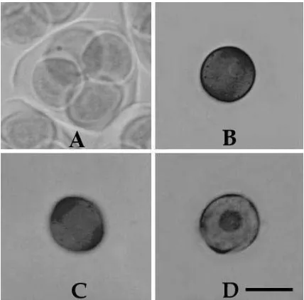
Ten anthers from each individual bud were bulk-squashed in a few drops of 1% aceto-orcein (Prakash, 2000). The microspore stage was determined based on the presence of a nuclear stage, i.e. tetrad, early-, mid- and late-uninucleate (Figure 1), from at least 100 observations under a light microscope (Zeiss Standard 20).
Figure 1. Cytological stages of Swainsona formosa microspore: A, tetrad; B, early-uninucleate; C, mid-uninucleate; D, late-uninucleate. Bar = 10 µm.
Since there could be microspores with various developmental stages within the anthers from the same bud, the stage of development was then determined based on at least by 50% observation. Data were analysed using analysis of variance with the aid of Microsoft Excel spreadsheet program (Microsoft-Corporation, 2000), followed by Fisher’s protected least significant difference (FPLSD) (Petersen, 1985) to separate the means, and the standard deviation (SD) was calculated.
Results obtained from this study indicated that the stage of microspore development in Sturt’s desert pea highly dependent on the size of floral buds (P = 0.00). The FPLSD test showed that the length of buds containing microspores at early-, mid- and late-uninucleate stages significantly differed from those containing tetrad microspores. In addition, significant difference in size was also recorded between buds containing early- and late-uninucleate microspores. However, the difference in size between buds containing early- and mid-, and between mid- and
late-uninucleate microspores were not significant (Table 1). The reason for this was the fact that these three stages lasted only for a short period, while the tetrad stage lasted longer. Tomasi et al. (1999) used a similar morphological predictor for determining the microspore developmental stage in Lasquerella sp. Floral bud length was then used as a morphological predictor in investigating the effect of the microspore developmental stage in subsequent anther culture, though no microspore-derived embryo was initiated. In Nicotiana
tabacum (Sunderland, 1974), Linum usitatissimum
(Nichterlein and Friedt, 1993) and Phleum pratense
(Guo et al., 1999), however, microspores at
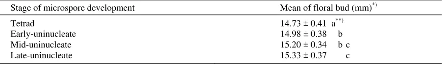
Table 1. The classification of microspore developmental stage based upon morphological measurements of floral bud length of glasshouse-grown Swainsona formosa
Stage of microspore development Mean of floral bud (mm)*)
Tetrad 14.73 ± 0.41 a**)
Early-uninucleate 14.98 ± 0.38 b
Mid-uninucleate 15.20 ± 0.34 b c
Late-uninucleate 15.33 ± 0.37 c
*)
Means ± Standard Deviation.
**)
Mean separation by FPLSD test at 0.05 protection level = 0.17.
Based on the work reported here, the floral bud length in S. formosa can be used as a reliable indicator of microspore developmental stage, eliminating the need for assessing every bud to ascertain its correct stage of microspore development at the time of culture initiation. This finding will also allow a quick staging
for future Swainsona formosa anther or microspore
culture.
ACKNOWLEDGEMENT
The author wish to thank Professor Acram Taji and Associate Professor Nalamilli Prakash of the University of New England for their invalueable guidance in conducting the research and comments on manuscript preparation.
REFERENCES
Barth, G. 1990. Cut flower potential of Sturt's Desert Pea. Australian Horticulture 88: 48-53.
Coumans, M., D. Zhong. 1995. Doubled haploid sunflower (Helianthus annuus) plant production by androgenesis: fact or artifact? Part 2. In vitro
isolated microspore culture. Plant Cell, Tissue and Organ Culture 41: 203-209.
Fan, Z., C. K. Armstrong, A. W. Keller. 1988. Development of microspores in vivo and in vitro in
Brassica napus L. Protoplasma 147: 191-199.
Guo, Y.-D., P. Sewón, S. Pulli. 1999. Improved embryogenesis from anther culture and plant regeneration in timothy. Plant Cell, Tissue and Organ Culture 57: 85-93.
Jusaitis, M. 1994. Floral development and breeding
system of Swainsona formosa (Leguminosae).
Hort. Sci. 29: 117-119.
Microsoft-Corporation. 2000. Microsoft Office 2000 Professional Edition. In: Microsoft Corporation, New York, USA.
Nichterlein, K., W. Friedt. 1993. Plant regeneration
from isolated microspores of linseed (Linum
usitatissimum L.). Plant Cell Reports 12: 426-430.
Petersen, R. G. 1985. Design and Analysis of Experiments. Marcerl Dekker, Inc., New York.
Prakash, N. 2000. Methods in Plant Microtechnique. University of New England, Armidale, Australia.
Romeijn, G., A. A. M. v. Lammeren. 1999. Plant regeneration through callus initiation from anthers and ovules of Scabiosa columbaria. Plant Cell, Tissue and Organ Culture 56: 169-177.
Sunderland, N. 1974. Anther Culture as a Means of Haploid Induction. In: Haploids in Higher Plants: Advances and Potential. Guelph, Canada. p. 91-122.
Tomasi, P., D. A. Dierig, R. A. Backhaus, K. B. Pigg. 1999. Floral bud and mean petal length as morphological predictors of microspore cytological stage in Lasquerella. Hort. Sci. 34: 1269-1270.
Williams, R. R., A. Taji. 1991. Sturt's Desert Pea in review. Australian Horticulture 89: 85-88.
Zhong, D., N. Michaux-Farriére, M. Coumans. 1995. Assay for doubled haploid sunflower (Helianthus
annuus) plant production by androgenesis: fact or
artifact? Part 1. In vitro anther culture. Plant Cell, Tissue and Organ Culture 41: 91-97.
Zulkarnain, Z., A. Taji, N. Prakash. 2002. Chromosome
number in Swainsona formosa (Fabaceae). New
1)
Fakultas Pertanian Universitas Jambi, Kampus Pinang Masak – Mendalo Darat, Jambi 36361 Email: [email protected]
RESEARCH NOTE
Floral Bud Length as Morphological Predictor for Microspore Developmental
Stage in Sturt’s Desert Pea (
Swainsona formosa
)
Zulkarnain1)
Diterima 7 Februari 2005 / Disetujui 6 Juli 2005
ABSTRACT
This work was conducted to establish the relationship between microspore developmental stage and length of the floral bud in glasshouse-grown Sturt’s desert pea, a native Australian legume. The stages of microspore development were segregated into tetrad, early-uninucleate, mid-uninucleate and late-uninucleate. The results showed that the stage of microspore development was highly dependent on the length of floral bud. The tetrad stage lasted longer than early-, mid- or late-uninucleate stages. The attempted induction of androgenesis in Sturt’s desert pea using anthers from floral buds with similar size, as in the present work, was unsuccessful. However, our work showed that the floral bud length can be used as a reliable predictor of microspore developmental stage in Sturt’s desert pea.
Key words: Sturt’s desert pea, Swainsona formosa, androgenesis, legume.
Sturt’s desert pea, Swainsona formosa (G.Don)
J.Thompson, is one of Australia's most spectacular wild flowers, and is the floral emblem of South Australia. It is a papilionoid legume with chromosome number of 2n = 16 (Zulkarnain et al., 2002), and self-compatible but self-pollination is often prevented by the presence of stigmatic cuticle that precluded pollen germination until ruptured (Jusaitis, 1994).
One of the economic importance of this plant is in its use as cut flower plant (Williams and Taji, 1991). However, its commercialisation is impeded by the production of a large amount of pollen grains that reduces flower quality (Barth, 1990) due to petal staining by pollen grains released by the anther during transportation. In addition, self pollination of the flowers during transportation would easily occur, especially by rough handling, resulting in rapid degeneration of flowers. Developing strategies to produce male-sterile plants is then becoming the most appropriate method to solve this problem. One approach to create such sterility is via androgenesis using anther culture method.
Androgenesis is determined by a number of factors, including the microspore developmental stage at the time of the introduction to the in vitro environment. Unfortunately, the exact stage for successful plant regeneration is species dependent. Romeijn and Lammeren (1999) found that first pollen mitosis was a suitable stage for the induction of androgenesis in
Scabiosa columbaria. Tetrad to mid-uninucleate stages
were found to be useful in androgenesis of Helianthus
annuus (Coumans and Zhong, 1995; Zhong et al.,
1995). Meanwhile, the late-uninucleate to early-binucleate stages were believed to be more responsive
in Brassica napus (Fan et al., 1988). As the
consequence, determining the correct stage of microspore development in Sturt’s desert pea is a crucial step before anther culture initiation.
Conventionally, the determination of microspore developmental stage of a given floral bud has been using aceto-orcein or aceto-carmine staining technique prior to observation under a light microscope (Prakash, 2000). This method, however, is impractical and time-consuming, particularly for a large sample size such as in a routine anther or microspore culture programme.
To our knowledge, no practical and quick microspore staging protocol has been developed for Sturt’s desert pea. The present study aimed at correlating the floral bud length as a morphometric attribute with microspore developmental stage for a better time prediction for Sturt’s desert pea anther culture.