248
Sjahrurachmanetal MedJ IndonesComparison of two dot
immunoassays
for
diagnosis
of
dengue
infection
Agus
Sjahrurachman,Betty Emawati,
Feralbrahim, Mardiastuti,
Tahjani Mirawati
Sudiro, Pratiu/i
SudarmonoAbstrak
Diagnosis definitif infelæi vints dengue memerlukan konfirmasi laboratorium, karena diagnosis tersebut sulwr sekali ditegak!'an bila
haiya berdasarlnn geiata dan tandà klinis. Oleh sebab itu pemertksaan laboratortum berperan penting dalam pengelolaan kasus dan pàberantasan peiyakitnya. Mengingat kebanyakan kasus infelcsi dengue terdapal
di
negara berkembang dimana sarana 'laborabrtumAaiya| yang tidak mimaàai, diperlukan cara pemeriksaan laborabrtum yang cePat dan sederhana. Dalam makalah
ini
dilaporkanperbaiaiigaiuit
dot imunoesei yang lcami kembangkan dengan salah satu kit dot imunoesei lamersil dengan pembandinguji'hambaian
hemaglutinasi. Hasilnya menunjuklan bahwadot
imunoesei yang kami lcembangkan mempunyai sensitifitas dan spesifisitas tinggi.Abstract
DeJinite diagnosis of dengue infection plays an important role of case management and disease control. Unfortunately, majoity of the injecrcd
"^âs
canior
bidiagposed solely on lhe basisofclinical
signs and symptoms. Laboratory investigalions are required' Sincedângue distribution mostly piesent in developing countries, appropiate laboralory diagnosis would be a rapid and simple one. Here,
weieport a preliminary'assessment of a dol immunoassay employing biotinylated-dengue antigens
for
detection of lgM-antidengue andb;dies which is simple in lerm of methodologt and rapid in term of the test result. This sudy was done by compartng out methodwith standard
H.I tesi
and commàrcially available dot immunoassay. The results indicate that thedol
immunoassay biotinylated-dengue (BDIA) is sensitive and speciJicfor diagnosis ofdengue infection.Keywords : qualitative immunoassay, diagnosis, dengue infection
the
most
showing
t
has beenestimated
that
more than
two
and
half billion
of
people are
at risk
of
infection.t't
It
it
estimated
thatthere
are
annually
20 million
cases
of
dengueinfections
all
over the world
resulting
in
around 24.000 deaths.2On the other
hand,it
well
known
that dengueinfection may
induce awide variety of
clinical
entity from
mild
undiffereptiated fever
to a
severe'often fatal dengue
shock syndrome. Further,
unusualdengue manifestation.
more
frequently
observedwithin
the last
decade.aDefinite
diagnosis
of
dengueinfection is
therefore
hardly
determined solely on
thebasis
of
clinical
symptoms
and
signs,
especially
whenever
other
arbovirus cocirculate.
Laboratory
confirmation is
require
in
majority
of
cases.2'5 SinceIgM-antidengue antibodies has been reported as
oneamong other immune
responsesthat
appearearlier
in
Department of Mitobiologt, Faculty of Medicine, University of Indonesia, Jakarta, Indonesia
the
courseofthe
diseases,short lasting
and relativelyspecific
for
dengue,2'5capturing lgM-anti-dengue in
human sera
for
rapid and
simple
diagnosis is
athacting
to be elaborated.In
previous report,
we
have
described
that
a dot
immunoassayemployi
engue@DIâ)
can
be
used
to
detect
antibodies.o"'oHere
we
report a comparative study
on
sensitivity
of
BDIA
and
commercially
available
dot immunoassay
(CDIA)
for
diagnosisof dengue
infection.
MATERHLS
AI\ID METHODS
Patient recruitment
Serum samples
were obtained
from
patients
clinically
diagnosed
as
dengue
haemorhagic
fever
and mild
undifferentiated fever.
Haemagglutination
inhibition
(HI)
test, according
to
Clarke and
Casals
method,eVol 9, No 4, October
-
December 2000immune
responseto
dengue
was
done according
toW.H.O
criteria.2Biotinylation of
dengueviruses
Laboratory
adapteddengue
1,2,3 and4 virus
infected
C6l36 monolayer
cell
culture
fluid
wasclarified from
cellular
debrisby low
speedcentrifugation. Viruses in
supematant
fluid
were precipitated
by polyethylene
glycol
andcentrifuged
at
10,000rpm for
30
minutes.The
virus in
thepellet
wasbiotinylated
by
biotinyl-e-amidocaproic acid N-hydroxy succinimide ester
aspreviously
described.6Dot
immunoassây
employing biotinylated
denguevirus (BDIA)
BDIA was
performed
as
previously
described.TBriefly,
anti human
IgM
was blotted on a piece
of
nitrocellulose
paper,blocked with
2%"skim milk
andair
dried.
Diluted
test serum was then applied
to
thepaper and incubated
at
room
temperature
for
two
hours. Reactions of
IgM
and
biotinylated
dengue aswell as
biotinylated dengue
and
horse-radishperoxidase-labelled
streptavidin were done
at
room
temperature
for
one
hour
each. Finally,
5-chloronaphthol substrate
was
added
for color
development.
After
30
minutes incubation
at
room
temperature,
the result
of
the test was
observed by
naked
eyesand
expressed aspositive
when
apurple-solid
color
appearedon the
blot
and negative
when theblot
was colorless. The test was donein
duplicate.Quality control of
the test was doneusing
reactive andnon reactive
control
serafrom
dengueblot kit.
Whenthe result
of
the
controls
were
inappropriate, the
test was repeated.IgM
anti-dengue
detection
by
commercially
available dot
immunoassay
(CDIA)
In
parallel
to
BDIA,
rapid
procedure assay
of
commercially available
DengueBlot IgM
(Diagnostic
Biotechnology
Ltd)
was
performed.
Briefly,
testsemm was
reacted
to
anti
p
chain
of
human IgM
which
has beenblotted
on a membranefor
onehour
at37oC.
Following that,
dengue antigens
were
addedand incubated
for two
hours
at
37oC; group-reactivemonoclonal
antibodiesto
dengue antigens were addedfor
two
hours
at
37oC;
working
conjugate solution
was added
for
onehour
at 37oC andworking
substratesolution was
addedfor
30 minutes at
37oC.Result of
the
tests
were
compared
with
naked eyes
with non
Diagnosis of dengue
infection
249reactive
control and
expressed
as positive when
apurple-ring
type
blot
appeared
on the blot
and negative when theblot
was colorless.Data
analysis
Sensitivity,
specificity and positive value
of
BDIA
and
CDIA
as
compared
to H.I.
were
calculated
asdescribed
previously.lo
Brief
description
is
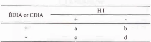
shownbelow:
BDIA OT CDIA
a
c
Sensitiviry is a divided by (a + c), specificity is d divided by (b + d), positive predictive value is a divided by (a + b).
The difference
betweenBDIA and
CDIA
is analyzed
by Mc
Nemar test applying Yates correction
and one degree offreedom.ll
When
p
value
is
lessthan
0.05,the BDIA
and
CDIA
is
scored
as
significantly
different.
RESULTS
Validity
of
BDIA
for
diagnosis
of
dengueinfection
Comparison
of H.I
test
andBDIA
to confirm
dengueinfection
on acute and convalescent sera are shown onTable
I
andTable 2. On
acute sera, 80 casescould
beconfirmed
as dengueinfection
casesby BDIA
and 24 casescould
not
be
confirmed.
In these
cases,BDIA
had
a sensitivity
of
'76.90Â,specificity
of 88.1%
andpositive predictive value
of
94.1%.
On
convalescent sera,96
casescould
beconfirmed
as dengueinfection
cases
by
BDIA
and
only
8 cases
could
not
beconfirmed.
BDIA
had
a
sensitivity
of
92.30Â,specificity
of
88.1%
andpositive predictive
value
of
95.0%
on
convalescent sera. Compared
to
blot of
acute
sera,
most
of
the
blot of convalescent
sera revealed darkercolor.
Typical
exampleof
theresult
is shown onFigure
l.
Table
l.
Comparison between H.I test and BDIA on acute seraDIA employing biotinylated dengue
(+)
(-)
H.t
b d
HI
(+)
(-)
805 [image:2.595.330.572.212.279.2]250
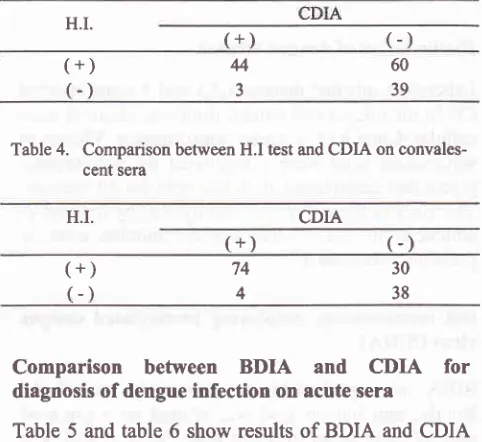
Sjahrurachmanetal Med J IndonesTable 3. Comparison between H.I test and CDIA on acute sera
Tablc2.
Comparison between H.I test and BDIA on convales-cent seraDIA employing biotinylated dengue H.I.
(+)
(-)
H.I.
(+)
(-)
(+)
(-)
60394
3
(+)
(-)
37896
5
N
SSS$
-YTable
4.
Comparison between H.I test and CDIA onconvales-cent sera
CDIA
(+)
Comparison between
BDIA and
CDIA
for
diagnosis of dengue infection
on acute sera
Table 5 and
table 6 show results of
BDIA
andCDIA
for
diagnosis
of
primary
and
secondary
dengueinfection
on
acute sera.
56.5%
primary
dengueinfection cases
could
be
confirmed
by
conducting
BDIA
on acute sera
while
those
by
CDIA
is
8.7%cases.
For
secondary
dengue infection,
BDIA
was ableto confirm
82.7%
caseswhile CDIA only 51.8%
cases.Result of Mc
Nemar test indicated that
p value
for
primary and secondary
infection are less
than 0.001 and less than 0.001,respectively.
Table
5. Comparison between
BDIA and CDIA for diagnosisof
primary dengue on acute sera
CDIA
BDIA
Total casesH.I.
(-)
.ii ii+ri,'r"rùtt3!+
74
4
(+)
(-)
3038+
40
2
27
12
67 t4 Total cases
Comparison between
BDIA
and CDIA
for
diagnosis
of dengue
infection
on conyalescent sera
Comparative
results
of
BDIA
and
CDIA
onconvalescent
sera showed
that
BDIA
was able to
confirm
the infection
in
82.6%oprimary dengue
caseswhile
those of CDIAin 52.2%o
primary
dengue cases.Positive results
of BDIA
and
CDIA
on convalescent
seraof
secondary dengue
infection were 95.1%
and76.5yo, respectively.
Result
of
Mc
Nemar
testindicated
that
p
value
for
primary and
secondaryinfection are less
than 0.05 and
less
than
0.001,respectively. Details
are shownin table
7 andtable
8.8l 42
$f
:'
t*,*
[image:3.595.78.316.90.537.2]{
Figure
I.
Typical results of BDIAPatients
and controls sera
were subjected
to
BDIA.
Positive result
is shown as solid dots. Negative result
is shown
as colorless dots. [image:3.595.332.573.113.334.2]Yol 9, No 4, October
-
December 2000Table
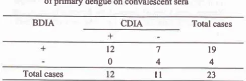
7.
Comparison betweenBDIA
and CDIA for diaposis ofprimary dengue on convalescent seraBDIA
CDIA
Total casest2 0
7
[image:4.595.70.314.102.183.2]4 Total cases
Table 8. Comparison between
BDIA
and CDIA for diagnosis ofsecondary dengue on convalescent seraBDIA Total cases
Diagnosis of dengue
infection
251
Ovqrall
results
indicate that sensitivity
of
BDIA
for
acute serais
almosttwo
times
comparedto
CDIA.
On
convalescent
sera,the sensitivity
of
BDIA
is
higher
than
CDIA,
though
not
as
high
as
on
acute sera.
Contrastly,
the specificity
of
both
techniques,
BDIA
and
CDIA,
are
very high
and
the
differences
of
specificity
betweenBDIA
andCDIA
isnot significant
comparedto
thoseof
sensitivity.
CONCLUSION
BDIA
has
a
high
sensitivity
and
specificity for
diagnosis
of
dengue
infection, especially
on
con-valescent sera. Comparative
assessmentwith
CDIA
indicate
that
BDIA
is
superior
to
CDIA.
Overall
results indicate that
BDIA
could
be used asa
simple and sensitiveroutine laboratory diagnostic
for dengue
infection. However,
further
clarification involving
larger sample is required.REFERENCES
l.
GublerDJ.
Resurgenceof
vector-bome diseases as aglobal health problem. Emerg Infect Dis 1998; 4:442-50.
2.
World Health Organization. Dengue Haemorhagic. Fever.Diagnosis, Treatrnen! Prevention
and Conhol.
WHOGeneva 1997, p. l-68.
3.
GreenbergRN,
Feinberg JE and PomeroyC.
The Hot Zone 1997: Emerging Infectious Diseases. Emerg Infect Dis 1998; 4:135'7.4.
Lam SK. Emerging infectious disease- South East Asia. Emerg Infect Dis 1998; 4:145-7.5.
Suvatte V, Immunological aspects of dengue haemonhagicfever studies
in Thailand. South East
AsiaJ
Trop Med Publ Health 1987; l8:312-5.6.
Agus S, Tallei T, Betty E, Amin S and pratiwi S. Biotin-labeled Dengue Virus for Detectionof IgM
anti DengueAntibodies. Indon J Clin Microb 1995;7:25-30.
7.
Agus S, BettyE, ljahjani
MS,Amin
S and pratiwi S.Development of Dot Enzyme Imrnunoassay for Detection of IgM antidengue. Indon J publ Health 1996; 3:208-14.
8.
Agus S, BettyE,
Fera I,Àlardiastuti, 1-ahjaniM
andPratiwi
S.
Semiquantitativedot
immunoassay for detectionof
IgM
anti-dengùé antibodiesin human
sera.1999. Submitted for publication.
9.
Clarke DH and Casals J. Technique for Haemaglutination. and Haemaglutination Inhibitionwith Arthropod-bome
Viruses. Am J Trop Med Hyg 1958; 7:561-73.10.
Illstrùp
DM. Statistical
Methodin
Microbiology. Clin Microb Rev 1990; 3:219-26.ll.
Zat JH. Biostatistical analysis. Prentice Hall Inc. 1984. p. 156-8.12.
Cardosa MJ and Tio PH. Dot Enzyme Immunoassay: AnAltemative
Diagnosticaid
for
Dengueand Dengue
Haemorhagic Fever. Bull WHO
l99l;
l2l:741-5.
l9
4 77 4 + 23ll
6tI
l6
3Total cases
DISCUSSION
Several
laboratory
approaches
to
confirm
dengueinfçgt!99-
in_ suspected
cases
have been
develop-ed.2'e'12'13't4't5
PoÎymerase
chain reaction and
virus
isolation
areamong the most
accurate techniques2in
term
that a
positive
result
of
the test
is
indisputable.Those
techniques,
however,
is not
suitable
to
beroutinely
used
in
developing countries
wherelaboratory
facilities
are
relatively
insuffrcient.
Therefore,
W.H.O
recommendedHI
test
as aroutine
testfor
diagnosisof
dengueinfections. Unfortunately,
interpretation
of
HI
testrequire
apair
of sera
taken
atleast
a week
apart. Therefore, though
the
HI
test
ispractical
in
term
of
themethodology, result
of
the testcan
not be drawn
soon
providing that the
test result
can
not
be used as
a
guidance
for
case management andprompt
diseasecontrol.
Previously,
we
havedeveloped
anew format of
DIA
for
detection
of
IgM-anti-dengue
antibodies.T Here,we report
acomparative study on sensitivity
of BDIA
and
CDIA
for
diagnosis
of
dengue
infection.
Theresults indicate that
on
acute seranumber
of primary
dengueinfected
cases diagnosedby
BDIA
is
at
leastsix
times more than those diagnosédby
CDIA. In case
of
secondary
infection,
the
proportion
of
cases diagnosedby BDIA
comparedto CDIA
is
almosttwo
times, indicating that
BDIA is
superior
to CDIA
for
determination
of
dengueinfection on
acute
sera.On
convalescent
sera,
BDIA
also
show
superiority
compared
to
CDIA
though
the
differences
is
not
ashigh
as on acute sera.Indication
thatBDIA
is superior
to
CDIA
wasconfirmed by Mc
Nemar test.8l
252 Sjahrurachman et al
Henchal
EA
and Putnak JR. The dengue viruses. Clin Microb Rev 1990; 3:376-96.Lam SK, Devi S and Pang T. Detection of Specilic IgM in Dengue Infection. South East
Asia
J
Trop Med
Publ Health 1987; l8:532-8.Med J Indones
15.
Lanciotti RS, CalisherCH,
Gubler DJ, Chang GJ andVomdam
AV.
Rapid Detection and Typing of Dengue Viruses from Clinical Samples by Reverse-Transcriptase Polymerase Chain Reaction. J Clin Microb1992;30:545-51.