Vrtl -1, No I, Juttuttry Murth t994 Anatomy of Hiccups
Anatomy
of
Hiccups and
its
Relations
to
its
Nonpharmacological
Manage-ment
Marjadi Hardjasudarma
Abstrak
Cegukan nasih merupakan teka-teki nedis karena tidak nempunyai nanfaat dan cara-cara pengobatannya jauh tebih banyak daripada penyebab-penyebabnya. Cara-cara pengobatan ini berkisar antara pengobatan-pengobatan tradisional yang telah terkenal sejak dahulu kala sampai obat- obat sintetik baru. Pengetahuan yang tepat dan rinci tentang asal dan sifat cegutrai belum banyak, meskipun akhir-akhir ini telah diusulkan suatu lengkung refleks cegukan dengan koordinasi supraspinal. Makalah ini dimal<sudkan untuk mengungkapkan aspek-aspek anatomi cegukan dikaitkan dengan mekanisne kerja penanganan-perutnganan non far-makologiknya.
Abstract
Hiccups remain a medical enigma, as they serve no useful purpose and have nuch nore cures than etiologies. These cures nay range fron universally known titne-honored remedies to new synthetic pharmacologic agents. Linte is known about the exact rnture of hiccups, although recent$ a hiccup reflexwithsupraspinal coordinationis proposed. The purpose ofthis paper is to revealthe anatomical aspects of hiccups and to relate thern to the mechanisnw of action of many non pharmacologic managitnents of hiccups.
Keywords : Hiccup reflex, Supraspinal coordinatiott, Nonphartnacological managetnent ofhiccups.
INTRODUCTION
Hiccup is usually a transient and benign annoyance experienced occasionally by most people as a sudden contraction of the inspiratory muscles terminated by abrupt closure of the glottis
to
produce the charac-teristic sound, from which it derives its onomatopoetic name ("hoquet" in French,"hik"
in Dutch, ,,Hipo,, in Spanish, "geehouk"in Hebrew, "hicka',
in
Swedish, "hikke" in Norwegian and Danish).1'2Most hiccups occur as brief, self-limited episodes lasting only a few seconds or minutes; they are often caused by overdistention of the stomach, excitenrent, a sudden change in temperature or alcohol ingestion.2
Hiccups that last more than 48 hours or recur at frequent intervals, are referred as ,,persistent,, and
serious
u
disorderrdial
infa
racranialOccasion
actable,,,months or years, causing nd dehisence after surgery, s resulting from inability to swallow fluids and food, and ventricular dysrhytmias as a result of disturbance in serum potassium;l'2 uil tt
i,
may result in high morbidity and even death.a
Al-though hiccups are a frequent and nearly universally experienced phenomenon, they serve no known useful or protective function. l'2In some cases, despite exhaustive investigations,
their
etiologies cannot
be determined,
so that
therapeutic recommendationsfor
management of hic-cups encompass a spectrum ofapproaches ranging from universallyknown
home remedies to new synthetic pharmacological agents.lNo wonder
that
Charles Mayo) in a discussion about 60 years ago commented: "Perhaps one is justifiedin saying that there is
no disease which has had more forms of treatments and fewer results from treatment that has persistent hiccup,,.Many of these home remedies are time-honored and although many
of
them are ancient and obscure, some have soundanatomical and physiological
bases.l'2On the other hand, the exact nature of hiccups is
still
not satisfactorily explained. On the anatomical part, the role of two nerves, the phrenic and the vagus, is firmly established; while on the physiological part there is little doubt that hiccups have a reflex ar",l'2'6l0
HardjasudannaIn this context, the author
tried
to
review theliteratures on the anatomical aspects
of hiccups
andrelate these to the many non pharmacological ap-proaches used in the management of hiccups'
DISCUSSION
Anatomical and physiological considerations
a) Anatomy of the phrenic and n"gu,
n"*"r7'8
The anatomy of these nerves
will
be briefly reviewed,as they are intimately related to the "hiccup reflex".
-
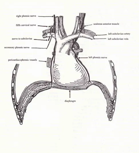
The phrenic nerve (seeFig.l)
usually arises fromthe third, fourth and
fifth
cervical nerves, so thatit
is derived from both cervical and brachial plexuses;
it passes downward in front of the anterior scalene
muscle, between the muscle and the prevertebral fascia, and
is commonly
attacked surgically asit
lies in this position.At operations, the vagus nerve and the cervical sympathetic chain have both been mistaken for the
phrenic nerve. The vagus nerve
lies within
thecarotid sheath, anterior to the phrenic nerve, and the
cervical sympathetic chain lies somewhat more medially, posterior
to the carotid
sheath, but not upon the anterior scalene muscle.At the root of the neck, the phrenic
nerve passes between the subclavian artery and thesub-clavian vein and so enters the thorax. Within the
thorax,
it
descends nearly vertically in front of theroot of the lung, and then between the pericardium and the mediastinal
pleura
(where
it is
accom-panied by the pericardiacophrenic vessels) to the
diaphragm which it innervates.ln30% of the cases
in which the phrenic nerve is crushed to alleviate intractable hiccups, there is an accessory phrenic nerve. It arises most frequently from the
fifth
cer-vical nerve (in common with the nerve to the
sub-clavius muscle) and runs usually lateral to the
normal phrenic to
join
it
at about the level of thefirst rib, but it may not unite with it until the level
of
the root of the lung or evenuntil
closeto
thediaphragm. The accessory phrenic nerve may
ac-count for the occasional failure to obtain diaphrag-matic paralysis after crushing of the phrenic nerve.
If
the phrenic nerve is avulsed from the well within the thorax, it is probable that all accessoryphrenic nerves
will
also be torn out. However, if thephrenic nerve is simply crushed in its normal
sur-gical position (as
it
lies on the anterior
scalenemuscle),
it
may be prudent to locate and crush the nerve to the subclavius muscle and to seek all other branches descendingfrom
the brachial
plexusMed J Univ Indon
along the lateral border
of the anterior
scalene muscle.-
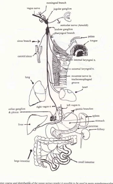
The vagus nerve (Latin : vagus = wanderer), or thetenth cranial nerve, has an extensive course and
distribution, since
it
passes through the neck and the thorax to the abdomen.The vagus nerve (see Fig.2) makes
its
exit through the jugular foramen; within the foramenit
bears its jugular (or superior) ganglion, from which arise the meningeal branch to the duramater in the
posterior cranial fossa, and the auricular branch (nerve of Arnold) which passes through the tym-panomastoid fissure to innervate parts of the exter-nal acoustic meatus and the outer surface
of
thetympanic membrane.
After its exit from
the jugular foramen, thevagus nerve enlarges into a second swelling, the nodose (or inferior) ganglion, from which arise the
pharyngeal branch (principal motor nerve
of
thepharynx), branches
to
the carotid body and thecarotid sinus, and
the superior laryngeal
nervewhich innervate the cricothyroid muscle and the mucous membrane of the larynx as far down as the
level of the vocal folds.
The right recurrent laryngeal nerve arises in
the neck, while the left one arises in the thorax; both
nerves ascend to the tracheoesophageal groove and
end in the larynx where they innervate almost all laryngeal intrinsic muscles and the mucous mem-brane
of
the larynx below the level of the vocal folds.Cardiac branches arise in the neck and in the
thorax,
to join
branchesfrom the cervical
sym-pathetic ganglions to form the cardiac plexus at the base of the heart.In the thorax, both vagus nerves pass behind the roots
of
the lungs, here they give off pul-monary branches toboth
lungs. Both nerves then descend on the esophagus, givingoff
esophageal branches and then enter the abdomen through theesophageal
opening
in
the
diaphragmto
the stomach. Hereit
distributes gastric branches forthe stomach and celiac branches
for the
celiac plexus and ganglion, from here twigsare
sent to the splenic, hepatic, renal, suprarenal and superiormesenteric plexuses, so the parasympathetic vagal
nerves can be traced in the large intestine as far as
the left colic (splenic) flexure.
b.
Anatomy of the "hiccup reflex"The precise pathophysiologic mechanisms responsible
for
hiccuping eluded anatomists, physiologists andcen-Vol 3, No l, January - March 1994
turies mentioned several pathological processes as-sociated
with
hiccups, suchas
inflammation of the liver and inflammationof
the stomach due to spoiled food.In
the latter, it was thought that hiccups occured as a convulsivemotion
of
the stomachto
dislodge what is impacted in its body.l,2The first anatomical structure related to hiccups was found by Shorte
in 1833, when
he recognized tÀe relationship between irritation of the phrenic nerve and hiccups.In
1943, Bailey l0 proposedthe
existenceof
a "hiccup reflex", a principal component of which was a "hiccup center" located somewherein the upper
cer-vical segments of the spinal cord. Since then Bailey's findings have been modified and expanded,it
is now known that hiccups result from stimulation of one or more components of the hiccup reflex arc.The afferent portion of the hiccup reflex are com-prised
of
the phrenic and vagus nerves and the sym-pathetic chain arisingfrom
the
thoracic segments T6-T12, while the efferent portion was then thought to be exclusively from the phrenic nerve.The central connections between the afferent and efferent limbs
of
the reflex, as well as the center of thereflex,
were anatomicallynot
so clear,but
it
was thought that the center was located somewhere in the spinal cord, between segments C3 and C5, which was the origin of the phrenic nerve.Bailey's findings were confirmed bv subsequent investigators, such as Salem and othersll, who con-tributed that besides the phrenic nerve as the primary efferent
limb
of
the reflex,
other
efferentsto
the glottis and accessory muscles of respiration were also thought to be involved in hiccuping. These contribu-tionswere
basedon
findings in patients in whom hiccups werestill
present even after transection of both phrenic nerves. Electromyographic studies done during hiccuping,have
demonstrated simultaneous firing of motor neurons to the anterior scalene muscles (C5-C7), external intercostal muscles(Tl-Tll)
and glottis (recurrent laryngeal nerve). It was also recentlynoted
that normal esophageal contractile tone andlower
esophageal sphincter pressure were reduced during hiccuping, suggesting the existenceof
a simul-taneous inhibitory autonomicp.o."rr.t
t,t2All
these findings and the finding of the bilateralhemi-diaphragm involvement
during
hiccuping, pointed to a complex supraspinal coordination of the efferent limb which included brainstem and midbrain areas such as the respiratory center, phrenic nervenuclei,
me.dullaryreticular
formation
and hypo-thalamus.l'l I'12Nathan and others6 went even further by propos-ing that the center of the hiccup reflex was in the brain
Anatorny of
Hiccups
I Istem itself, independent
of
the respiratory center, its afferentlimbs
consistedof the
pharyngeal plexus, branchesof
sympathetic thoracic ganglions, phrenic and recurrent laryngeal (vagus) nerves, while the ef-ferentlimb
consistedof
the nervesto
the anterior scalene muscle, and externaI intercostal muscles, the phrenic and the recurrent laryngeal (vagus) nerves.So, in this theory, the vagus and phrenic nerves supply both afferent and efferent pathways to and from the hemi diaphragms and the laryngeal musculature (see Fig.3).
It
was thought for many years, that the originof
the hiccup reflex was respiratory in nature (hence the term "hiccough"): This thinking was recently chal-lenged, by Davisl3 who monitoràd diaphragmatic and intercostal electromyographic recordings andpul-monary spirometric
function
simultaneously, to demonstrate that hiccups have only a minimal effect on ventilation. He arrived at this conclusion by deter-mining that glottic closure was noted to be very tran-sient and occur only 35 ms after the onset of respiratory muscle motor discharge.Mechanisms involved
in
the non pharmacological managements of hiccupsMost mechanisms of action of the many time-honored remedies still used in the non pharmacological mana-gement of hiccups are readily explained by anatomical knowledge of the hiccup reflex arc.
Some of these mechanisms are :
l.
Stimulation of vagal afferents of the hiccup reflex arc, thereby interrupting the vagal limb of the ur".t'2 According to the "gate control,, theory,6'14 impul-ses arising in response to vagal stimulation (e.g by pharyngeal stimulation), may block or inhibit other afferent impulses being transmitted through the vagus and thus interrupting the hiccup reflex.Hiccup
remedies
which
usethis
mechanism ua.' l'2'15a.
manipulationof
an aberrant hairirritating
the tympanic membrane (mediated via the auricular branch of the vagus nerve).t2 Hardjasudarma
scalenus anterior muscle
Med J Univ Indon
left subclavian artery
left subclavian vein right phrenic nerve
fifth cervical nerve
-.
.
nerve to subclavius
-{
accessory phrenic nerve
-
left phrenic nerve pericardiacophrenic vessels
ï
diaphragm
Fig t. The course and distribution ofthe phrenic nerves. The phrenic nerve is conntonly attacked surgically as it lies infront of the scalenus anterior muscle, beneath the prevertebralfascia. On the right side, an accessory phrenic nerve arisesfrorn the ftfth
[image:4.595.74.560.90.626.2]Vol -1. No I. Jurrtrttrt' Murch 1994
vagus nerve
sinus branch
7r-)
jugular ganglion
auricular nerve (Arnoldi) nodose ganglion
pharyngeal branch
heart
ar/
-
tongueintemal laryngeal n.
external laryngeal n.
recurrent nerve in tracheoesophageal groove
right vagus n left vagus n.
gastric branches
[image:5.595.116.490.110.716.2]large intest
Fig 2. The extensive course and distribution ofthe vagus tremes render it possible to be used in nany nonphannacological renedies of hiccups, by stimulating branches of the nerves at dffirent locntiorc (see arrows : the pharyngeal, sinus, auricular and gastric branches).
Anatonry of
Hiccups
l3I4 Hardjasudartna Med J Unit, Indon
Anterior Scalene
Muscle
HICCOUGH CENTER
INSPIRATION CENTER
c5,6,
Tl-ll
-->,/
%t
EXPIRATIONCENTER
Vagus Nerve
Pharyngeal Plexus
c2-4
c3,4,5
Glottic Closure
External Intercostal Muscle
Sympathetic Ganglion
T6-12.
D
Afferentm
EfferentEH
Both afferent and efferentFig3. Schematicrepresentation of the hiccup reflex are as proposedbyNathanetal.6.ThecenterofthereJlexis located inthe
brain stem, independent ofthe respiratory center. Brancltes of the pharyngeal plexus and the thoracic sytnpathetic chain supply afferent pathways,whiletheefferentonesconsistofnemes to the anteriorscaleneandintercostal muscles. The phrenic and the
vagus nerves supply both afferent and efferent pathways to andfrotn the henidiaphragms and the laryngeal nusculature.
Vol 3, No l, Jurtuary - March 1994
remedies
are
mediatedvia
the
pharyngealbranch of the vagus nerve).
c.
nasogastric suction andiced
gastric lavage(these remedies are mediated
via
the gastric branches of the vagus nerve, but some authorsbelieve
that
direct diaphragmatic stimulationmay play a role).
d.
other vagomimetic procedures such as carotidmassage and digital ocular globe pressure.
2.
Interruption of the phrenic limb of the hiccup reflext-2'
are :
Remedies using this mechanism are :
Cervical
(C5)
dermatome stimulation, done byrhytmic tapping over the
fifth
cervical vertebralevel at the origin ofthe phrenic nerve, spraying the
area
with
vapocoolants, acupuncture, galvanic(electric) stimulation, direct phrenic nerve
stimula-tion by operative placement of electrodes and
sur-gical
phrenic nerve interruption
(crushing ortransection of the nerve).
Some remedies are explained on a physiological
basis, such as creating a respiratory acidosis which
inhibits the diaphragm contractility by :
sneezing or coughing by various means, breath
hold-ing, hyperventilation, grasping (like that precipitated
by fright or sudden pain) and breathing in a paper bag
(use of a plastic bag is not recommended and is in fact
dangerous, as
it
may stickto
the face causing as-phyxia).r'2Despite
all
effortsto
explain these mechanismson
an anatomico-physiologicalbasis, for some
re-medies there
is
still
no satisfying explanation avail-able, e.g. :a.
auriculo-acupuncture and auriculo-pressure:
ac-cording
to
TCM
(Traditional Chinese Medicine),in hiccups the normal stomach function of
send-ing food down is disturbed, resultsend-ing in reversed
upward surge
of
stomach
Qi to
attack
thediaphragm.l6
b.
hypnosis:
although Kirkner and WestlTfelt
thathypnosis may inhibit the cerebral cortex (as seen by
the influence
of
voluntary action, relaxation andsleep
on the
magnitudeof
reflexes), the exactmechanism by which hypnosis cures hiccups is still
obscure.
c.
some bizarre remedies, such as :-
prayersto
St.Jude, the patronof
lost causes,which were ultimately successfull
in
a patientwho hiccuped for more than 8 years and
repor-tedly
received 60.000 letters containing"pos-sible cures.""Anatomy of
Hiccups
t5-
a
financial
rewardof
US
$
10offered
byKirkmanl8 to a hiccuper
if
he could voluntarilycontinue to hiccup on demand. Kirkman stated,
that this approach worked among his family,
friends and a few patients; leading him to
pos-tulate that the "neural energy" was suddenly
drained from the hiccup pathway after he
of-fered the reward.
Acknowledgement
The author wishes
to
thankDr.
MardjohanHarda-sudarma (Shreveport-LA, USA)
for
providing somereferences used in this review,
REFERENCES
l.
Kolodzik PW, Eiters MA. Hiccups (singultus) : Review andapproach to management. Ann Emerg Med
l99l;20(5):565-'73.
2. Lewis JH. Hiccups, causes and cures. J Clin Gastroenterol
1985;7(6):539-52.
3. Swan HR, Simson LH. Hiccups complicating myocardial
infarction. N Eng J Med 1952;247:726-8.
4. Fleet WF, Morgan HJ, Morello PJ. A fatal case of hiccups
(Vanderbilt molning report). J Tenn Med Assoc 1990:
Febr;79-80.
5. Mayo CW. Hiccup. Surg Gynecol Obstet 1932;55:700-8.
6. Nathan MD, Leshner RT, Keller Jr AP. Intractable hiccups
(singultus). Laryngoscope 1980;90:1612-8.
7. Hollinshead HW. Anatomy for surgeons. Tokyo : Hoeber
Harper first reprint edition, 1966;(l):496-8.
8. Johnston TB, Davies DV, Davies F. Gray's Anatomy,
descriptive and apllied. Glasgow : Longman's Green & Co
32nd. edition, 1958; I 130- 6.
9. l.ewis JH. Hiccup, its causes and cure. J Clin Gastroenterol
1985;7(6):539-52.
10. I-ewis JH. Persistent hiccup. J Clin Gastroenterol 1985;
7(6):539- 52.
11. Salem MR, Baraka A, Rattenborg CC, Holaday DA.
Treat-ment of hiccups by pharyngeal stimulation in anesthetized
and conscious objects. JAMA 1967;202:126-3O.
12. Rosenberg J, Hansen BJ. Possible role of autonomic
af-ferents
in
tleatmentof
postoperative hiccups. Lancet 1989:Oct;7:873.13. Kolodzki PW, Eilers MA. An experimental study of hiccup.
Ann Emerg Med l99t;20(5):565-73.
14. Wall PD, Sweet WH. Temporary abolition of pain in man.
Science 1967;155:08.
15. Cardi E. Hiccups associated with hair in extemal auditory
canal-successful treatment by manipulation. N Eng J Med
t96l;265-59.
16.
Li
X, Yi J, Qi B. Treatment of hiccough withauriculo-acupuncture and auriculo-pressure.
I
Tradit Chin Medl99O;rO(4):257-9.
17. Bendersky G, Baren M. Hypnotic treatment of persistent
hiccup; a case report. Retracted by Arch Int Med 1959;
lO4:417-20.
18. Kirkman R. Discussion in Nathan et al. Intractable hiccups