Lymphokine activated killer cells from peripheral blood mononuclear cells of
endometriosis of patients improve cytotoxicity to endometriosis cell culture
Muharam Natadisastra,1 Arleni A. Bustami,2 Indra G. Mansur,2 Teuku Z. Jacoeb,2 Jerome Giustiniani,3 Valerie Schiavon,3 Armand Bensussan3
1 Doctoral Program Faculty of Medicine, University of Indonesia, Jakarta, Indonesia 2 Faculty of Medicine University of Indonesia, Jakarta, Indonesia
3 INSERM U976, Saint-Louis Hospital-Paris VII, Paris, France
Abstrak
Latar belakang: Untuk menilai peningkatan imunitas selular terhadap biakan sel endometriosis dari sel LAK hasil perangsangan Sel Mononuklir Darah Tepi (SMDT) penderita endometriosis dengan IL-2.
Metode: Studi ini merupakan penelitian kuasi-eksperimental pra dan pasca-perlakuan dengan menggunakan pembanding (kontrol). Dilakukan pemeriksaan fenotip CD3+ CD4+, CD3+ CD8+ dan CD56+ sel efektor dari SMDT endometriosis dan
kontrol. Dilakukan pula uji sitotoksisititas SMDT penderita endometriosis dan kontrol terhadap lini-sel Daudi, K562, dan biakan sel endometriosis dengan menggunakan 51 Chromium pada teknik teraradioimun (radioimmunoassay, RIA)
Hasil: Pada pemeriksaan fenotip SMDT dari 10 pasien endometriosis dan 6 pasien kontrol pada sebelum dan sesudah perangsangan dengan IL-2 ditemukan bahwa sebelum perangsangan dengan IL-2 ditemukan CD3+CD4+, CD56+
pada kelompok endometriosis lebih rendah daripada kelompok kontrol (p>0,05); fenotip CD3+ CD8+ pada kelompok
endometriosis lebih tinggi daripada kelompok kontrol. Setelah perangsangan dengan IL-2 ditemukan CD3+CD8+,
CD56+ dari SMDT kelompok endometriosis lebih tinggi daripada sebelum perangsangan dengan IL-2 dan ditemukan
perbedaan yang bermakna (p < 0,05). Pada uji sitotoksisitas ditemukan peningkatan bermakna (p < 0,05) sitotoksisitas sel efektor baik pada SMDT endometriosis maupun SMDT kontrol terhadap sasaran (target) lini-sel Daudi dan K562 setelah perangsangan IL-2. Sitotoksisitas sel efektor baik pada SMDT endometriosis maupun SMDT kontrol terhadap sasaran biakan sel endometriosis setelah perangsangan IL-2 tampak meningkat
Kesimpulan: Sel LAK hasil perangsangan SDMT penderita endometriosis dengan IL-2 meningkatkan imunitas selular terhadap biakan sel endometriosis. (Med J Indones 2011; 20:87-93 )
Abstract
Background: To assess the increased cellular immunity of Peripheral Blood Mononuclear Cells (PBMC) derived LAK cells from endometriosis patients towards endometriosis cell cultures after stimulation with IL-2.
Methods: This study is a quasi-experimental study of pre and post treatment using controls. Phenotype evaluation of CD3+CD4+, CD3+CD8+ and CD56+ effector cells of PBMC from endometriosis patients and controls was performed.
Cytotoxicity test of PBMC from endometriosis patients and control towards Daudi, K562 cell line and endometriosis cell cultures using 51Chromium release assay was also carried out.
Results: Phenotype evaluation of PBMC from endometriosis patients (n=10) and controls (n=6) were done prior to and after IL-2 stimulation. Before IL-2 stimulation, CD3+CD4+, CD56+ from endometriosis group (n=10) tend to be lower
than control (n=6) whereas CD3+CD8 + were higher in endometriosis group than controls. After IL-2 stimulation, CD3+
CD8+, CD56+ of PBMC from endometriosis group were signifi cantly increased (p <0.05).Cytotoxicity test revealed
a signifi cant increase (p <0.05) in both PBMC’s effector cells from endometriosis and control group towards target cells, Daudi, and K562 cell lines after IL-2 stimulation. PBMC’s effector cells cytotoxicity from both endometriosis and control towards target endometriosis cell cultures were also elevated after IL-2 stimulation.
Conclusion: LAK cells derived IL-2 stimulated PBMC from endometriosis patients increased cellular immunity towards endometriosis cell cultures. (Med J Indones 2011; 20:87-93 )
Key words: Daudi cell line, endometriosis, endometriosis cell cultures, K562 cell line, LAK cells, NK cells
Correspondence email to: [email protected]
Endometriosis is an endometrial-like benign disorder which grows outside the uterine cavity, and commonly triggers chronic infl ammatory reaction.1-3 Chronic pelvic pain, infertility, and menstrual disorders are among the most common symptoms related to endometriosis. Endometriosis associated internal genitalia adhesion is often ended up with hysterectomy. Recurrence rate after surgery and hormonal treatment is 33.3 to 40.3%. Furthermore, endometriosis
could disrupt women’s productivity and is also related with a high cost treatment.2, 3
Indonesia, an estimation of 6.1 million women were with this disorder.4,5 Since
fi rst described by Austrian Karl Freiherr von Rokitansky (1860), various theories have been proposed to explain the pathogenesis of endometriosis, including involvement of immune system.4,6-8 Immune related theory in endometriosis is probably most attractive when by connected with other theories thus might be potential to develop a strategy in endometriosis therapy.
The presence of immunological defects in endometriosis, especially impairment of natural killer (NK) cell function and activity has been widely described.9 Decreased activity of NK cells is a qualitative defect thus not caused by lack of NK cell numbers.10 Decrease in NK cell activity resulted in disruption of endometrial clearance including those in the reversed fl ow into the peritoneal cavity.11, 12 Decreased activity of NK cells in peripheral blood mononuclear cells (PBMC) has also been described in patients with endometriosis.13,14
NK cells cytotoxicity can be enhanced after stimulation of interleukin (IL)-2 into lymphokine activated killer (LAK) cells. LAK cells are leukocytes stimulated tumor killer cells which are associated with non restricted major histocompatibility complex (MHC) antigens.15 The cytotoxic capacity of LAK cells can be determined by using Daudi, K562 cell lines, and endometrial cell culture. Destruction of K562 cell line would only be caused by NK cells while Daudi cell line is a target of cytotoxic T and LAK cells can destruct both of two cell lines.9,16
Oosterlynck, et al17 found an elevation of PBMC’s cytotoxic activity after stimulation with IL-2 for three days on endometrial cultured and uncultured cells, taken from the uterus of endometriosis patients controls and non endometriosis controls. To our knowledge, there is no endometriosis study using IL-2 stimulated LAK cells from PBMC’s endometriosis patients with IL-2 against endometriosis cell cultures, this study is different from study done by Oosterlynck, et.al. who used target cells endometrial carcinoma (AN3CA and RL95) and endometrial cells.16, 18
METHODS
This study is a pre and post-treatment quasi experi-ment versus controls, assessing the role of LAK cells from IL-2 stimulated PBMC of endometriosis patients on endometriosis cell cultures. Samples were collected from PBMC of endometriosi and non endometriosis patients (controls).
Samples for this study were taken from dr. Cipto Man-gunkusumo General Hospital Jakarta, and Fatmawati Hospital Jakarta, from November 2009 to June 2010.
Samples were processed and assessed in Makmal In-tegrated Immunoendocrinology Faculty of Medicine, University of Indonesia (MTIE-FMUI) and the IN-SERM U976 Saint-Louis Hospital, Paris VII, Paris, France. Approval for the study was obtained from the Institutional Review Board of the Faculty of Medicine, University of Indonesia.
Subjects and samples
The subjects of this study were women undergoing laparoscopy for endometriosis treatment (n=10) and control (n=6) in the Division of Reproductive Immunoendocrinology, Department of Obstetrics and Gynecology, dr. Cipto Mangunkusumo Hospital Jakarta, and Fatmawati Hospital Jakarta. None of the patients received any hormonal treatment during the 3 months prior to the intervention. Endometriosis scoring was determined according to the revised American Fertility Society Classifi cation. Endometriosis was diagnosed as class III (n = 2) and class IV (n = 8). The diagnosis was further confi rmed by histopathology examination. Endometriosis cells were obtained from endometriosis cysts taken from each patient after laparoscopy.
Venous blood samples were taken from healthy volunteers (n=6) and endometriosis patients (n=10) for evaluation of NK cells, cytotoxic T cells, PBMC and LAK cells. Blood samples were collected into heparinized tubes and delivered to the laboratory. All subjects signed a consent form approved by the FMUI. PBMC were isolated by Ficoll-Hypaque gradient centrifugation. PBMC were then recovered, washed in phosphate buffered saline (PBS), counted, and assessed for viability in a trypan blue dye (0.2% (v/v) in PBS), and promptly used for fl ow cytometry and Chromium Release Assay (CRA).
Cell targets
K562, a human chronic myelogenous leukemia cell line, was cultured in RPMI 1640 medium supplemented with 10% (v/v) heat-inactivated fetal calf serum (FCS), 2 mM L-glutamine, 100 U/mL penicillin, and 100 μg/ mL streptomycin (complete medium, CM) at 37°C, 5% CO2. Daudi, a space human lymphoma cell line, and primary endometriosis culture were cultured in culture medium and used as targets for NK cell, cytotoxic T cell, PBMC and LAK cell assays. Target cells were harvested on day4, washed in PBS and counted in a trypan blue dye prior to examination. K562 and Daudi cell lines were kindly provided by INSERM.
LAK cell generation
provided by INSERM). Cells were cultured in triplicate in a 24 wells plate at 37°C, 5% CO2 for 4 days. On day 4, LAK cells were harvested, counted, and evaluated for cytotoxic activity towards Daudi, K562 cell lines and endometriosis cells.
51Cr-release assay
K-562, Daudi cell lines and endometriosis cells were
labeled with 100 μCi of 51Cr mCi/mg (Perkin Elmer,
Billerica, MA 400-1200Ci) for 1h at 37°C and used as target cells. The labeled cells were washed twice, resuspended in complete medium, and counted for the viable cells. Cells were co-incubated at a ratio 1:20; 1:5 between target to effector (T:E) cells. NK-cell, T-cell, PBMC and LAK-cell assays were done with 5x10³ labeled target cells per well in triplicate using 96-wells V-bottom microtiter plates.
The fi nal volume of culture medium with 10% FCS was 200 μl in each well. After 4 h of incubation, 100 μl of medium was removed from each well in a γ counter to determine 51Cr release. Radioactivity was measured by Trilux Micro Beta Automatic Gamma Counter. This procedur was performed at INSERM U976 Saint-Louis Hospital, Paris VII, Paris, France.
The percentage of cytotoxic activity was calculated using the formula:
% specifi c lysis = (sample cpm - spontaneous cpm)/ (maximal cpm-spontaneous cpm) × 100%.
Immunofl urescence Assay
In parallel to measurement of cytotoxicity, phenotypic analyses were performed with Ficoll/hypaque peri pheral blood leukocyte gated on lymphocytes. For immu-nofl uorescence experiment, cells were simultaneously incubated for 30 min with a PE-conjugated mAB, PCy5-conjugated mAB and FITC-labeled BY55 mAB. Indirect immunofluorescence assay was perfomed using a PE-conjugated goat anti mouse IgM from Beckman Coulter. Analysis was which performed on a FACScan microfl uorometer (Beckman Coulter FC50) after compensation among PE, PCy5 and FITC. This
procedur was performed at INSERM U976 Saint-Louis Hospital, Paris VII, Paris, France.
Data analysis
Data was recorded on a special form and analyzed statistically using SPSS 12.0. Descriptive data was displayed to see the phenotype characteristics of CD3+CD4+, CD3+CD8+, CD56 and cytotoxicity of NK cells, cytotoxic T cells, and PBMC’s LAK cells from endometriosis patients and controls towards Daudi, K562 cell lines and endometriosis cell cultures. Normality test was performed using Kolmogorov-Smirnov, univariate analysis and followed by bivariate analysis. Normally distributed data was tested using unpaired t-test. Pre-and post-treatment data was analyzed using a parametric test (pair test). On the contrary, abnormal data pattern was analyzed using Mann-Whitney test on unpaired groups and Wilcoxon test for pre-and post-treatment pairs.
RESULTS
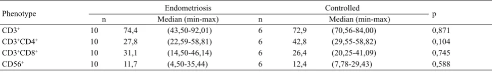
In this study, samples consisted of PBMC from endo-metriosis patients (n=10; 2 moderate and 8 severe stage), and control (n=6). Before stimulation with IL-2 on PBMC from endometriosis and controls, CD3+CD4+ (helper T cells), CD56+ (NK cells) phenotype in endometriosis group was lower than the control group although there was no signifi cant difference. Furthermore, the CD3+CD8+ (cytotoxic T cells) of endometriosis group was higher than the control group without any signifi cant difference. It appears that the ratio of CD4+(helper T cells) to CD8+ (cytotoxic T cells) in the endometriosis group is less than one (Table 1).
Cytotoxicity test before IL-2 stimulation revealed that
cytotoxic T cells in endometriosis group were lower than the ineffective control group. Moreover, the NK cells in endometriosis group were signifi cantly lower than those in control group. The cytotoxicity of PBMC from endometriosis group was also lower compared to the control group. It appears that even though the toxicity was lower than control, PBMC from the endometriosis group could react effectively towards endometrial cell cultures before IL-2 stimulation (Table 2).
Phenotype Endometriosis Controlled p
n Median (min-max) n Median (min-max)
CD3+ 10 74,4 (43,50-92,01) 6 72,9 (70,56-84,00) 0,871
CD3+CD4+ 10 27,8 (22,59-58,81) 6 42,8 (29,55-58,82) 0,104
CD3+CD8+ 10 31,1 (14,50-46,14) 6 26,4 (20,25-41,09) 0,745
CD56+ 10 11,7 (4,50-35,44) 6 12,4 (7,78-29,43) 0,588
Table 1. Characteristics of controlled PBMC and endometriosis phenotype
The phenotypes of CD3+CD4+, CD3+CD8+and CD56+ from both endometriosis and control group were increased after stimulation with IL-2. Elevation of CD3+CD4+(helper T cells) and CD3+CD8+ (T cytotoxic cells) after stimulation with IL-2 were signifi cantly higher (p = 0.005 and p =0.007, respectively) than before treatment. It has also been found that the ratio of CD4+ (helper T cells) to CD8+ (T cytotoxic cells) in PBMC from endometriosis group was greater than 1after stimulation with IL-2 (Table 3, Figure 1, Figure 2, and Figure 3).
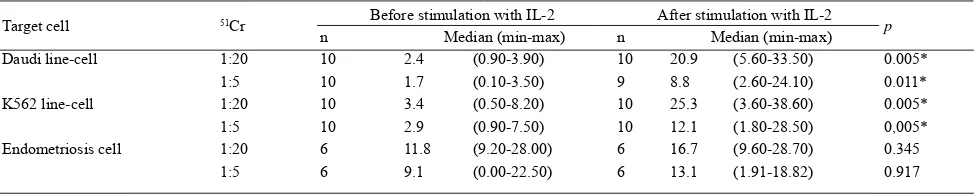
The cytotoxicity of LAK cells from endometriosis group
towards Daudi, K562cell lines, and endometriosis cell cultures was increased after IL-2 stimulation. It seems that cytotoxic T cells from endometriosis group could response effectively towards Daudi cell line after IL-2 stimulation by activation into LAK cells. Likewise, cytotoxic T cells in control group could also react towards Daudi cell line after IL-2 stimulation with higher effi ciency than the endometriosis group. Furthermore, NK cells in endometriosis group could respond more potently towards K562 cell line after IL-2 stimulation. However, its toxicity was lower than control group.
n Phenotype Before stimulation with IL-2 After stimulation with IL-2 p
Median (min-max) Median (min-max)
Control group:
6 CD3+ 72.9 (70.56-84) 73.1 (63.62-88.61) 0,753
6 CD3+CD4+ 42.8 (29.55-58,82) 62.6 (33.4-70.26) 0.028*
6 CD3+CD8+ 26.4 (20.25-41.09) 27.8 (13.77-40.82) 0.917
6 CD56+ 12.4 (7.78-29.43) 15.1 (4.88-33.48) 0,173
Endometriosis group:
10 CD3+ 74.4 (43.50-92.01) 81.4 (70.78-96.63) 0.007*
10 CD3+CD4+ 27.8 (22.59-58.81) 48,5 (29.14-65.78) 0.005*
10 CD3+CD8+ 31.1 (14.50-46.14) 39,3 (27.28-59.63) 0.007*
10 CD56+ 11.7 (4.50-35.44) 15,4 (7.04-42.20) 0.007*
Table 3. PBMC phenotype characteristic groups before and after stimulation with IL-2
Wilcoxon test; phenotype unit (%); CD3+ (T cells); CD3+CD4+ (helper T cells); CD3+CD8+ (cytotoxic T cells); CD56+ (NK cells); signifi cant if p<0,05
Target cell 51Cr Before stimulation with IL-2 After stimulation with IL-2 p
n Median (min-max) n Median (min-max)
Daudi line-cell 1:20 10 2.4 (0.90-3.90) 10 20.9 (5.60-33.50) 0.005*
1:5 10 1.7 (0.10-3.50) 9 8.8 (2.60-24.10) 0.011*
K562 line-cell 1:20 10 3.4 (0.50-8.20) 10 25.3 (3.60-38.60) 0.005*
1:5 10 2.9 (0.90-7.50) 10 12.1 (1.80-28.50) 0,005*
Endometriosis cell 1:20 6 11.8 (9.20-28.00) 6 16.7 (9.60-28.70) 0.345
1:5 6 9.1 (0.00-22.50) 6 13.1 (1.91-18.82) 0.917
Wilcoxon test; Toxiticity value in %; Cytotoxicity value if effective if more than 10%, and ineffective if less than 10%); Signifi cant if p<0,05; PBMC = peripheral blood mononuclear cell
Table 4. Cytotoxicity test with 51Cr 1:20 and 51Cr 1:5 calibration in PBMC endometriosis group before and after stimulation with IL-2 against Daudi line-cells, K562 line-cells, and cultured endometriosis cell
Target cell 51Cr Endometriosis Control p
n Median (min-max) n Median (min-max)
Daudi cell line 1:20 10 2.4 (0.90-3.90) 5 6.0 (1.80-9.30) 0.125
1:5 10 1.7 (0.10-3.50) 5 2.5 (0.60-5.60) 0.297
K562 cell line 1:20 10 3.4 (0.50-8.20) 5 24.1 (1.30-38.20) 0.050*
1:5 10 2.9 (0.90-7.50) 5 9.7 (0.80-18.30) 0.066
Endometriosis cells 1:20 6 11.8 (9.20-28.00) 4 21.7 (13.50-28,30) 0.136
1:5 6 9.1 (0.00-22.50) 4 17.4 (12.10-22.00) 0.286
Table 2. Cytotoxicity test with 51Cr 1:20 and 51Cr 1:5 calibration on PBMC in endometriosis and control groups before to stimulation with IL-2 on Daudi, K562 cell lines and endometriosis cell cultures
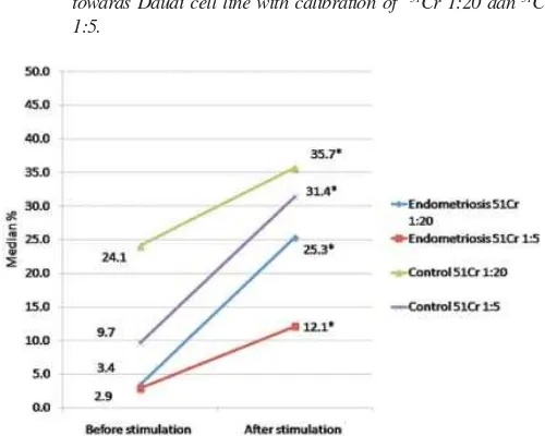
Figure 1. Endometriosis and control group before and after IL-2 stimulation towards Daudi cell line with calibration of 51Cr 1:20 dan 51Cr
1:5.
Figure 2. PBMC cytotoxicity and endometriosis LAK cells group and the control before and after stimulation with IL-2 towards K562 cells-line with calibration of 51Cr 1:20 and 51Cr 1:5.
DISCUSSION
Endometriosis is an endometrial-like benign disorder which grows outside the uterine cavity, and could trigger chronic infl ammatory reaction.1-4 Theories have been proposed to explain the pathogenesis of endometriosis. Immune related theory was one of the most attractive theories which could interrelate with other theories. Thus, immune factors may play an important role to prevent and overcome endometriosis regardless its pathogenesis. Endometriosis associated immunological defect, particularly in NK cells function and activity, has been widely reported. In this study we demonstrate a defect in PBMC’s NK cells from endometriosis group before IL-2 stimulation towards K562 cell line. Impairment of NK cells function from endometriosis group was also supported by the fact that cytotoxicity of LAK cells after IL-2 stimulation was lower than control group.
Impairment of NK cells function could be repaired by stimulating PBMC from endometriosis patients with IL-2 to activate them into LAK cells. Study by Roussel19 showed an increased proliferation of CD3+ after IL-2 stimulation related to the formation of LAK cells. Furthermore, Oosterlynck et al17 did not
fi nd any signifi cant difference between the endometriosis (n=13) and control group (n=5) without IL-2 stimulation.
Difference in immune response is also affected by genetic factors. Hsieh et al20 reported an association of homozygous polymorphism of IL-2 receptor beta (IL-2Rbeta)-627*C with endometriosis. Moreover, Kitawaki et al21 demonstrated that the genotype of killer cells immunoglobulin-like receptors (KIRs) is correlated with susceptibility towards endometriosis. Our study found a signifi cant increase of the phenotype CD3+CD4+ (helper T cells), CD56+ (NK cells) after IL-2 stimulation in PBMC of endometriosis group. There was also elevation in LAK cell cytotoxicity from endometriosis group towards Daudi, K562 cell lines and endometriosis cell cultures after IL-2 stimulation. From this study, we assume that IL-2 can stimulate activation of PBMC into LAK cells which are more potent in cytotoxicity and not associated with MHC antigens.15
In this study, the increase of LAK cells cytotoxicity from endometriosis group is lower than control. Szyllo et al22 reported that IL-2 concentration in PBMC from endometriosis patients was higher (8.3±7.13) than control (2.8 ±1.28) after stimulation with phytohemagglutinin (PHA). Oosterlynck et al17stated that there is no intrinsic abnormality found in lymphocyte cells of endometriosis patients since they are still capable to respond after IL-2 stimulation. Tsudo et al23 described an important role of p75 peptide as receptor of IL-2 on large granular lymphocytes in the activation of these cells into LAK cells. Disruption
in affi nity between p75 and IL-2 would cause a decrease in proliferation response after IL-2 stimulation in patients with leukemia. Therefore, the role of p75 in pathogenesis of endometriosis should be further elaborated.
In conclusion, defect of cellular immunity in endo-metriosis could be repaired by stimulating PBMC from endometriosis patients with IL-2 into LAK cells. Activation of PBMC into LAK cells after IL-2 stimulation in endometriosis group resulted in increased cellular immunity towards endometriosis cell cultures.
Acknowledgments
We thank the staffs of INSERM U976 Saint Louis Hospital-Paris VII, for their support in conducting this research. We also thank MTIE FMUI and Division of Reproductive Immunoendocrinology FMUI, especially to Professors Endy M. Moegni, Ali Baziad, Soetarti Eko Prasetyaningsih, Eva, Nilda, and Neneng.
REFERENCES
1. Haney AF. Etiology and histogenesis of endometriosis. Prog Clin Biol Res. 1990;323:1-14.
2. Jacoeb TZ. Faktor imunoendokrinologis dan seluler lingkungan mikro zalir peritoneal pada infertilitas idiopatik wanita. [disertasi]. Jakarta: Universitas Indonesia; 1990. 3. Missmer SA, Cramer DW. Epidemiology of endometriosis.
In: Olive DL, editor. Endometriosis in clinical practice. London: Taylor & Francis; 2005. p. 49-60.
4. Olive DL, Schwartz LB. Endometriosis. N Engl J Med. 1993;328(24):1759-69.
5. Oepomo TD. Peran interleukin-6 serta interleukin-8 dalam zalir peritoneal pada infertilitas disertai endometriosis dalam proses apoptosis sel granulosa ovarii yang patologis (suatu pendekatan imunopatologi). Maj Obstet Ginekol Indones. 2005;16:26. 6. Witz CA, Allsup KT, Montoya-Rodriguez IA, Vaughan SL,
Centonze VE, Schenken RS. Pathogenesis of endometriosis--current research. Hum Fertil (Camb). 2003;6(1):34-40. 7. Bulun SE, Fang Z, Imir G, Gurates B, Tamura M, Yilmaz
B, et al. Aromatase and endometriosis. Semin Reprod Med. 2004;22(1):45-50.
8. Kitawaki J, Kado N, Ishihara H, Koshiba H, Kitaoka Y, Honjo H. Endometriosis: the pathophysiology as an estrogen-dependent disease. J Steroid Biochem Mol Biol. 2002;83(1-5):149-55.
9. Lebovic DI, Mueller MD, Taylor RN. Immunobiology of endometriosis. Fertil Steril. 2001;75(1):1-10.
10. Gagne D, Rivard M, Page M, Lepine M, Platon C, Shazand K, et al. Development of a nonsurgical diagnostic tool for endometriosis based on the detection of endometrial leukocyte subsets and serum CA-125 levels. Fertil Steril. 2003;80(4):876-85.
11. Claus M, Greil J, Watzl C. Comprehensive analysis of NK cell function in whole blood samples. J Immunol Methods. 2009;341:154–64.
receptors on peritoneal natural killer cells in women with endometriosis. Fertil Steril. 2000;74(6):1187-91.
13. D’Hooghe TM. Clinical relevance of the baboon as a model for the study of endometriosis. Fertil Steril. 1997;68(4):613-25. 14. Farag SS, Caligiuri MA. Human natural killer cell
development and biology. Blood Reviews. 2006;20:123–37. 15. Matsubara K, Nagamatsu T, Fuji T, Kozuma S, Taketani Y.
Lymphokine-activated killer cells induced from decidual lymphocytes reduce the angiogenic activity of trophoblasts by enhancing the release of soluble fms-like tyrosine kinase-1 from trophoblasts: An implication for the pathophysiology of preeclampsia. J Reprod Immunol. 2005;68:27–37.
16. Lucidi RS, Witz CA, Chrisco M, Binkley PA, Shain SA, Schenken RS. A novel in vitro model of the early endometriotic lesion demonstrates that attachment of endometrial cells to mesothelial cells is dependent on the source of endometrial cells. Fertil Steril. 2005;84(1):16-21.
17. Oosterlynck DJ, Lacquet FA, Waer M, Koninckx PR. Lymphokine-activated killer activity in women with endometriosis. Gynecol Obstet Invest. 1994;37(3):185-90. 18. Oner Ozdemira SS. Combinational IL-2/IL-15 induction does
not further enhance IL-15-induced lymphokine-activated killer cell cytotoxicity against human leukemia/lymphoma cells. Clin Immunol. 2005;115:240–9.
19. Roussel E, Gerrard JM, Greenberg AH. Long-term cultures of human peripheral blood lymphocytes with recombinant human interleukin-2 generate a population of virtually pure CD3+CD16-CD56- large granular lymphocyte LAK cells. Clin exp Immunol. 1990;82:416-21.
20. Hsieh YY, Chang CC, Tsai FJ, Hsu CM, Lin CC, Tsai CH. Interleukin-2 receptor beta (IL-2R beta)-627*C homozygote but not IL-12R beta 1 codon 378 or IL-18 105 polymorphism is associated with higher susceptibility to endometriosis. Fertil Steril. 2005;84(2):510-2.
21. Kitawaki J, Obayashi H, Kado N, Ishihara H, Koshiba H, Maruya E, et al. Association of HLA class I and class II alleles with susceptibility to endometriosis. Hum Immunol. 2002;63(11):1033-8.
22. Szyllo K, Tchorzewski H, Banasik M, Glowacka E, Lewkowicz P, Kamer-Bartosinska A. The involvement of T lymphocytes in the pathogenesis of endometriotic tissues overgrowth in women with endometriosis. Mediators Infl amm. 2003;12(3):131-8. 23. Tsudo M, Goldman CK, Bongiovanni KF, Chan WC,