BRAIN LESION CLASSIFICATION BASED ON STATISTICAL DISCRIMINATION FOR MAGNETIC RESONANCE IMAGING (MRI) IMAGE
SYED MOHD SYAFIQ BIN ABU BAKAR
This Report Is Submitted In Partial Fulfilment Of Requirements For The Bachelor Degree of Electronic Engineering (Electronic Engineering)
Fakulti Kejuruteraan Elektronik Dan Kejuruteraan Komputer Universiti Teknikal Malaysia Melaka
UNIVERSTI TEKNIKAL MALAYSIA MELAKA FAKULTI KEJURUTERAAN ELEKTRONIK DAN KEJURUTERAAN
KOMPUTER
BORANG PENGESAHAN STATUS LAPORAN
PROJEK SARJANA MUDA II
Tajuk Projek :
BRAIN LESION CLASSIFICATION BASED ON STATISTICAL DISCRIMINATION FOR MAGNETIC RESONANCE IMAGING (MRI) IMAGE.
Sesi
Pengajian : 1 3 / 1 4
Saya SYED MOHD SYAFIQ BIN ABU BAKAR mengaku membenarkan Laporan Projek Sarjana Muda ini disimpan di Perpustakaan dengan syarat-syarat kegunaan seperti berikut:
1. Laporan adalah hakmilik Universiti Teknikal Malaysia Melaka.
2. Perpustakaan dibenarkan membuat salinan untuk tujuan pengajian sahaja.
3. Perpustakaan dibenarkan membuat salinan laporan ini sebagai bahan pertukaran antara institusi
pengajian tinggi.
4. Sila tandakan ( √ ) :
SULIT* *(Mengandungi maklumat yang berdarjah keselamatan atau kepentingan Malaysia seperti yang termaktub di dalam AKTA RAHSIA RASMI 1972)
TERHAD** **(Mengandungi maklumat terhad yang telah ditentukan oleh
organisasi/badan di mana penyelidikan dijalankan)
TIDAK TERHAD
Disahkan oleh:
__________________________ ___________________________________ (TANDATANGAN PENULIS) (COP DAN TANDATANGAN PENYELIA)
TABLE OF CONTENTS
CHAPTER TITLE PAGE
PROJECT TITLE i
REPORT STATUS VERIFICATION FORM ii
DECLARATION iii
SUPERVISOR DECLARATION iv
DEDICATION v
ACKNOWLEDGEMENT vi
ABSTRACT vii
ABSTRAK viii
TABLE OF CONTENTS ix
LIST OF TABLE xii
LIST OF FIGURE xiv
LIST OF ABBREVIATIONS xvi
CHAPTER 1 INTRODUCTION
1.1 Project background 1
1.2 Problem statements 2
1.3 Objective 3
1.4 Scope of project 3
1.5 Thesis Methodology 3
CHAPTER 2 LITERATURE REVIEW
2.1 Magnetic Resonance Imaging (MRI) 6
2.2 Funtional Block Diagram (MRI) 8
2.2.1 Static Magnetic Field 9
2.2.2 Gradient Coil 9
2.2.3 Transmit/Receive Coil 11
2.2.4 RF Reciever 11
2.2.5 Transmitter 12
2.3 Types Brain Disease 14
2.3.1 Stroke 14
2.3.2 Tumor 15
2.3.3 Chronic Stroke 16
2.3.4 Necrosis 17
2.4 Current technology medical imaging software 18
2.4.1 Osirix 18
2.4.2 3D Slicer 21
CHAPTER 3 METHODOLOGY
3.1 Introduction 24
3.2 Project flow chart 25
3.3 MRI Image 27
3.4 Image Preprocessing 29
3.5 Segmentation 30
3.6 Feature Extraction 31
CHAPTER 4 RESULT & DISCUSSION
4.1 Introduction 34
4.2 Graphical User Interface (GUI) 35
4.3 Region Of Interest (ROI) 36
4.4 Feature Extraction 38
4.5 Range Of Feature 42
4.6 Rule Based Classifier 45
4.7 Classification Performance Analysis 51 4.7.1 Training Sample For 30% 51 4.7.2 Training Sample For 50% 52 4.7.3 Training Sample For 70% 52 4.7.4 Training Sample For 90% 53
CHAPTER 5 CONCLUSION
5.1 Conclusion 55
5.2 Future Work 56
REFERENCES 57
LIST OF TABLE
No TITLE PAGE
2.1 OsiriX Current Features 19
4.1 MRI Segmentation image 37
4.2 Input classifier 42
4.3 Input Classify (30%) 43
4.4 Input Classify (50%) 43
4.5 Input Classify (70%) 43
4.6 Input Classify (90%) 44
4.7 Stroke Feature 46
4.8 Stroke Input Classify 46
4.9 Stroke Result 47
4.10 Tumor Feature 47
4.11 Tumor Input Classify 47
4.12 Tumor Result 48
4.13 Chronic Stroke Feature 48
4.14 Chronic Stroke Input Classify 48
4.15 Chronic Stroke Result 49
4.16 Necrosis Feature 49
4.18 Necrosis Result 50
4.19 Result Classification for 30% 51
4.20 Result Classification for 50% 52
4.21 Result Classification for 70% 53
LIST OF FIGURE
No TITLE PAGE
2.1 MRI Scanner. 7
2.2 Block Diagram MRI 8
2.3 Brain Stroke. 13
2.4 Brain Tumor 15
2.5 Brain hemorrhagic stroke. 16
2.6 Brain Necrosis 17
2.7 Osirix User Interface 20
2.8 3D Slicer User Interface 23
3.1 Flow Chart 25
3.2 Graphical Classification Flow 26
3.3 Stroke 28
3.4 Tumor 28
3.5 Chronic Stroke 28
3.6 Necrosis 28
3.7 Original Image 29
3.7 Pre-processing Image 29
4.2 Mode versus Standard Deviation 38
4.3 Compactness versus Mean Boundary 39
4.4 Median versus Mean 40
4.5 Perimeter versus Area 41
4.6 Characteristic ROI features 45
LIST OF ABBREVIATIONS
MRI - Magnetic Resonance Imaging ROI - Region Of Interest
I declare that this project report entitled “Brain lesion classification based on statistical
discrimination for magnetic resonance imaging (MRI) image”, is the result of my own
research except as cited in the references.
Signature: ……… Name: SYED MOHD SYAFIQ BIN ABU BAKAR
I hereby declare that I have read this project report and in my opinion this thesis is sufficient in terms of scope and quality for the award of degree of Bachelor of Electronic
Engineering (Computer Engineering).
ACKNOWLEDGEMENT
Alhamdulillah. ‘In the name of Allah, most gracious, most merciful’. Firstly, I would like to extend my deep gratitude towards the almighty Allah S.W.T because of His mercy and kindness, I was able to complete my Final Year Project and thesis. In this final year project, I have learned a lot of valuable knowledge. On this occasion, I would like to thanks to all parties involved to complete this project.
I would like to express profound gratitude to my final year project supervisor, Norhashimah Binti Mohd Saad for her invaluable support, encouragement, supervision and useful knowledge throughout this duration of my project.
I would like to express my appreciation especially to my late father Abu Bakar Bin Habib Jaafar and my beloved mother Nurbibah Bt Kandiluk because they always support me and was give me a lot of motivation and pray for my success to complete the task of Final Year Project. For my little sister, thanks a lot because give me a spirit and support everytime.
Finally, I am also thankful to my colleagues of Electronic and Computer Engineering and to all my friends in Universiti Teknikal Malaysia Melaka for their assistance and understanding.
Thank you so much,
ABSTRACT
ABSTRAK
CHAPTER 1
INTRODUCTION
1.1 Project Background
In this project, brain MRI images are analyzed to develop a brain MRI classification system. This project aim is to discriminate brain lesions and diseases in MRI brain images using statistical analysis and digital image processing algorithm. Types of diseases are solid tumor, stroke, chronic stroke and necrosis.
Brain lesion such as tumor is one of the most brain lesions that are affected in the brain cerebrum. Nowadays, tumor and stroke diseases were the third and fourth leading cause of death in Malaysia. Detection and diagnosis of brain lesion is the key for implementing successful therapy and treatment planning. By using the Magnetic Resonance Imaging MRI, the image will show clearly.
The purpose of this project is to classify the lesion based on statistical discrimination of abnormalities. There are many techniques in order to detect types of lesion such as support vector machine (SVM), K-Mean Clustering, Principle Component Analysis (PCA) and etc. In this project, technique that is used to classify the lesion is based on ruled based classifier. From the image, the information will be obtained by taking the statistical calculation of the Region Of Interest (ROI). The information is used as feature and also as input to the classifier.
1.2 Problem Statement
1.3 Objective
The objectives in this project is :
1. To design method of brain MRI features extraction and classification system. 2. To analyze statistical value of different abnormalities.
3. To verify the performance of brain classification system.
1.4 Scope Of Project
There are 3 scopes for this project:
1. The segmentation Region Of Interest (ROI) of brain lesion is draw manually and not fully automatic.
2. The classification technique focus only on statistical discrimination technique of different brain lesion.
3. The proposed algorithm of MRI brain classification system will be written in MATLAB software.
1.5 Thesis Methodology
• MRI brain images
• Image Pre-processing
Image database are original captured from MRI. It means that the images are not process yet. In this section, the images was normalize before proceed to
segmentation.
• Segmentation
Segmentation purpose is to obtain the Region Of Interest (ROI). This process are done manually using handfree tool from MATLAB software.
• Feature Extraction
Feature extraction process is to obtain the information from ROI image. This process was done using statistical approach.
• Classification
The statistic value was used as input to the classifier. The classification technique that be used is rule based classifier.
1.6 Organization
Chapter two is the literature review about the Magnetic Resonance Imaging (MRI) and functional block diagram MRI. This chapter also reviews on types of disease that effected in human brain. Then, review on current technology medical imaging.
Chapter three explains about methodology of the project. In this project, there are five stages to complete the classification. Begin with MRI image, pre-processing, manual segmentation, feature extraction and lastly classification. The flow chart of the project and the algorithms use is explained in this chapter.
CHAPTER 2
BACKGROUND STUDY
2.1 Magnetic Resonance Imaging (MRI)
The image and resolution produced by MRI is quite detailed and can detect tiny changes of structures within the body. For some procedures, contrast agents, such as gadolinium, are used to increase the accuracy of the images[2].
An MRI scan can be used as an extremely accurate method of disease detection throughout the body and is most often used after the other testing fails to provide sufficient information to confirm a patient's diagnosis. In the head, trauma to the brain can be seen as bleeding or swelling. Other abnormalities often found include brain aneurysms, stroke, tumors of the brain, as well as tumors or inflammation of the spine.
After the MRI scanning is completed, the computer generates images of the area of the body that was scanned. These images can be transferred to film. A radiologist who is specially trained will interpret and diagnose images. The interpretation is transmitted in the form of a report to the practitioner who requested the scan. The diagnosis results will be used for further treatment of the diagnosis. Figure 2.1 below show the MRI scanner hardware capture the brain images.
2.2 Funtional Block Diagram (MRI)
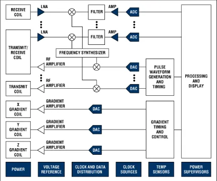
Figure 2.2 Block Diagram MRI