Yol 9, No 4, October
-
December 2000 Cytoloxicity ofJatropha curcaslatex
253
Cytotoxicity
of
Jatropha
curcas
@uphorbiaceae) latex
on
fïbroblast by MTT
assay
Fazwishni Siregar,* Siti Mardewi
SoeronoAkb#
Abstract
The latet ofJatropha carcas (Euphorbiaceae) had been used as traditional plant medicine among others to cure toothache. Despite its long time use, not many scientific research on this lalex were reporled. The aim
of
this study was to evaluate its cytotoricity on cell culture. Lalex was lyophilized and storedû
-2dCfor
standardization. Freshlaiet
was aiound l|%freeze-drted latex. Fibroblast L929 cell line were exposed to 37-10.0001q/nl
latet in mediumforI,
2, and 3 days and the cytotoxicity was evaluated by MTT assay.The result showed that the number of cells was
haf
of contrbl at concentralionof
625 14/ml and no viable cells were found at concentralion of 2500 1q/ml freeze dried latex. Lower concenlralion of latex was needed to yield similar eîect to human gingival Jibroblast primary cells. After 2 days the number of gingival libroblast cells was nearly hatf of that of controlar
150 14/ml tatexsolutions.
It
is concluded, that J. curcas latet was q)totoxic to Fib L929 and gingivalfibroblasl cells.Abstrak
Getah Jalropha curcas (getahjarak, Euphorbiaceae) telah digunakan sebagai obat tradisional antara lain untuk menanggulangi nyert
pulpa
ifianini
adalah untuk mengetahui syaTlttotottstiitasgetah
dan disimpan pada-2dC.
Xonseniasi getah segaradala
n gelah 37-10.000 14/ml medium kuhur selamat,2,
dan 3 hàn.Kemudian
sdat
sitotolæisitas getah dievaluasi dengan assay MTT.Hasil
nenunjukkan bahwa absorbansi sel Fib L929 menjadisetengah
dai
absorbansi kelompok kontrol pada 625g/ml,
dan tidak ada sel yang hidup pada konsentrasi gelah 2500 1q/ml. Efek yang sama terhadapJibroblast gingiva manusia ditemukan pada ladar getah yang lebih rendah. Sesudah 2 hirt, jumlah sellarobiast gingiva manusia mendekati setengah iumlah sel kontrol pada larutan getah 15014/ml.
Kesimpulan adalah gelah J. carcas bersifat sitotolrsik terhadap sel Fib L929 danJibroblast gingiva manusia.Kqmords: Jatropha curcas latex, cytotoxicity, MTT assay, Jibroblast.
Jatropha curcas)
an Euphorbiaceae,is
a shrubor
treefound
in
Indonesia
andother
tropical
areas.Its local
names
are
balacai
(Sulawesi),
naïvaih
nawas (Sumatera Barat),jarak
(Jawa Barat),jarak
pagar andmany
others.
Latex of
Jatropha curcas
containstannins, saponin,
wal(, and resin.l2 From the latex
were
isolated aproteolytic
enrqecalled
curcain3 anda
cyclic
octapeptide named curcacyclin
A
which
inhibits the classical pathway
of
human complement
andproliferation of
human T-cells.aLatex of,/.
curcas showed ananti
against Staphylococcus aureus 5 anda
,rity itt tn ittio
test.6In
tropical
Africa
and SoutheastAsia
thelatex is
used asa
hemostatic, wound dressing,
and
is
said
to
be'Orol
Biologt, Department of Oral Biologt, Facultyof
Dentistry, Universitlt of Indonesia, Jakarta, Indonesia * Conservative Denlistry, Faculty of Dentistry, Universityof
Indone s ia, Jalcart a, Indonesia
effective
in
treating
scabies,
eczema,and ringworm.
Furthermore,
it
is
used
as
mouth rinse to
heat
bleeding
gums,to touch
ababy's inflamed
tongue and to cure tootache.l' 2' 7' 8'MTT is
amicroplate
assayrequiring no cell tansfers.
This method was
adaptedfor measuring proliferation
andcytotoxicity in
sensitive,rapid,
and semiautomatic mannerwithout
radioactive
isotope.l0 TheMTT
assay254
Siregar and AkbarThe
solubility of
MTT-reducedformazan product
waspoor in
acid isopropyl alcohol
(as usedin
the methodof Mosmann)ll while
better absorption
characteristicswere
observed
with
mineral
oil
and
DMSO
as solvents.l3The
amountof
formazanproduct
generatedand then
measured
after solubilization
in
DMSO
isproportional to cell number, although absolute absorbance
for
agiven cell
number varies betweencell
lines.l3Despite
the
long time use
of J.
curcas as
a plant
medicine
among
other
to
cure
toothache,
not
many
scientific
researchswere
conducted.
After
the
latex
wasput
in
direct
contactwith
the dentalpulp,
thepain
would
disappear.
The
purpose
of
the study was
to
explore
the
mechanism
of
pain relief
and
onepossibility is
that the latex
is cytotoxic. In this
study,cytotoxicity
of
latex
of
J.
curcas was
evaluated by
MTT
assay
on Fib
L929
and
human
gingival
fibroblast primary culture.
METHODS
RPMI
I 640(Sigma), Penicillin-streptomycin
(Sigma),MTT
(Sigma),
fetal
bovine
serum
(FBS,
Gibco),
Fungizone
(Gibco),
Hepes
(Gibco)
and
DMSO
wereused
in
this
study. Fibroblast
L929
cell
line
andhuman gingival fibroblast were
kindly donated
by
Moekti
G.,
ResearchInstitute for Veterinary
Sciences,Bogor,
and LeyhausenG., Hannover, respectively. To
standardizethe
sample,latex
of Jatropha
curcas was
obtained
from
./.
curcas trees grown
in
ResearchInstitute
for
Plant Medicine
and
Spices,
Bogor.
Subsequently, thelatex were
lyophilized
50 hours andstored
at
-200C.
V/e
found
that
fresh latex
wasequivalent
with
15%solution
of
freeze-dried latex.Cell
culture
Fibroblast
L929
andhuman
gingival fibroblast
werecultured
as monolayers
in
RPMI
1640
medium
andDMEM
respectively,
supplemented
with
l0%
(vlv)
fetal bovine serum (FBS),
25
mM
Hepes,
100.000IUL
penicillin,
100
mg/L
streptomycin,
and, 2,5mglL
fungizone.Growing
cultures weremaintained in
25
cm2
culture flasks
at
370C
in a
humidified
atmosphere
of
5%o COzin
air. Medium
was changed 2times
aweek or when
there was apH
decrease shownby
colored
changes
in
the medium.
At
confluency,
they were
harvested
by trypsination
and counted
by
trypan
blue
staining.Med J Indones
MTT
assayInto
each well
of
monolayer fibroblast
in
96-wells
microplate,
20
pl
of
5
mg/ml
MTT,
was
added andincubated at
37uCfor
a further
4
hours.Medium
wasthen aspirated and
50
pl DMSO
were
addedinto
eachwell.
After
15minutes
of
incubation, the
absorbance(OD) were
determined
by ELISA
microplate
reader(Titertek Multiskan
MCC)
at
wavelength
540
nm substractedby
690 nm.Optimalization
To
evaluatethe cytotoxicity
of
latex on cell
culture,
certain amount
of
latex was
added
into the
medium
which
was
supplementedwith FBS.
In
our work
wefound precipitation formed
in
medium when
addedwith
latex. The higher theFBS
and latex concentration, the more precipitation formed. Therefore, optimalizationof
FBS
and
latex
concentrations used in
experiment should be done. The amountof
FBSin
medium assayedwas
0olo,50Â and
l0%,
while
latex
concentration assayedwas
from
150.000
p{ml
(15%)
and diluted
byhalf
until
37.5p{nl
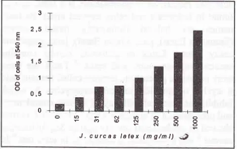
medium (0.003%).We
also
found that diluted latex
in
medium
had
itsown optical
density
(OD).
At
37.5
p{mt
to
150.000pglml
the
OD
at 540
nm
ranged
from
0.080
to
1.288(figure
l).
Consequently,
the cytotoxicity
data
wereobtained
from the OD
of
cells after
exposureto
thediluted latex
substractedby
theOD
of
latexin
mediumwith
the same concentration as those usedfor
cells.E
c
o
t
o I oo o oo
3 2,5
2
1,5
1
0,5
0
OOFNAOôoFo@NOOO
FNOo
[image:2.595.339.573.494.641.2]J. curcas
latsx
(mg/nl)
".?Figure
l.
OD at 540 nm ofseveral concentrationofJ.
curcas latex in medium. J. curcas solution has its own absorbanceCytotoxicity
testof latex
ofJatropha
curcasOne hundred
and
fifty
microlitre
of 40.000 cells/ml
Yol 9, No 4, Oclober
-
December 2000confluency, the medium was
aspirated
and
changedwith
a
new
medium
addedby
several concentrationsof
latex
supplementedby FBS
and
antibiotics.
Sameconcentrations
of
latex
in
medium (without
cells)
were
also
put into
other wells
and treated as
thosewells
with
cells. Three-fold replication
lvas conductedthrought
out the
experiment.
After
certain
exposuretime
with
latex, the
cytotoxicity
of
latex
wasevaluated by MTT
assay.The cytotoxicity
data
was obtainedfrom
the OD
of
cells
which
were
exposedto
diluted latex
substractedby
theOD of diluted
latex.RESULT
Optimalization
of
FBS
concentration
in
medium
showed that
with l0%
FBS
there was moreprecipitate
formed than
with
5% FBS, while 0% FBS
showed lessercell
number
comparedto
those
of
l0%o.OD
at540
nm
of
cells
in
medium
supplementedwith
10%FBS
and treatedwith latex
15to
1000pglml
rangedfrom
0.648
to 0.351.
While
for
the cells
in medium
treated
with
5YoFCS,
the OD
ranged
from
0.730 to
0.407.
Moreover,
sampleswith
5% FBS was
easierto
manipulate, because there was less
precipitate
formed.Therefore,
medium
supplementedwith
5% FBS
was usedfor
experiment. Optimalization
for
concentrationof latex
from
36 to
150.000pglml
showedthat
therewere significant
difference between 1100
pglml
and2300
p,g/mL
The
OD
was nearly
0
at
2300 pglml
latex,
while at
ll00
the
OD
was around 0.500.
Thenwe
chose5000
pglml
latex
in
medium
asthe
highestconcentrations
for experiment.
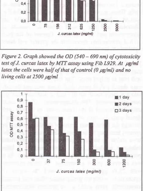
MTT
assay
of
Fib
L929 cells after
exposureto
different concentration
of
latex
showedthat the
OD
was lowered
at 312
pg/ml
latex and
became half
of that of control (0 pglml
latex)
at
625
ltg/ml. No living
cells were
observed atlatex of 2500 pglml (figure 2). The
various
time of
exposure
which was
l,
2,
and
3 days
at
the
sameconcentration
showed
no difference
at OD
values.Clotoxicity
in
human
gingival fibroblast
showed noliving
cells
at
1200pglml
latex,
andOD
becamehalf
of
thatof control at
150pglml
(figure
3).DISCUSSION
The
aim
of
this
study was
to
explore the
mechanismof
how dental
pulpal
pain
disappeared
with
norecrrrence
if
J. curcas
latex is
put into
the
cavity
formed in
dental caries tohave a direct contact with
Cytotoxicity of Jatropha carcas latet
255
1,4
12
-
1,0 3I
o,aF
E 0,e
ôo0,4
o2
0,0
ORESR388
Fo6NRg J.cæætdax(nO/H)
Figure 2. Graph showed the OD (540
-
690 nn) olcytotoxicitytest of J. curcas
latq
by MTT assay using Fib L929.At
tA/ml
latex the cells were half of that of control (0 1tg/ml) and no
[image:3.595.335.567.88.251.2] [image:3.595.335.569.170.483.2]living cells at 2500 1q/ml
Figure 3. Graph of OD (540
-
690 nm) of cytotoxicity test of J.curcas latex by MTT assay using human gingival cells. At day
2, the amount of cell was
haf
ol that of control at I 5014/nl
late+ while was no living cells was observed at 1200 1q/ml latex
the
pulp.
Onepossibility
is that the latex is cytotoxic,
hence, the
pulp in
contact will
benecrotized,
and as a consequence therewill
be no more pain.The
experiment
showed that the latex wascytotoxic to
fibroblast
cell line
and primary
cell culture.
Thecytotoxic effect was
obtained
in
low
concentration, 1250- 2500
pglml,
comparedto
the concentration
of
fresh
lateï
which
is
150.000
pglml. Besides,
it
isfound
that
the latex
has
the ability to
precipitate
theprotein
component
of the medium. However,
othercytotoxic
experiments
using agar overlay
techniqueshowed
that
the
cytotoxic effect was
limited
to
acertain zone
surounding
thelatex,
and doesnot cover
109 08
à
07H
ooF
05Ë
ono
03o2
01
0
oo ôo
@N
Yol 9, No 4, October
-
December 20(NKone-Bamba
D,
PelissierY,
OzoukouZF,
Kouao D. Haemostatic activityof fifteen
medicinal plantsof
thetraditional medecine
of
The Ivory Cost.
plant
MedPhytother 1987 ; 2lQ):122-30.
Burkill
IH.
A
dictionary of the eèonomic productsof
Malay Peninsula. London: The crown agents for colonies;
1935.
Heyne
K.
Tumbuhan berguna Indonesia.Vol II. lst
ed. Translated by Litbang Kehutanan: Jakarta; 1987.Suwondo S. Upaya meningkatkan manfaat tumbuhan obat
tradisional
Indonesiauntuk
pencegahalrkaries
dangingivitis berdasarkan
uji
aktivitas anti bakteri terhadapSfeptococcus mutans dan
uji
klinik
terhadap gingivitis [dissertation]. Bandung: Padjadjaran Univ.;
I 993.van de
LoosdrechtAA, Nennie E,
Ossenkoppele GJ,Beelen
RÉU, Langenhuijsen MAC.
Cell
mediatedCytotoxicity ofJatropha carcas
latu
257cytotoxicity against U937 cells by human monocytes and
macrophages
in
a
modified colorimetricMTT
assay.'J Immnnol Methl99l;
l4l:
15-22.Mosmann T. Rapid colorimetric assay for cellular growth and survival: application to proliferation and cytotoxicity assays. J Immunol Meth 1983; 65:55-63.
Berridge MV,
Tan
AS,
McCoy
KD, Wang
R.
Thebiochemical and cellular basis of cell proliferation assays that use tetazolium salts. Biochemica 1996;4:15-20. Carmichael
J, DeGraff
WG,
Gazdar A-F,Minna
JD,Mtchell JB. Evaluation of a tetazolium-based seniautomated
colorimetric assay:
assessmentof
chemosensitivity testing. Cancer Res 1987 ; 47 : 93642.Smulson
MH,
Sieraski SM. Histopathology and diseasesof
the
dentalpulp.
In: Weine
FS,
editor. Endodontic therapy. 4th ed. St Louis; The Mosby Co; 1989, p. 95-l 16.lt.
12
13.
14. 7.
8.