Jurnal
AgroBJogen
Vol. 8, No. 1, April 2012Jurnal AgroBiogen memuat artikel primer dan tinjauan hasil penelitian bioteknologi dan sumber daya genetik tanaman, serangga, dan mikroba pertanian. Jurnal ini diterbitkan tiga kali setahun pada bulan April, Agustus, dan Desember oleh Balai Besar Penelitian dan Pengembangan Bioteknologi dan Sumberdaya Genetik Pertanian . Jurnal ini sudah mendapat akreditasi ulang pada 24 April 2012.
SK Kepala LIPI Nomor: 395/D/2012 Tanggal 24 April 2012
Penanggung jawab
Kepala Balai Besar Penelitian dan Pengembangan Bioteknologi dan Sumberdaya Genetik Pertanian
Dewan Redaksi
BahagiawatiBioteknologi Pertanian
Karden MulyaHama dan Penyakit Tanaman
AsadiPemuliaan dan Cenetika Tanaman
I Made TasmaBioteknologi Pertanian
lswari S. DewiBioteknologi Pertanian
Mitra Bestari
I Cede SuastikaBiomolekuler dan Virologi
Ni Made Armini WiendiBioteknologi Tanaman
Bambang S. PurwokoBioteknologi Tanaman/Fisiologi
SutrisnoBioteknologi Pertanian
M . MachmudHama dan Penyakit Tanaman Pangan dan Hortikultura
Redaksi Pelaksana
Tri Puji PriyatnoJoko Prasetiyono Ida N. Orbani
Alamat Penerbit
Balai Besar Penelitian dan Pengembangan Bioteknologi dan Sumberdaya Genetik Pertanian Jalan Tentara Pelajar 3A, Bogor 16111, Indonesia
E-mail : [email protected]
Telepon: (0251) 8339793, 8337975 Faksimile: (0251) 8338820
Kala Terbit
Jurnal AgroBiogen 8(1 ):8-18
Indirect Organogenesis and Somatic Embryogenesis of
Pineapple Induced by
DichlorophenoxyAcetic Acid
Ika Roostika1
*, Ika Mariska
1, Nurul Khumaida2, and Gustaaf A. Wattimena3
1
1ndonesian Center for Agricultural Biotechnology and Genetic Resources Research and Development, Jl. Tentara Pelajar 3A, Bogar 16111 Phone (0251) 8337975; Facs. (0251) 8338820; *E-mail: [email protected]
2
Department of Agronomy and Horticulture, Bogar Agricultural University, Jl. Meranti, Campus Darmaga Bogar 16680
3Professor Emeritus in Department of Agronomy and Horticulture, Bogar Agricultural University, Jl. Meranti, Campus Darmaga Bogar 16680
Submitted: 29 September 2011; Accepted: 17 February 2012
ABSTRAK
Organogenesis dan Embriogenesis Somatik Tak Lang-sung pada Nenas yang Diinduksi oleh Dichlorophenoxy Acetic Acid. lka Roostika, Ika Mariska, Nurul Khumaida, dan Gustaaf A. Wattimena. Penelitian ini bertujuan untuk mengetahui pengaruh 2,4-D, AdS, dan media dasar dalam regenerasi tanaman nenas secara organogenesis dan embriogenesis somatik tak langsung. Induksi kalus dilaku-kan dengan perlakuan 2,4-D (21, 41, dan 62 11M) dengan penambahan TDZ 9 11M. Sel-sel yang non embriogenik di-regenerasikan secara organogenesis dengan memindahkan-nya ke media yang mengandung kinetin 4,65 11M. lnduksi kalus embriogenik dilakukan pada media MS atau Bac yang diperkaya dengan senyawa N-organik (Gin 6,8 11M, CH 500 mg 1·1
, Arg 0,69 mM, dan Gly 0,027 11M) dengan penambahan
AdS (0; 0,05; dan 0,1 11M). Kalus embriogenik diregenerasi-kan pada dua macam media. Media pertama adalah media MS modifikasi dengan penambahan IBA 0,9 11M, BA 1,1 11M, GA3 0,09 11M sedangkan yang kedua adalah media MS
de-ngan penambahan BA 0,018 mM. Hasil penelitian membukti-kan bahwa penggunaan 2,4-D secara tunggal tidak mampu menginduksi sel embriogenik sehingga sebagian kalus non embriogenik diregenerasikan secara organogenesis. Media terbaik untuk organogenesis adalah 2,4-D 21 11M karena menghasilkan persentase pembentukan kalus yang tinggi (80%), bobot basah kalus tertinggi (0,2 g/eksplan), dan jum-lah tunas tertinggi (sekitar 25 tunas/eksplan/2 bulan). Kalus embriogenik terbentuk ketika kalus non embriogenik diper-lakukan dengan senyawa N-organik. Media regenerasi me-rupakan faktor yang berpengaruh nyata terhadap tingkat pencoklatan. Media MS dengan penambahan BA 0,018 mM mampu menekan pencoklatan. Terdapat interaksi yang nyata antara media induksi kalus embriogenik dengan media regenerasi terhadap jumlah embrio somatik dewasa. Media MS yang diperkaya dengan senyawa N-organik dan AdS 0,05 11M yang dikombinasikan dengan media MS dengan penambahan BA 0,018 mM merupakan perlakuan terbaik untuk induksi kalus embriogenik dan regenerasi embrio somatik nenas (17 embrio dewasa/eksplan).
Kata kunci: Organogenesis, embriogenesis somatik, nenas, 2,4-D, adenin sulfat.
Hak Cipta © 2012, BB Biogen
ABSTRACT
Indirect Organogenesis and Somatic Embryogenesis of Pineapple Induced by Dichlorophenoxy Acetic Acid. Ika Roostika, lka Mariska, Nurul Khumaida, and Gustaaf A. Wattimena. This research aimed to study the effect of 2,4-D, AdS, and basal media to the regeneration of pineapple through indirect organogenesis and somatic embryogenesis, and to study the complete event of somatic embryogenesis. Callus formation was induced by 21, 41, and 62 11M 2,4-D with addition of 9 11M TDZ. The non embryogenic calli were transferred onto 4.65 11M Kn containing medium. Embryogenic callus formation was induced on MS or Bac basal media consisted of N-organic compounds with addition of AdS (0, 0.05 and 0.1 11M). The embryogenic calli were regenerated on modified MS medium with addition of 0.9 11M IBA, 1.1 11M BA, 0.09 11M GA3 or MS medium
supplemented with 0.018 mM BA. The result proved that the single auxin of 2,4-D was not enough to induce embryogenic cells. Therefore the non embryogenic calli were regenerated through organogenesis. The 21 11M 2,4-D yielded high level of callus formation (80%), higher fresh weight (0.2 g/explant) and higher number of shoot (25 shoots/explant in two months). Embryogenic calli were produced on N-organic compounds enriched media. The regeneration medium significantly affected the level of browning, where the MS medium with addition of 0.018 mM BA yielded lower level of browning. There was an interaction of embryogenic callus induction medium and regeneration medium to the number of mature somatic embryos. The embryogenic callus induction on MS medium enriched with N-organic compounds and 0.05 11M AdS followed by the regeneration of somatic embryos on MS medium with addition of 0.018 mM BA was the best treatment which yielded 1 7 mature somatic embryos/explant.
Keywords: Organogenesis, somatic embryogenesis, pineapple, 2,4-D, adenine sulphate.
INTRODUCTION
2012 I. ROOSTIKA ET AL.: Indirect Organogenesis and Somatic Embryogenesis 9
but their reproductive time are not uniform. Unfortunately, the vegetative propagule availability is commonly limited in cultivar Smooth Cayenne then the conventional method need to be supported by the other method for large scale seedling production.
Today micropropagation is being used
commercially in the pineapple industry in abroad
(Smith et al., 2003) but in Indonesia, the industry
survives with conventional vegetative propagation such as Great Giant Pineapple Company. However, this method should be applied to provide a large number of seedlings of important new cultivars resulted from hybridization, selection, mutation, and genetic engineering (Firoozabady and May, 2004;
Nursandi eta/., 2005). In practice, micropropagation is
used for the establishment of multiplication blocks which then provide conventional planting material for
larger production blocks. Furthermore, the
micropropagation method allows screening of variants on fruit characteristic before conventional method of
propagation is used (Smith eta/., 2003).
In Indonesia, the in vitro culture of pineapple
cultivar Smooth Cayenne is still limited to discus shoot
proliferation (Purnamaningsih et al., 2009), shoot
etiolation (Nursandi et a/., 2003), and mutagenesis
(Suminar, 2010). There is no report concerning on organogenesis and somatic embryogenesis for mass propagation of cultivar Smooth Cayenne.
Organogenesis is a direct formation of
adventitious shoot from the explant yielding unipolar structure, whereas somatic embryogenesis is a regeneration process of somatic cells without vascular connection with the original tissue that are developed by cell divisions to form bipolar structure as complete embryos. Unipolar structures contains of shoot or root, while bipolar structure of the somatic embryos
contains of both shoot and root meristem (Phillips et
a/., 1995; Arnold eta/., 2002). Unfortunately, there is no report of the complete event of pineapple somatic
embryogenesis worldwide. An understanding of
embryogenesis is important to provide an excellent morphogenic system by investigating the cellular
process underlying differentiation (Corredoira et a/.,
2006). It will be benefit to provide an establish method
of somatic embryogenesis that can be applied for large-scale clonal propagation, cryopreservation, and tranformation.
Among synthetic auxins, 2,4-D is commonly used at relatively low concentration. According to Kelley and Riechers (2007), 2,4-D is an auxinic herbicide that possess similar hormonal properties to natural auxin. For instance Srivastava (2002) reviewed that this
compound is an effective inducer for cell proliferation in tissue and cell culture.
The aim of this research was to study the effect of
dichlorophenoxyacetic acid (2,4-D), adenine sulphate
(AdS), and basal media to the regeneration of pineapple cultivar Smooth Cayenne through indirect organogenesis and somatic embryogenesis, and to study the complete event of somatic embryogenesis started from embryogenic cells initiation to somatic embryo development.
MATERIALS AND METHODS
The plant material used was in vitro culture of
pineapple cultivar Smooth Cayenne clone Simadu
from Subang, West Java. The cultures were
maintained on MS (Murashige and Skoog, 1962) salts and vitamins, supplemented with 3o/o sucrose, 2.21 11M
(0.5 mg J·1
) benzyl adenine (BA) and 4.65 11M (1 mg J·1) kinetin (Kn). The pH of the medium was adjusted to
5. 7 ±0.1 before autoclaving. The cultures were
illuminated 16 h per day with 1.000 lux irradiance provided by neon light and exposed to a temperature of 25±2°C. The research was divided into two regen-eration methods that were indirect organogenesis and indirect somatic embryogenesis.
Indirect Organogenesis Regeneration
In vitro shoots about 4-6 em height were cut longitudinally become two sections. The sections were placed horizontally on callus induction media ( 1 0 sections per bottle) and incubated under dim light (below 500 lux). Callus induction media were MS basal media containing of 2,4-D at the rate of 21, 41,
and 62 11M (4.6, 9.2, and 13.7 mg 1·1
) with 9 11M (2 mg
J-1
) thidiazuron (TDZ). The explants were incubated
under 25±2°C in the dark condition. After 3 weeks
(explants grew larger), leaves were isolated
individually and they were plated on the same medium and incubated at the same condition as
previous step. The experimental design was
Completely Randomized Design with 5 replications (bottles). Each bottle contained of 15 leaf base explants. The callus formation was observed 4 times at 3, 6, 8, and 12 weeks of incubation periods. The fresh weight of the calli was measured at the end of observation. The data was analyzed using SPSS Version 19 (IBM Company) and the differences between the means were compared by Duncans's multiple range tests at the 0.05 level. Visual performance of the calli (compact or friable, white or yellow or green, easy to disperse or not, watery or
10 JURNAL AGROBIOGEN VOL. 8 No.1
examination was also conducted to check the type of cell division. If there was an asymmetrical division, the calli were then transferred onto somatic embryo regeneration medium but if there was a symmetrical division, the calli were subcultured onto shoot regeneration medium (MS basal media with addition
of 4.65 uM (I mg J·1
) Kn) to regenerate and elongate
the shoots, at least 1 em height. The variables observed were number of normal and abnormal shoots. The shoot abnormality was observed visually and determined as imbalance axiality. The data was presented as averages and deviation standards.
Indirect Somatic Embryogenesis Regeneration
Firstly, callus was induced from leaf base explants by planting the explants on MS basal medium containing of the best rate of 2,4-D, based on the previous experiment. After callus formation, the cultures were subcultured onto embryogenic callus induction media. The experimental design was factorial in Completely Randomized Design with 1 0 replications. The first factor was basal media (MS and
Bac) and the second factor was adenine sulphate
(AdS) at the rate of 0, 0.05, and 0.1 uM (0, 20, and 40
mg J·1
). Both of basal media consisted of N-organic
compounds which were 6.8 J..LM (I mg J·1
) glutamine
(Gin), 500 mg I-1 casein hydrolysate (CH), 0.69 mM (120
mg 1'1
) arginine (Arg), and 0.027 J..LM (2 mg J·1) glycine
(Gly). Bac medium is similar with MS medium excepted in the composition of nitrogen source in the
form of KN03 (2528 mg J·
1
), NH4Cl (535 mg J·1) which
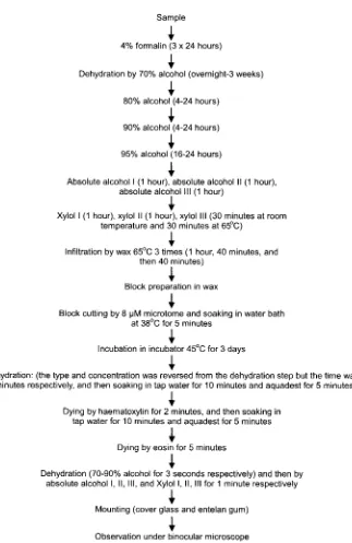
was used by Firoozabady and Moy (2004). The calli including original explants were placed in the room culture with 25±2°C, in the dark condition for 3 weeks. The variable observed were the longest and shortest diameter of the calli. Microscopical and histological examination was also conducted to check the type of the cell division and the development of somatic embryo. The histological analysis was modified method of Kiernan (1990), presented in Figure 1.
The factorial Completely Randomized Design was also used in the experiment of somatic embryo development. The first factor was embryogenic callus induction media and the second factor was regeneration media. The embryogenic callus induction media were MS+ or Bac+ with addition of AdS as mentioned above. The first regeneration medium was coded as Reg A which was MS basal medium without nicotinic acid and pyridoxine HCI with addition of 0.9
J..LM (0.18 mg
n
IBA, 1.1 J..LM (0.25 mg 1"1) 8A, 0.09 J.1M
(0.03 mg
n
GA3 according to Perez et al. (2009). Thesecond regeneration medium was coded as Reg 8 which was MS basal medium supplemented with
0.018 mM (4 mg J·1
) BA according to Firoozabady and
Moy (2004). The treatments were replicated 6 times. The explants were illuminated 16 h per day with 1.000 lux irradiance provided by neon light and exposed to a temperature 25±2°C. The variables observed were percentage of browning and the number of mature somatic embryos.
RESULT AND DISCUSSION
Indirect Organogenesis Regeneration
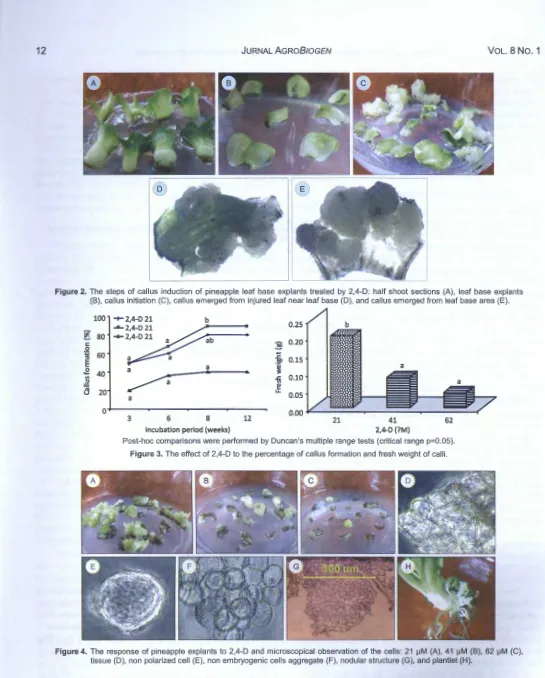
After 1-2 weeks, the shoot sections were swollen (Figure 2A), and then several leaves opened in consequence of the activity of plant growth regulator (Figure 28). The callus formation was initiated 3 weeks after planting (Figure 2C). The most of calli initiated from leaf base area and partly initiated from injured leaf area near leaf base (Figure 2D-E). Firoozabady and Moy (2004) believed that leaf bases may either contain meristematic regions or possess newly developed tissue with rapidly dividing cells that are amenable to morphogenesis in the tissue culture.
Suryowinoto (1996) explained that Bromeliaceae has
adventitious meristem in the leaf base. The emergence of calli from injured leaf may be due to the effect of TDZ in the callus induction media. TDZ is a
phenylurea-type compound which has high
physiological activity like cytokinin (Sakakibara 2004). Figure 3 showed that callus formation increased from 3 weeks to 8 weeks, and then the growth was stopped. The highest callus formation (90%) resulted from 41 J..LM 2,4-D but it was not significantly different with the result of 21 J..LM 2,4-D (Figure 3A). However, the 21 J..LM 2,4-D yielded the highest fresh weight significantly different with the other treatment (Figure
38). It is indicated that the calli grew faster in 21 J..LM
2,4-D than the other treatments. Thus, 21 J..LM 2,4-D was the best treatment for inducing callus formation. This result was better than the previous research which
conducted by Sripaoraya et a/. (2003) who provided
the lower frequency of callus formation of pineapple cultivar Phuket (58%) and also by Firoozabady and Moy (2004) who harvested 55% callus formation of pineapple cultivar Smooth Cayenne.
2012 I. RoosTIKA ET AL.: Indirect Organogenesis and Somatic Embryogenesis 11
Sample
•
4% formalin (3 x 24 hours)
•
Dehydration by 70% alcohol (ovemight-3 weeks)
•
80% alcohol (4-24 hours)
•
90% alcohol (4-24 hours)
•
95% alcohol (16-24 hours)
•
Absolute alcohol I (1 hour), absolute alcohol II (1 hour), absolute alcohol Ill (1 hour)
•
Xylol I (1 hour), xylol II (1 hour), xylol Ill (30 minutes at room temperature and 30 minutes at 65'C)
•
Infiltration by wax 65°C 3 times (1 hour, 40 minutes, and then 40 minutes)
•
Block preparation in wax
•
Block cutting by 8 iJM microtome and soaking in water bath at 38°C for 5 minutes
•
Incubation in incubator 45°C for 3 days
•
Rehydration: (the type and concentration was reversed from the dehydration step but the time was 5 minutes respectively, and then soaking in tap water for 10 minutes and aquadest for 5 minutes)
•
Dying by haematoxylin for 2 minutes, and then soaking in tap water for 10 minutes and aquadest for 5 minutes
•
Dying by eosin for 5 minutes
•
Dehydration (70-90% alcohol for 3 seconds respectively) and then by absolute alcohol I, II, Ill, and Xylol I, II, Ill for 1 minute respectively
•
Mounting (cover glass and entelan gum)
•
[image:6.614.158.481.79.576.2]Observation under binocular microscope
Figure 1. The step of histological analysis of pineapple embryogenic callus.
The microscopical examination showed that 2,4-D-treated cells still bound in the tissue (Figure 40) and the dispersed cells even did not show polarization
(Figure 4E). The cell aggregate showed the
symmetrical cell division (Figure 4F). This indicated
that the cells were not embryogenic. It was explained
that the non embryogenic cells can be determined by symmetrical division (Abrash and Bergmann, 2009; Paciorek and Bergmann, 2010). Furthermore, the non embryogenic cell aggregate grew to become nodular
structure (Figure 4G) and finally to form plantlet which consisted of several roots and sometimes with little buds (Figure 4H), typically different with the characteristic of bipolar structure of somatic embryo.
12 JURNAL AGR08/0GEN VoL. 8 No. 1
Figure 2. The steps of callus induction of pineapple leaf base explants treated by 2,4-D : half shoot sections (A), leaf base explants (B), callus initiation (C), callus emerged from injured leaf near leaf base (D), and callus emerged from leaf base area (E).
100 -r 2,4-D 21 b
..._ 2,4-D 21 0.25 b
セ@ 80 -+-2,4-D 21 ab
0.20
c: :§
0
.... 60
t
"'
0.15E
セ@
'i
0 40
...
"' -5i 0.10
セ@ セ@
a
20 u..0.05
a
a
0 0.00
3 6 8 12 21 41 62
Incubation period (weeks) 2,4-D (?M)
Post-hoc comparisons were performed by Duncan's multiple range tests (critical range p=O.OS).
Figure 3. The effect of 2,4-D to the percentage of callus formation and fresh weight of calli.
Figure 4. The response of pineapple explants to 2,4-D and microscopical observation of the cells: 21 iJM (A), 41 iJM (B), 62 iJM (C), tissue (D), non polarized cell (E), non embryogenic cells aggregate (F), nodular structure (G), and plantlet (H).
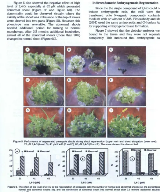
shown in the treatment of 62 J..LM 2,4-D (Figure SC). The high level of shoot regeneration (about 25 shoots/
2012 I. ROOSTIKA ET AL.: Indirect Organogenesis and Somatic Embryogenesis 13
Figure 5 also showed the negative effect of high level of 2,4-D, especially at 62 I!M which generated abnormal shoot (Figure SF and Figure 68). The abnormality could be observed visually where the axiality of the shoot was imbalance or the top of leaves were cleaved into two parts (Figure 51). However, this phenotype was reversible. The abnormal shoots needed additional period for turning to normal morphology. After 3.5 months additional incubation, almost all of the abnormal shoots (more than 90%) changed to normal shoot (Figure 6C).
Indirect Somatic Embryogenesis Regeneration
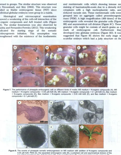
Since the the single compound of 2,4-D could not induce embryogenic cells, the calli were then transferred onto N-organic compounds containing medium with or without of AdS. Firoozabady and May (2004) used the same amino acids and CH unless AdS for supporting embryogenic tissue formation.
Figure 7 showed that the globular embryos were bound in the tissue and they were not separated completely. This indicated that embryogenic calli
Figure 5. Performance of regenerated pineapple shoots during shoot regeneration (upper row) and shoot elongation (lower row): 21 セm@ 2,4-D (A and D), 41 セm@ 2,4-D (B and E), 62 セm@ 2,4-D (C and F). The arrow showed the cleaved leaf.
25
ZQセセMMセMᄋQ@
100 10080 80
60 60
·:l
rlJ
セ@
J
*- 40 20 *- 40 20I I I 0 0
21 41 62 21 41 62 21 41 62
2,4-D(IlM) 2,4:0 (11M) 2,4-D(I.lM)
[image:8.612.38.562.86.715.2]14 JURNAL AGR08/0GEN VOL. 8 No.1
formed in groups. The similar structure was observed
by Firoozabady and Moy (2004). This structure was
called as friable embryogenic tissue (FET) since individual globular embryos were group in a matrix.
Histological and microscopical examination showed a weakening of the cell-cell interaction of the N-organic compounds and AdS treated cells (Figure 8A) . The similar fenomenon was also observed by
Cunha and Fernandez-Ferreira (2012) . This weakening
indicated the starting stage of the somatic
embryogenesis initiation. This assumption was
strengthened with the existence of the isodiametric
and meristematic cells which showing intense red staining of haematoxylin-eosin due to a densely rich cytoplasm with a high nucleoplasmic ratio, and reduced vacuole size. These meristematic cells were grouped into an aggregate called proembryogenic
mass (PEM) . A high magnification (400 times) of the
embryogenic cells revealed the granular cells (Figure 88) and asymmetrical cell division (Figure 8C). These granular cells might be contain of starch grains as a
mark of embryogenic cell. Furthermore they
developed into globular embryos (Figure 80). It was
[image:9.612.52.551.84.722.2]suggested that Figure 8E shown the early stage of scutellar embryo which had a jutty structure on the
Figure 7. The performance of pineapple embryogenic calli on different kinds of media: MS medium + N-organic compounds (A), MS medium+ N-organic compounds+ 0.05 1-1M AdS (B), MS medium+ N-organic compounds+ 0.1 1-1M AdS (C), Bac medium + N-organic compounds (D), Bac medium + N-organic compounds + 0.05 mM AdS (E), and Bac medium + N-organic compounds + 0.1 1-1M AdS (F).
2012 I. RoosTIKA Er AL.: Indirect Organogenesis and Somatic Embryogenesis 15
top. Unfortunately, the coleoptilar stage could not be observed in this study. Finally, the mature embryos developed into germinated embryos or plantlets (Figure 8F) which clearly showed bipolarity (shoot and root). After 5 months, the embryogenic cells turned to dark brown and mixed with the non embryogenic
cells. It indicated that the embryogenic competent lost
accompanied with long period of the culture and they need to be subcultured immediately before they turn to brown color completely.
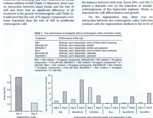
Generally, all of embryogenic callus induction media yielded globular and semi dispersible calli. However, the browning calli were observed on media
without addition of AdS (Table 1). Moreover, there was
no interaction between basal media and the rate of AdS and there was no significant difference of all treatment to the growth of embryogenic calli (Table 2). It indicated that the role of N-organic compounds were more important than the role of AdS to proliferate embryogenic calli.
As reported by many researchers, nitrogen (N) is also fundamental element for plant cell and tissue culture, being essential for the synthesis of DNA, RNA, and protein. When MS medium compared to Bac
medium, the total N in MS medium is higher (60 mM)
than that in Bac medium (35 mM). However, the ratio
between N-oxidized (N03·) and N-reduced (NH4 +) is
higher in Bac medium (2.5 : 1) than that in MS medium
(2 : 1). It means that the ratio of oxidized and
N-reduced in both media is rather not different. Therefore, there was not difference response resulted from all of media to the growth of calli (Table 2).
Cunha and Fernandez-Ferreira (2012) mentioned that
the balance between both ionic forms (N03· and NH4 +)
played a dramatic role on the induction of somatic embryogenesis of flax hypocotyls explants. Nitrate is important for calli differentiation and growth.
On the regeneration step, there was no interaction between the embryogenic callus induction medium with the regeneration medium to the level of
Table 1. The performance of pineapple calli on embryogenic callus formation media.
Treatment Performance of the calli
1' 0.81
£. 0.6
.!!f
c
セ@ 0.4
dl
PNRセ@
0 J
セ@
I
セ@
I
Reg. A
MS+ MSAdS0.05 MSAdS0.1 Bac+ BacAdS0.05 BacAdS0.1
Globular, semi dispersible, some of them were browning Globular, semi dispersible, whitish
Globular, semi dispersible , whitish and greenish Globular, semi dispersible, some of them were browning Globular, semi dispersible, yellowish
Globular, semi dispersible, yellowish
MS+ = MS medium + N-organic compounds , MSAdS0.05 = MS medium + N-organic compounds + 0.05 セma、s L@ MSAdS0.1
=
MS medium + N-organic compounds + 0.1セm@ AdS, Bac+
=
Bac medium + N-organic compounds , BacAdS0.05=
Bac medium + N-organic compounds + 0.05 セm@ AdS, BacAdS0.1=
Bac medium + N-organic compounds + 0.1 セma、s N@20
V>
0
セ@
.c
E 16
QJ
u
"ji; 12 E セ@ セ@ 8 ::;J セ@ "'
E 4
... 0
HMZM[ゥセセQ@ セ@
セ@ 0
E
::;J
,.,.,.,
..
I z
Reg. B I MS+ I MSAdSO.OS I MSAdS0.1 Bac+ BacAdSO.OS Regeneration media Embryogenic callus induction media and regeneration media
Reg B BacAdSO.l
Reg. A= modified MS medium without nicotinic acid and pyridoxine HCI with addition of 0.9 セm@ (0.18 mg 1-1) IBA, 1.1 セm@ (0.25 mg 1"1) BA, 0.09
セm@ (0.03 mg 1"1) gセ N@Reg. B = MS medium with addition of 0.018 mM (4 mg 1"1) BA, MS+ = MS medium+ N-organic compounds, MSAdS0.05 =
MS medium + N-organic compounds + 0.05 セma、sL@ MSAdS0.1
=
MS medium + N-organic compounds + 0.1 セma、s L@ Bac+=
Bac medium + N-organic compounds , BacAdS0.05 = Bac medium + N-N-organic compounds + 0.05 セma、s L@ BacAdS0.1=
Bac medium + N-organic compounds + 0.1 セma、sN@ [image:10.612.49.571.241.645.2]16 JURNAL AGR08/0GEN VOL. 8 No.1
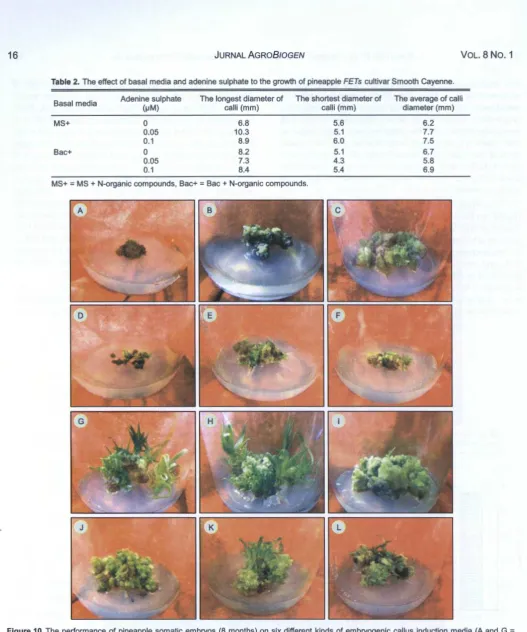
Table 2. The effect of basal media and adenine sulphate to the growth of pineapple FETs cultivar Smooth Cayenne.
Basal media Adenine sulphate The longest diameter of The shortest diameter of The average of calli
HセmI@ calli (mm) calli (mm) diameter (mm)
MS+ 0 6.8 5.6 6.2
0.05 10.3 5.1 7.7
0.1 8.9 6.0 7.5
Bac+ 0 8.2 5.1 6.7
0.05 7.3 4 .3 5.8
0.1 8.4 5.4 6.9
MS+ = MS + N-organic compounds, Bac+ = Bac + N-organic compounds.
Figure 10. The performance of pineapple somatic embryos (8 months) on six different kinds of embryogenic callus induction media (A and G = MS medium + N-organic compounds, B and H = MS medium + N-organic compounds + 0.05 セma、sL@ C and I = MS medium + N-organic compounds + 0.1 セm@ AdS , D and J = Bac medium + N-organic compounds , E and K = Bac medium + N-organic compounds + 0.05 セm@
AdS , and F and L = Bac medium + N-organic compounds + 0.1 セm@ AdS} and two different kinds of regeneration media (the upper rows= MS + 0.9 セm@ IBA + 1.1 セmbaK@ 0.09 セm@ GA3 and the lower rows= MS basal medium supplemented with 0.018 mM BA).
browning but the single factor of regeneration medium affected that variable. The MS medium containing of
[image:11.612.35.563.18.650.2]2012 I. ROOSTIKA ET AL.: Indirect Organogenesis and Somatic Embryogenesis 17
As mentioned above, that AdS did not affect the proliferation of embryogenic calli. Interestingly, there was interaction between the embryogenic callus induction medium with the regeneration medium to the number of mature somatic embryos (Figure 9 and 10). On this medium, the number of mature somatic
embryos reached about 17 embryos/explant.
Structurally, AdS is similar with cytokinin compounds since it has an adenine ring which has a rich of nitrogen compound. The AdS may act as nitrogen source in the synthesis of RNA, DNA, and protein for cell division. Therefore it may play important role in the embryo development.
CONCLUSION
The single auxin of 2,4-D was not enough to induce embryogenic cells. Therefore a part non embryogenic calli were regenerated through indirect organogenesis. The best treatment for organogenesis was 21 J.lM 2,4-D because it provided high level of callus formation (80%), the highest fresh weight (0.2 g/explant), and the highest number of shoot (about 25
shoots/explant). The friable embryogenic callus
formation could be induced by N-organic compounds with or without addition of AdS. There was interaction between the embryogenic callus induction medium with the regeneration medium to the number of mature somatic embryos. The combination treatment of MS medium enriched with N-organic compounds and 0.05 J.lM AdS as embryogenic callus induction medium and MS medium with addition of BA 0.018 mM BA as regeneration medium was the best treatment which yielded about 17 embryos/explant.
ACKNOWLEDGEMENTS
The authors acknowledge Indonesian Agency for Agricultural Research and Development (IAARD) which supporting the fund through KKP3T program 2011.
REFERENCES
Abrash, E.B. and D.C. Bergmann. 2009. Asymmetric cell
divisions: A view from plant development.
Developmental Cell 16:783-796.
Arnold, S., I. Sabala. P. Bozhkov, J. Dyachok, and L. Filonova. 2002. Developmental pathways of somatic embryogenesis. Plant Cell Tiss. Org. Cult. 69:233-249.
Coppens d'Eeckenbrugge, G. and F. Leal. 2003.
Morphology, anatomy, and taxonomy. In D.P.
Bartholomew, R.E. Pauli, and K.G. Rohrbach ( eds.) The
Pineapple Botany: Production and Uses. CABI
Publishing.Wallingford. p. 13-32.
Corredoira, E., S. Valladares, and A.M. Vieitez. 2006.
Morphohistological analysis of the ong1n and
development of somatic embryos from leaves of mature
Quercus robur. In Vitro Cell. Dev. Bioi. Plant 42:525-533.
Cunha, A. and M. Fernandez-Ferreira. 2012. Influence of medium parameters on somatic embryogenesis from
hypocotyl explants of flax (Unum usitatissimum L.):
Effect of carbon source, total inorganic nitrogen and balance between ionic forms and interaction between calcium and zeatin. http://dx.doi.org/1 0.1 016/S0176-1617(99)80059-5.
Firoozabady, E. and Y. Moy. 2004. Regeneration of
pineapple via somatic embryogenesis and
organogenesis. In Vitro Cell. Dev. Bioi. Plant 40:67-74.
International Board for Plant Genetic Resources [IBPGR]. 1991. Descriptors for Pineapple. IBPGR Headquarters, Rome.
Kelley, K.B. and D.E. Riechers. 2007. Recent development in auxin biology and new properties for auxinic herbicide
research. Pesticide Biochemistry and Physiology
89:1-11.
Kiernan, J.A. 1990. Histological and Histochemical Methods: Theory and Practice. 2"d edition. Pergamon Press.
Murashige, T. and F. Skoog. 1962. A revised medium for
rapid growth and bioassays with tobacco tissue culture. Physiol. Plant 15:4 73-497.
Nursandi, F., R. Poerwanto, Sobir, S. Sujiprihati, dan A.
Purwito. 2005. Studi pertumbuhan nenas (Ananas
comosus L. Merr) kultivar Queen hasil in vitro dan
analisis kestabilan genetik berdasarkan karakter
morfologi dan RAPD. Simposium dan Konggres Peripi. 25-27 Agustus 2005. Universitas Jenderal Soedirman. Purwokerto.
Nursandi, F., R. Poerwanto, Sobir, dan S. Sujiprihati. 2003.
Perbanyakan in vitro nenas (Ananas comas us (L.)
Merr.) dengan menggunakan BAP dan TDZ. Simposium Nasional dan Konggres VIII. Bandar Lampung.
Paciorek, T. and D.C Bergmann. 2010. The secret to life is
being different: asymmetric divisions in plant
development. Current Opinion in Plant Bioi. 13:661-669.
Perez, G., E. Yanes, M. lsidron, J.C. Lorenzo. 2009. Phenotypic and AFLP characterization of two new
pineapple somaclones derived from in vitro culture.
PCTOC 96:113-116.
Phillips, G.C., J.F. Hubstenberger, and E.E. Hansen. 1995. Plant regeneration from callus and cell suspension
cultures by somatic embryogenesis. In O.L. Gamborg
and G.C. Phillips (eds.) Plant Cell Tissue and Organ
Culture: Fundamental Methods. Springer. Berlin.
p. 81-90.
Purnamaningsih, R., I. Mariska, dan Y. Supriati. 2009. Peng-gunaan ABA dan paklobutrazol dalam perbanyakan
nenas Simadu melalui kultur in vitro. Berita Biologi
18 JURNAL AGR08JOGEN VOL. 8 No.1
Sakakibara. H. 2004. Cytokinin biosynthesis and
metabolism. In P.J. Davies ( ed.) Plant Hormones:
Biosynthesis, Signal, Action. 3'd Edition. Kluwer Academic Publishers. London. p. 95-114.
Smith, M.K., H.-L. Ko. S.D. Hamil, G.M. Sanewski, and M.W.
Graham. 2003. Biotechnology. In D.P. Bartholomew,
R.E. Pauli, and K.G. Rohrbach (eds.) The Pineapple
Botany: Production and Uses. CABI Publishing. Wallingford. p. 57-68.
Sripaoraya, S., R. Marchamt, J.B. Power, and M.R. Davey. 2003. Plant regeneration by somatic embryogenesis
and organogenesis in commercial pineapple (Ananas
comosus L.). In Vitro Cell. Dev. Bioi. Plant 39:450-454.
Srivastava, L.M. 2002. Plant Growth and Development. Academic Press. New York. 772 p.
Suminar, E. 2010. lnduksi keragaman genetik dengan
mutagen sinar gamma pada nenas (Ananas comosus
(L.) Merr.) secara in vitro. Seminar Sekolah
Pascasarjana, lnstitut Pertanian Bogor.
Suryowinoto, M. 1996. Pemuliaan Tanaman secara In Vitro.
Pusat Antar Universitas-Bioteknologi Universitas