Hospital Acquired Bacterial Infection in Burns Unit
at Cipto Mangunkusumo Hospital, Jakarta
PRATIWI SUDARMONO∗ & VERONICA WIWING
Department of Microbiology, Medical Faculty, Universitas Indonesia, Jalan Pegangsaan Timur 16, Jakarta 10320, Indonesia
Burn injury causes mechanical disruption to the skin, which allows environmental microbes to invade the deeper tissues. A prospective study of infections in burn patients has shown that the incidence of hospital acquired bacterial infection in burn wounds was high. In the Burns Unit, Cipto Mangunkusumo Hospital, Jakarta, 94 patients were hospitalized from January to July 2004. The objective of this study was to evaluate the hospital acquired infections in burn wounds. Using a cross sectional study, 49 patients were included. The specimens for bacterial investigation were obtained from clean eschar which has healthy tissue taken at day 1, day 5 and day 10. At the same time, bacterial investigations were conducted from the air and the water, as well as from the hand and nasal swabs of hospital personnel. The results show that Klebsiella pneumoniae is the most prominent bacterium found in the wounds, but it is also found in the air. Pseudomonas aeruginosa was the number two causative bacteria which caused a change of the bacterial infectivity on day 5 and 10. These bacteria were always found when we conducted bacterial investigations from the water resource of the burns unit. Methicillin Resistant Staphylococcus aureus is also found in the nasal swab of hospital personnel. Using the antibiogram pattern, there were similarities between bacteria found in the wounds and in bacteria found in the air and water. In conclusion, hospital acquired burn wound infection in Burns Unit, Cipto Mangunkusumo Hospital is as high as 62%. The surveillance data are very important for developing good clinical practice guidelines in burn injury treatment and management.
Key words: burn wound infection, burn injury management
_____________________________________________
_________________
∗Corresponding author, Phone: +62-21 3160492 ext. 10, Fax: +62-21 3100810, E-mail: pratiwi@cbn.net.id
Burns are one of the most common and devastating forms of trauma. Patients with serious thermal injury require immediate specialized care in order to minimize morbidity and mortality (Church et al. 2006). Burn injury causes
mechanical disruption at the skin, which allows environmental microbes to invade the deeper tissues. The usual skin barrier to microbes is replaced by a moist, protein-rich, avascular eschar that fosters microbial growth. The burn wound surface is sterile immediately following injury; however, it is repopulated quickly with gram-positive organisms from hair follicles, skin appendages, and the environment during the first 48 hours. More virulent gram-negative organisms replace the gram-positive organisms after 5-7 days. Gram-negative organisms have greater motility, possess many antibiotic resistance mechanisms, and have the ability to secrete collagenases, proteases, lipases, and elastases, enabling them to proliferate and penetrate into the subeschar space. The risk of burn wound infection is directly correlated to the extent of the burn and is related to impaired resistance resulting from disruption of the skin’s mechanical integrity and generalized immune suppression. (Bowler et al. 2001;
Agnihotri et al. 2004). The criteria for admission to the burn
care unit are : children with burns involving at least 10% or adults with burns involving at least 20% of their total body surface; burns affecting face, perineum or feet; suspected or proven airway injury; electric or chemical burns; age less than one year or more than 50; or pre-existing disease regardless of the extent of the burns (Santucci et al. 2003).
When the burn is very large, the patient will be seriously ill, and usually will die after seven days. For those surviving, the fiercest fight is against bacterial pathogens, which usually will increase the duration of hospitalization to 40-148 days (Schlager et al. 1994; Nasser et al. 2003;
Taneja et al. 2004).During hospitalization, the nosocomial
infection occurs, and the pathogens can come from endogenous as well as exogenous sites (Chambers 1997). The bacteria can infect the wound by the airborne route, direct contact from the hands of paramedics or contamination by non sterile equipment (Samy et al. 2003).
There is very limited data in this hospital about the sources of bacterial infection causing nosocomial infection. Although the nosocomial infection rate in any hospital indicates good clinical practice in patient management, even if the rate is low, the burns unit needs a very specific condition since all patients are immunocompromised.
In Cipto Mangunkusumo Hospital, Jakarta, no data are available about the existence of pathogens in the environment, making it very difficult when a doctor must choose the right antibiotic if infection is detected in burn wounds. Besides, any data on burn infections will be very useful, because burn wound treatment is very complex and varies from one patient to another. The objective of this study was to describe infections in a specialized burns intensive care unit in Cipto Mangunkusumo Hospital, Jakarta, from January to July 2004. This study was conducted mainly to determine the source of infection from the air and the water which had been used by the patients during their hospitalization and the characteristics of secondary bacterial infection, together with its antibiotic resistance pattern. The data will be very useful for prevention of hospital acquired bacterial infection at the Burns Unit of Cipto Mangunkusumo Hospital, and to review the standard operational procedures for burn treatment management.
MATERIALS AND METHODS
Materials. We used the observational cross-sectional method, so a control group is not necessary. The population comprised all patients with burn injuries hospitalized in this
Burns Unit, Cipto Mangunkusumo Hospital from January-July 2004. For statistical calculation, we used the formula:
N = Zα2 x P x Q where :
d2
P = the condition which infection is likely happened = 50%; Q is 1-P = 50%; d is precision absolute = 15%, and Zα = 1.96;
43 patients were used in this study.
The study had also been conducted to identify the bacterial pathogens from the air, instruments, linen, gloves, and from the skin and nasal swab of the paramedics.
Samples. Patient samples were obtained from burn wound eschar which still contains healthy tissues. The samples were taken aseptically using a sharp scalpel after wound cleansing in day 1, day 5, and day 10 of hospitalization. Samples from day 1 were taken to prove the existence of bacteria at the time of injury. Those from day 5 were taken to look for a changing microbial population in the wound and those from day 10 were taken to understand the continuity of infection. The samples were excluded from the study if the specimens were taken using cotton swab (because there will be no healthy tissues) or when the specimens were contaminated by pus. All specimens were transported to The Department of Microbiology for bacterial culture and identification using brain heart infusion broth (Difco, USA) and when bacteria were identified, the antibiograms were conducted following NCCLS standard.
Bacterial Examination from Air. Airborne specimens were obtained using an air sampler “MAS”100 (Merck’s Air Sampler MAS-100) which has been set to take the samples from 10% of room volume. The Burns Unit at Cipto Mangunkusumo Hospital has one large room whith a volume of 150 m3so the air sampler was set to take samples from 15
positions in the room. The air samples captured were inoculated in blood agar and Endo agar (Difco, USA) and incubated at 36 oC for 24 hours. The growing bacteria were
Gram stained, and then identified and characterized. Bacterial Examination from Water. Bacterial examination was conducted to search for bacterial contamination in the water used for washing and cleaning patients’ bodies. A 1.0 l aliquot of water was obtained from previously sterilized tap
water. The method used to calculate the bacterial count was by most probable number(MPN). The water specimens were 10 times serial diluted, and inoculated in triplicate with 10 ml lactose broth agar followed by incubation at 35 oC for 24
hours (presumptive test). When fermentation was detected, the specimens were continuously inoculated in brilliant green lactose broth (BGLB) as a confirmation test. This was followed by inoculation of 0.5 ml aliquots in Endo agar for species identification.
Linen, Instrument, and Gloves. Linen and gloves were cut using sterile scissors and placed in a glass tube contained thioglycolate solution. Instruments such as scalpels and pinsets, which has been used to treat the wound, were also dipped in a flask containing the thioglycolate solution. All tubes and containers were incubated at 35 oC for 24 hours,
and the bacteria grown in the solution were then identified further.
Hands and Nasal Swabs. The nasal swabs and the swabs from hands of doctors and paramedics were taken and cultured directly in the blood agar for further bacterial identification.
Ethical Approval. This research had been approved by the Internal Review Board of the Medical Faculty, University of Indonesia. Informed consent was clearly obtained from each patient.
RESULTS
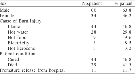
Analysis of Surviving and Terminal Patients. From January to July 2004 there were 94 patients who had been hospitalized in the Burns Unit, Cipto Mangunkusumo Hospital, Jakarta. Some 60 persons were men and 34 were women. The causes of burns were fire, hot water, hot food, electricity and hot kerosene. Fortyfour (46.8%) patients were cured and 39 (41.5%) patients died while the remainder left hospital on their own before treatment was complete. Data were analyzed using the Kruskal-Wallis test for three variables and the Mann-Whitney test for two variables (Tables 1 & 2). Using the Kruskal-Wallis test, the data showed a positive correlation between the age of patients and the cure rate. The average age of the patients who died was higher than the average age of the cured patients group (p = 0.003). The level of burn injury of the terminally-ill patients was significantly larger than that of the cured patients (p = 0.000).The length of hospitalization for patients who died was significantly shorter than for patient who recovered (p = 0.017). The second analysis was performed using the Mann-Whitney test. Using this method the average age of the terminal patients was higher than for the patients who recovered (p < 0.006). The extent of burn injury of the patients who died was significantly larger than for the cured patients group (p < 0.000). The length of hospitalization for patients who died was significantly shorter compare to the patients who survived (p < 0.006).
Table 1 Characteristics of patients hospitalized in the Burns Unit, Cipto Mangunkusumo Hospital, from January to July 2004 Sex No.patient % patient Male
Female
Cause of Burn Injury Flame
Hot water Hot food Electricity Hot kerosene Patient condition
Cured Died
Premature release from hospital
6 0 3 4
4 4 2 8 9 8 3
4 4 3 9 1 1
63.8 36.2
46.8 29.8 9.6 8.5 3.2
46.8 41.5 11.7
Table 2 Data analysis using Kruskal-Wallis method
Patients
Cured Premature release from hospital Died
Variable p
Age
The width of injury Days of hospitalization
14.9 + 12.3b 24.1 + 13.2c 32.9 + 20.0d
19.1 + 12.3 20.7 + 10.1 31.1 + 9.5
24.3 + 15.3b 46.1 + 19.1b 21.9 + 14.7d
0.003 0.000 0.017
Since Cipto Mangunkusumo Hospital, Jakarta is the national referral hospital; the patients who were hospitalized did not always arrive on the first day of their injury. Forty two patients (85.7%) were referred by other hospitals, and these arrived mainly on the second or third days of injury (65.4%). In two cases patient arrived on day 10 or day 11, both with very large burn injuries.
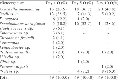
Infecting Bacteria and their Sources. Forty nine patients were included in this study on day 1, but four patients had died by day 5, and another four patients had died by day 10. Thus, only 41 patients completed this study. From the eschar culture, Klebsiella pneumoniae was the most prominent
bacterium isolated from day 1, day 5, and day 10, from 26.5, 36.7, and 40.8% patients respectively. The second most prominent bacterium isolated was Pseudomonasaeruginosa.
This was found from only 5 (10.2%) patients on day one, but on day 5 it was isolated from 16 (32.7%) patients, and on day 10 it was isolated from 14 (28.6%) patients (Table 3). The increase in P. aeruginosa isolates during hospitalization may
be caused by the use of water which was also contaminated with the same bacterium (data not shown). Of the other
bacteria species isolated, one was from water (Citrobacter freundii), and one from linen/bedsheets(Bacillus sp.). From
the air, K. pneumoniae was dominant, followed by the growth
of Staphylococcus aureus, Enterobacter sp., Acinetobacter
sp., and Bacillus sp. There were no bacteria isolated from
gloves and medical instruments. Of the more significant findings was the isolation of bacteria such as Bacillus sp.,
C. freundii, and Staphylococcus sp. from the hands of
doctors and paramedics. Even worse, bacteria were isolated from nasal swabs of some hospital personnel: 15 persons were contaminated with S. aureus and 8 with Methicillin ResistantS. aureus (MRSA).
Changes in Bacterial Population Over Time. Cross tabulation was conducted for all bacteria obtained on day 1, day 5, and day 10. Only in 19 (37.9%) patients, the burn wounds were inhabited by the same bacteria, while in 30 (62.1%) patients, the bacteria obtained in day 1, day 5 and day 10 had changed or were different. Only 13 patients on day 1 were infected with K.pneumoniae but on day 5, K. pneumoniae was identified in 18 patients which were
originally (day 1) inhabited by Staphylococcus sp., K. oxytoca, P. aeruginosa, Enterococcus sp., and Aeromonas sp. For 2
patients whose wounds were initially (day 1) inhabited by
K. pneumoniae, by day 5 superinfection occurred being
caused by P. aeruginosa. To prove that K. pneumoniae
isolated from the wound on the day 5 was from airborne contamination, the patterns of antibiograms were compared. This showed that the antibiogram patterns of K. pneumoniae
isolated both from the air and from the wounds were similar. For P. aeruginosa a similar phenomenon was observed. On
day 1, only 5 patients were infected by this bacteria, but by day 5 some 12 patients, which on day 1 were infected by
S. aureus, K. oxytoca, Enterococcus sp., Aeromonas sp., Acinetobacter sp., C. freundii, Proteus mirabilis, Shigella
sp., and Bacillus sp., were now superinfected by P. aeruginosa. The bacteria colonies isolated from the water
used in the Burns Unit were also identified as P. aeruginosa.
Moreover, both groups showed similar antibiogram patterns. All antibiograms were compared using 23 different antibiotics (Tables 4 & 5).
DISCUSSION
The burn wound surface is sterile immediately following injury; however, it is repopulated quickly with gram-positive organisms from hair follicles, skin appendages, and the environment during the first 48 hours. More virulent gram-negative organisms replace the gram-positive organisms after 5-7 days. In this study we took specimens from eschars at day 1, day 5, and day 10 to detect any changes in bacterial infection or superinfection of the burn wounds. Santucci et al. (2003) surveyed 320 patients admitted to a
burns-intensive-Table 3 Bacteria from burn wounds in patients at Burns Unit, Cipto Mangunkusumo Hospital isolated at day 1, day 5, and day 10 from hospitalization
Microorganism Day 1 Ó (%) Day 5 Ó (%) Day 10 Ó(%) Klebsiella pneumoniae
Bacillus sp. K. oxytoca
Pseudomonas aeruginosa Staphylococcus sp. Enterococcus sp. Citrobacter freundii Aeromonas sp. Acinetobacter sp. Proteus mirabilis Shigella sp. Vibrio sp. Proteus vulgaris Proteus sp. Total
Table 4 The similarity in antibiogram of Klebsiella pneumoniae isolated from burn wounds compared to K. pneumoniae isolated from the air
care unit in Brazil, of whom one hundred and seventy-five (55%) developed hospital-acquired infections. The microorganisms causing infections were S. aureus (24%), P. aeruginosa (18%), Acinetobacter spp. (14%), and coagulase-negative staphylococci (12%). During the first
three days of hospitalization in the burns intensive care unit there were eight infections caused by S. aureus and three of
these were resistant to oxacillin (MRSA). Those data were almost similar with the data obtained from the present study conducted at The Burns Unit at Cipto Mangunkusumo Hospitals, Jakarta. The rate of hospital-acquired infections in our study is 62.1%, somewhat higher than in Brazil. The patterns of bacterial infections are almost the same, except for K. pneumoniae which was consistently isolated from the
air of our Burn Intensive Care Units as well as from the burn wounds. In Sweden, Appelgren et al. (2002) also conducted
a 3-year prospective study of all infections presented in the burns unit of a university hospital. Some 230 adult patients were included. Of these 83 patients had a total of 176 infections, giving an infection rate of 48 per 1000 patient days including both nosocomial and community-acquired infections. The most common microorganisms were coagulase-negative staphylococci and methicillin-sensitive
S. aureus. Seventy-two patients had 107 burn wound
infections. Comparison of data from Indonesia, Brazil and Sweden showed that hospital-acquired burn wound infection occurred, although with a different level of prevalence. The very high prevalence in Indonesia showed that potential infection did not come solely from the endogenous bacteria, but also from bacteria from the air and water which played an important role in secondary infection. Antibiotics in burn therapy can play a double role, as prophylaxis as well as for treatment. In Indonesia, all patients in our Burns Unit received antibiotics, while in Sweden antibiotics were given to only 50% of the burns patients, including 96% of the patients with infection and 26% of those without infection. The overuse of antibiotics in Indonesia indicates the overanxiety of the doctors, since the quality of water and air in the hospital, as well as the hygiene of the medical personnel, is not controllable by their hospital management. However, as
Table 5 The similarity in antibiogram of Pseudomonas aeruginosa isolated from burn wounds compared to P. aeruginosa isolated from the water 1 = AML: Amoxycilin, 2 = AMC: Amoxycillin + clav. acid, 3 = TIC: Carbenicillin, 4 = CE: Cephradine, 5 = CL: Cephalexin, 6 = CTM: Cephotiam, 7 = CXM: Cephuroxime, 8 = CEC: Cephaclor, 9 = CRO: Cephtriaxone, 10 = CTX: Cephotaxime, 11 = CFM: Cefixime, 12 = FEP: Cefepime, 13 = CPO: Cefpirome, 14 = CN: Gentamisin, 15 = AK: Amikacin, 16 = C: Chloramphenicol, 17 = SXT: Cotrimoxazole, 18 = CIP: Ciprofloxacin, 19 = MXF: Moxifloxacin, 20 = GAT: Gatifloxacin, 21 = VA: Vancomycin, 22 = IEM: Imipenem, 23 = OX: Oxacillin.
mentioned above, since Cipto Mangunkusumo Hospital in Jakarta has no database about the clinical epidemiology of nosocomial infection, any new data will be very useful. The database can be used to evaluate the effects of changes in burn treatment, staffing and design of burn units, and antimicrobial resistance development in relation to antibiotic usage. These data provide background information regarding extensive burn patients on which decisions for control and prevention of hospital-acquired infections can be made.
ACKNOWLEDGEMENT
We are most grateful to Yefta Moenadjat, the head of The Burns Unit Cipto Mangunkusumo Hospital, for their guidance and attention which made this study possible.
REFERENCES
Agnihotri N, Gupta V, Joshi RM. 2004. Aerobic bacterial isolates from burn wound infections and their antibiogram, a five-year-study. Burns 30:241-243.
Applegren P, Bjornhagen V, Bragderyd K, Jonsson CE, Ransjo U. 2002. Prospective study of infections in burn patients. Burns 28:39-46.
Bowler PG, Duerden BI, Armstrong DG. 2001. Wound microbiology and associated approaches to wound management. Clin Microbiol Rev 14:244-269.
Chambers HF. 1997. Methicillin Resistant Staphylococcus aureus: Molecular and biochemical basis and clinical implications. Clin Microbiol Rev 4:781-791.
Church D, El-Sayed S, Reid O, Winston B, Lindsay R. 2006. Burn wound infection. Clin Microbiol Rev 19:403-434.
Nasser S, Mabrouk A, Maher A. 2003. Colonization of burn wound in Ain Shams University Burn Unit. Burns 29:229-233.
Samy A, Shehab E, El-Sayed I, Mohammad R. 2003. Methicillin Resistant Staphylococcus aureus, A problem in the burn unit. Egypt J Plast Reconstr Surg 27:1-10.
Santucci SG, Gobara S, Santos CR, Fontana C, Levin AS. 2003. Infections in a burn intensive care unit: experience of seven years. J Hosp Infect 53:6-13.
Schlager T, Sadler J, Weber D, Donowitz L, Lohr J. 1994. Hospital-acquired infections in pediatric burn patients. South Med J 87:481-4. Taneja N, Emmanuel R, Chari PS, Sharma M. 2004. A prospective