I nt roduc t ion t o Bioinorga nic Che m ist ry
University of Lund, May/June 2008
Lecture notes
Dieter Rehder
1. Scope and Introduction
“Bioinorganic Chemistry“ is at the gate-way of inorganic chemistry and biochemistry, i.e. it describes the mutual relationship between these two sub-disciplines, with focus upon the function of inorganic “substances“ in living systems, including the transport, speciation and, eventually, mineralisation of inorganic materials, and including the use of inorganics in medicinal therapy and diagnosis. These “substances” can be metal ions (such as K+, ferrous and ferric), composite ions (e.g. molybdate), coordination compounds1 (like cisplatin and
carbonyltechnetium), or inorganic molecules such as CO, NO, O3. Medicinal inorganic
chemistry on the one hand, and biomineralisation on the other hand, are important integral parts.
Inorganic reactions have possibly played an important role in the formation and development of organic “life molecules” in the prebiotic area (terrestrial and/or extraterrestrial), and from the very beginning of life on Earth. Inorganic chemistry is involved in structure and function of all life forms present nowadays on Earth, belonging to one of the three main branches, viz. bacteria, archaea and eucarya (Fig. 1). Life started ca 3.5 billion years ago with LUCA, the first uniform (and unknown) common ancestor. At that time, our planet was already covered by oceans. The overall situation was, however, completely different from that of today: The primordial atmosphere (also referred to as “primordial broth”) contained CO2, N2 and H2O as
the main components, and trace amounts of gases like H2, CO, COS, H2S, NH3 and CH4 from
volcanic exhalations, and trace amounts of oxygen from the decomposition of water by electric discharges, cosmic rays and radioactivity. The Earth’s crust was essentially unstable due to wide-spread volcanism and bombardment by debris (meteorites), remainders from the constitution of the solar system some 4.5 billion years ago.
A key reaction at that time was the conversion of ferrous sulfide to ferrous disulfide (pyrite, FeS2) (eqn. 1), accompanied by a reduction potential of -620 mV, enough to enable
reductive carbon fixation, including reductive C-C coupling, and thus to allow entrance into the world of organic compounds. Eqns. (2) (formation of thiomethanol as a key compound) and (3) (formation of thioacetic acid) are examples. Of particular interest is the formation of “active acetic acid methylester“ (eqn. 3b), which is an essential constituent of acetyl-coenzyme-A, a focal product in biological carbon cycling, the synthesis of which is catalysed by an
acetylcoenzyme-M synthase, a iron-nickel-sulphur enzyme.
FeS + HS-→ FeS2 + 2[H] , ∆E0 = - 620 mV (1)
COS + 6[H] → CH3SH + H2O (2)
CH3SH + CO → CH3COSH (3a)
2CH3SH + CO2 + FeS → CH3CO(SCH3) + H2O + FeS2 (3b)
LUCA
109 a 0.5
1.2 2.1 3.0 3.5 E u c a r y a
Lichen Chlorophyta
Rhodophyta P l a n t a e
F u n g i
Basidiomycota Ascomycota Phaeophyta B a c t e r i a
A r c h a e a Proteobacteria
Cyanobacteria
A n i m a l e s
4.7 Planet Earth
Figure 1. Phylogenetic tree. Time scale in billion years. LUCA = last uniform common ancestor.
“Active acetic acid” readily reacts with amino acids (formed in the primordial broth by electric discharge; and/or in interstellar clouds by irradiation and carried to Earth confined in the ice cores of comets) to form peptides, which chiral selection and further polymerise on chiral matrices provided by certain clays and quartz minerals. Concomitantly, nucleobases can form under primordial and interstellar conditions, and polymerise to RNA, unique molecules which not only store information and transform this information into proteins, but also can act – like proteins – as enzymes (so-called ribozymes). The first life forms, primitive cellular organisms capable of self-sustenance and self-replication, are actually believed to have been members of an “RNA world”, which later has been replaced by our DNA world.
Fig. 2 classifies the bio-elements: Along with the “organic elements” (C, H, O, N, S), building up bio-mass, many “inorganic elements” play an important role in the physiological context. Some of these inorganic elements, such as Fe, Cu and Zn, are present in (practically) all
organisms, others are important for a restricted number of organisms only. An additional group of elements are used for diagnostic or therapeutic applications.
Significance of biologically important elements (selection)
Na+ and K+: Most important „free“ intra- and extracellular cations. Regulation of the osmotic
pressure, membrane potentials, enzyme activity, signalling. Mg2+: Chlorophyll; anaerobic energy metabolism (ATP → ATP).
Ca2+: Signalling, muscle contraction, enzyme regulation. Main inorganic part of the endoskeletons (bones, teeth, enamel: hydroxyapatite; Ca5(PO4)3(OH)). Exoskeletons of
mussels, shells, corals, sea urchins etc: aragonite or calcite; CaCO3).
VIV/V, MoIV/VI, WIV/VI, MnII/III/IV, FeII/III, NiI/II/III, CuI/II: active centres in electron-transport (redox) enzymes, oxygenases, dismutases.
Fe and Cu: Transport of oxygen. FeIII: Iron-storage proteins (ferritins).
FeII + FeIII in magnetite (Fe3O4): orientation of magnetobacteria, pigeons, bees in Earth’s
magnetic field.
Co: Synthases and isomerases (cobalamines, e.g. vitamin-B12); methylation of inorganics.
Zn2+: In the active centre of hydrolases, carboanhydrase, alcohol dehydrogenase, synthases;
genetic transciption (zinc fingers), stabilisation of tertiary and quartary structures of proteins; repair enzymes.
SiIV (“silicate“): Involved in the built-up of bones. In the form of SiO2/silica-gels as support in
monocotyledonous plants (like grass) and the shells of diatoms.
PV (phosphate): Constituent in hydroxi- and fluorapatite (Ca5(PO4)3(OH/F)); energy
metabolism (ATP), NADPH, activation of organic substrate; phospholipids in cell membranes; phosphate esters (DNA, RNA,…).
Se-II: Selenocystein in special enzymes (e.g. glutathionperoxidase) F-: Fluorapatite (Ca
5(PO4)3F) in dental enamel
Cl-: Along with hydrogencarbonate the most important free anion. I: Constituent of thyroid hormones (such as thyroxine).
Medicinal relevant elements (selection):
Li+: Treatment of bipolar disorder (maniac depression) and hypertension. Gd3+: Contrast agent in magnetic resonance tomography of soft tissues.
BaSO4: Contrast agent for X-ray tomography. Sun protection. 99mTc (a metastable γ-emitter; t
PtII: Chemotherapy (e.g. with cisplatin cis-[Pt(NH3)2Cl2]) of cancer (ovaria, testes)
AuI: Therapy of rheumatic arthritis.
SbIII: Treatment of inflammatory skin pimples like acne. BiIII: Treatment of gastritis.
Transition metal ions commonly are not present in a free form, but rather coordinated
(complexed) to ligands. In particular, this applies to metal ions in the active centres of enzymes or otherwise integrated into peptides and proteins. Examples for the respective ligands are listed below (N-functional: peptide moiety, porphinogenes, histidine; O-functional: tyrosinate, serinate, glutamate and aspartate; S-functional: cysteinate and methionine):
Additional inorganic ligands:
2. Iron
Iron takes over a central role in biological events (see also its role in the primordial synthesis of organic compounds; ch. 1). On the one hand, this is due to its general availability (iron is abundant and ubiquitous in the geo- and biospheres), on the other hand, iron has specific and “biologically suitable” properties otherwise not (or less) available with other transition metals:
(1) Ease of change between the oxidation states +II and +III (and disposability also of the oxidation states +IV and +V);
(2) Formation of hexaaqua cations in water; these hexaaqua cations are Brønsted acids;
-C H
R
N C O
H -C
H
R
N C H
O
-C H
R
N C O
-H+
N N
-CH2 -H+
N
NH R = -CH2
NH
N
-CH2
δ
ε
C O
O M C
O
O M C
O
O
M M end-on
monodentate
side-on
bidentate (chelate) end-on bridging
CH2 O- Tyr (Y) -(CH2)n C
O
O- n = 1: Asp (D); n = 2: Glu (E)
O-
CH2 S
CH2 CH2 Se- CH2 S CH3
Ser (S) Cys (C) Selenocysteinate Met (M)
N N
N
N
H
O
H
O H O 2
Hydroxido Oxido [Oxo] Aqua
O-O O-O S 2
(3) Tendency to form oligo- and polymers by condensation;
(4) Easy change between high- and low-spin states in ligand fields of medium strength (spin cross-over; see inset on p. 7, upper part);
(5) Flexibility with respect to the nature of the donating ligand function (see inset on p. 7, lower part, for ligand classification), the coordination number and coordination geometry.
The average amount of iron in the human body (70 kg) is ca. 5 g; iron is thus the most abundant transition metal in our organism. About 70% of this amount is used for oxygen transport and storage (haemoglobin, myoglobin), almost 30% are stored in ferritins (iron storage proteins), and about 1% is bound to the transport protein transferrin and to various iron-dependent enzymes; cf. the rough classification to the right.
Aqueous iron chemistry
The redox potential for the pair Fe2+/Fe3+ at pH = 7 demonstrates that FeII is easily oxidised to FeIII under aerobic conditions (cf. also the tutorial on oxidation and reduction on p 16):
Fe2+ ' Fe3+ + e-; E = -0.23 V (at pH 7)
compare: 2H2O ' O2 + 4H+ + 4e-; E (pH 7) = +0.82 V
NADH + H+ ' NAD+ + 2H+ + 2e-; E(pH 7) = -0.32 V (NADH = nicotine-adenine-dinucleotide in its reduced form) Tutorial: Coordination compounds (1): Definition
Coordination compounds, or complexes, are integral molecular or ionic units consisting of a central metal ion (or atom), bonded to a defined number of ligands in a defined geometrical arrangement. The ligands can be ions or (induced) dipolar molecules. Each ligand provides a free electron pair, i.e. the ligands are Lewis bases, while the metal in the coordination centre is the Lewis acid. The bonding can thus be described in terms of Lewis acid/Lewis base interaction. Other descriptions of the bonding situation are: (i) donor bond; (ii) coordinative covalent bond, often denoted by L→M, where L = ligand and M = metal. Complexes tend to be stable when the overall electron configuration at the metal centre (the sum of metal valence electrons plus electron pairs provided by the ligands) is 18 (or 16 for the late transition metals).
M + nL ' [MLn]q (n = number of ligands, q = charge of the complex) c(MLn)
c(M) cn(L) = K
K is the stability constant or complex formation constant (pK = -logK); its inverse, K-1, is
termed dissociation constant.
F F
/
/
H /
/
F F
F
L
L
Haeme-type
(e.g. cytochromes, haemoglobin)
Iron-sulfur proteins
(e.g. ferredoxins, Rieske proteins)
Two-iron centres
Hexaaquairon(III) ions are cationic Brønstedt acids:
[Fe(H2O)6]3+ + H2O ' [Fe(H2O)5OH]2+ + H3O+ pKS1 = 2.2
[Fe(H2O)5(OH)]2+ + H2O ' [Fe(H2O)4(OH)2]+ + H3O+ pKS2 = 3.5
[Fe(H2O)4(OH)2]+ + H2O ' [Fe(H2O)3(OH)3] (= Fe(OH)3·aq) + H3O+ pKS3 = 6.0
The formation of ferric hydroxide Fe(OH)3·aq hence already starts in weakly acidic media. The
protolytic reactions are accompanied by condensation reactions, leading to hydroxido- and oxido-bridged aggregates and finally to colloids and hardly soluble ferric oxide hydrates. The colloids can be spheroids of molecular mass M ca. 1.5⋅105 Da and ca. 70 Å diameter, or
needles/rods (M = 1.9·106, length up to 500 Å). The composition of these ironoxide hydrates
correspond to that of the minerals FeO(OH) (goethite) and 5Fe2O3·9H2O (ferrihydrite).
Mobilisation of Fe3+ by siderophores
The extremely low solubility of Fe(OH)3 [solubility product L = 2·10-39, solubility (pH 7) l =
10-18 mol·l-1], and thus the unavailability of iron in aqueous media under oxic conditions, has forced many groups of organisms to develop suitable systems for the mobilisation of iron. These systems, so-called siderophores (Greek for iron transporter), excreted by the organisms, are multidentate anionic ligands which form extremely stable complexes with Fe3+ (complex formation constants up to 1050 M-1). The functional groups of these ligands are, in many cases, catecholates (o
-hydroxyquinolates), as in the case of enterobactin, or hydroxamates
(ferrioxamines and ferriochromes). The
complexes are more or less globular, with the outer sphere furnished with hydrophilic groups (amide and ester groups), allowing for the water solubility and easy transport in the aquatic medium. Internalisation of the iron-loaded siderophore by the organism typically
takes place by endocytosis; the cytosolic remobilisation of the iron either by reduction of Fe3+ to Fe2+ and recycling of the siderophore, or by oxidative destruction of the siderophore.
Ferrichrom
N H N H
N H N H
N H N H
O O O
O O O
(C H2)3 (C H2)3 (C H2)3
N N N
C C C
C H3 C H3 C H3
O O O
O- O- O
-Enterobaktin -H N O N H O O O O N H O O O O -O O O -O -O -O Catecholate O -O -R Hydroxamate R -C O
N O -R ' O O O O O O F e
3-Fe 3+ -Enterobaktin-Komplex
H
Fe H2O
H2O OH2 OH2 O O Fe OH2 OH2 OH2 OH2 H Fe O Fe O O Fe O Fe H H 2+ -2H+ 4+ Fe H2O
H2O OH2 OH2 O O Fe OH2 OH2 OH2 OH2 -H2O, -2H+
2[Fe(H2O)6]3+
Tutorial: Coordination compounds (2): Ligand-field splitting and spin state
Example: FeII (d6): In an octahedral (Oh) field, the degeneracy of the five d-orbitals is lifted.
Depending on the strength of the ligand field, the ligand field stabilisation energy (i.e. the energy set free as all of the electrons are accommodated in the orbitals of lower energy) can be
(i) less and (ii) more than the energy needed for electron pairing. In the case of (i), i.e. aqua
ligands, a high-spin complex is formed; in the case of (ii), i.e. cyanido ligands, a low-spin complex is formed. Asymmetrically occupied orbital sets, as in the case of [Fe(H2O)6]2+, result
in further stabilisation through symmetry lowering: Jahn-Teller distortion.
Energy
perturbation
under D4h strong
perturbation under Oh weak
perturbation under Oh
undisturbed spheric disturbance
Jahn-Teller distortion
[Fe(CN)
6]
4-[Fe(H
2O)
6]
2+CN CN NC
NC CN
CN
OH2 OH2
H2O
H2O OH2 OH2
Tutorial: Coordination compounds (3): Classification of ligands;and the chelate effect
Series of ligand strengths: Halides ≈ {S} < {O} < {N} < CN- < NO+≈ CO
Pearson classification (soft and hard): hard metal centres (usually early and transient transition metals in high oxidation states, e.g. Mo6+ and Fe3+) prefer hard ligand (i.e. more electronegative ones, such as oxygen-based donors), soft metal centres (late transition metals, e.g. Cu+) prefer soft ligands (such as cysteinate). There are many exceptions from this “rule“.
Chelate effect: Stabilisation of a complex by multidentate ligands. The chelate effect is an entropic effect (high entropy = high disorder [increase of particle number]). Example: The complex formed between the siderophore enterobactin (ent6-, a hexadentate ligand) and Fe3+ is
Uptake, transport and storage of iron
Iron, when taken up with the food and processed in the mouth (chewing, admixture of saliva) is mostly present in its ferric (Fe3+) form and thus gets into the gastro-intestinal tract as Fe3+. In case of an intact milieu in the small intestines, ferric iron is reduced to its ferrous form (Fe2+). Only in this oxidation state can iron be absorbed by the epithelium cells of the mucosa. For transfer to the blood serum, reoxidation to Fe3+ is necessary. The oxidation Fe2+→ Fe3+ in the mucosa is catalysed by a copper enzyme (ceruloplasmin, containing 7 copper centres: Cu+→
Cu2+). The Fe3+ ions are then taken up by apotransferrin (H2Tf); simultaneously, carbonate is
coordinated to iron. Fe3+-Tf is the transport form for iron. The iron-loaded transferrin, Fig. 3) delivers iron to sites of potential use (e.g. incorporation into protoporphyrin IX and generation of haemoglobin), or stored in iron storage proteins (ferritins). The delivery of iron affords reduction from the ferric to the ferrous state; a reductant employed here is ascorbate (vitamin C):
uptake: H2Tf + Fe3+ + HCO3-→ [(Tf)FeIII(CO3)]- + 3 H+
release: [(Tf)FeIII(CO3)]- + e- + 3 H+→ H2Tf + HCO3- + Fe2+
usage: Fe2+ + (protoporphyrin-IX) + globin → haemoglobin + 2H+
The daily absorption rate of iron supplied by food amounts to ca. 1 mg. Within our organism, about 40 mg of Fe are mobilised and transported by Tf into the spinal marrow for the
haemoglobin synthesis, and about 6 mg are stored within or mobilised from the ferritins (vide infra). Transferrin is a glycoproteid of molecular weight 80 kDa (containing ca. 6%
carbohydrate), having available two almost equivalent binding sites for iron(III), in the C- and N-teminal lobes, respectively. The pK (K = stability constant; see inset on p. 5) at pH 7.4 (the
pH of blood) is -20.2. Transferrin is also an effective transporter for other tri- and divalent metal cations, and even for anions (e.g. vanadate). Since its loading capacity for iron commonly is only ca. 40%, other ions can be transported simultaneously.
Figure 3: The Fe3+-carbonate-transferrin complex. Coordination of carbonate(2-) is supported by salt interaction with an arginine residue in the protein pocket.
Ferritins (Fig. 4) are iron storage proteins, built up of a hollow protein sphere (apo-ferritin, M =
450 kDa, 24 subunits of 163 amino acids each) with an outer diameter of 130 and an inner diameter of 70 Å. The inner surface of this capsule is lined with carboxylate functions, which can coordinate Fe3+. Up to 4500 Fe3+ can be taken up. The various iron centres are connected
by bridging oxido and hydroxido groups very much as in the colloidal form of ferric hydroxide (see above) or the mineral goethite. The overall composition of the iron nucleus is
8FeO(OH)·FeO(H2PO4). Channels of threefold symmetry and a width of 10 Å allow for an
exchange of Fe3+ between the interior and exterior. For the primary uptake process, iron has to be in the oxidation state +II. Its transport along the channels and built-in into the core is accompanied by oxidation to the +III state:
N NH
O O
O
O O O
Fe O
NH2 NH2
HN Arg
His
Tyr Tyr
2Fe2+ + O
2→ Fe3+(µ-O2)Fe3+; Fe3+(µ-O2)Fe3+ + 2H2O + 2 [H] → 2FeO(OH) + 4H+
Figure 4. The iron storage protein ferritin. Left: Apoferritin (the inside of the hollow sphere is lined with carboxylates); centre: subunit structure and channels of C2, C3 (for iron exchange
with the surroundings) and C4 symmetry; right: one of the subunits.
Ferritins – like many other proteins – exhibit high symmetry. High symmetry (also found with higher organised forms of life such as viruses, bacteria and even proteins in plants and animals) makes less reactive – as a consequence of minimised overall polarity – and thus has “a
protective function”. For some basic considerations on symmetry, also of relevance in the context of the electronic configuration of metal ions in coordination compounds (and thus for oxygen binding by haeme; next chapter), see the inset on page 10.
3. Oxygen transport
Dry air contains 20.96 vol.-% of O2; 1 L of water at 20 °C can dissolve 31 ml of O2; with
increasing temperature, the solubility decreases (23 ml at 40 °C). In the pulmonary alveoli, O2
is taken up by haemoglobin (Hb, M = 65 kDa; Fig. 5); at saturation, 1 L of blood can dissolve
ca. 200 ml of oxygen. Simultaneously, hydrogencarbonate is converted to carbonic acid, which in turn is catalytically degraded into CO2 und H2O (by the zinc enzyme carbonic anhydrase):
Hb·H+ + O2 + HCO3- ' Hb·O2 + H2CO3
Desoxi-Hb Oxi-Hb H2CO3 ' H2O + CO2
Figure 5. Left: Schematic view of haemoglobin (a tetramer, mainly α2β2 in adults; there is one
haeme group per subunit). Myoglobin is a monomer. Right: Affinity of haemoglobin and myoglobin to oxygen. The O2 partial pressure at saturation (100%) is ca. 100 Torr (ca. 0.13 bar
2
1 4
3 2
1 4
3
2
1 4
3 C4 C4 2
1 4
3
C4 C4
2
1 4
3 C2 2
1 4
3
C2
2
1 4
3 C2 2
4
2
1 4
3
C2
2
1 4
3 σh 2
1 4
3
2
1 4
3 2
1 4
3
σd σd
2
1 4
3 2
1 4
3
i i
σh
C8
σv
σv 2
1 4
3 2
1 4
3 2
3 4
1
Digression: Symmetry operations
Unit operation
I
Rotation around 4-fold axis (90°) (C4)4 = I
(C2)2 = I
Rotation around 2-fold axis (180°)
Rotation around diagonal 2-fold axis (180°)
Reflexion at horizontal mirror plane
Reflexion at
dihedral mirror plane
Reflexion at
vertical mirror plane
Rotatory reflexion
Inversion (σd)2 = I
(σv)2 = I
After transport of O2 by haemoglobin in the blood stream, the oxygen is transferred to tissue
myoglobin (Mb). As shown in Fig. 5, Mb has a higher affinity to O2 than Hb.
In the desoxy form of Hb, Fe2+ is in its high-spin state (cf. the inset on p. 7, top) and thus exhibits a paramagnetism corresponding to four unpaired electrons. The diameter of high-spin Fe2+ is 92 pm; the Fe2+ ion thus is too large to fit into the space left by the four
N-functions of the protoporhyrin. Actually, Fe2+ is displaced from the plane spanned by the porphyrin by 40 pm towards the proximal His; cf. Fig. 6; resulting in a watchglass bulge of the porphyrin, i.e. a tensed situation. Consequently, desoxy-Hb is termed T (for tensed) form. On uptake of oxygen, the iron spin state converts to low-spin, resulting in a reduction of its diameter to 75 pm (Fe2+, no unpaired electrons) or 69 pm (Fe3+, 1 unpaired electron),
respectively. The iron ion now moves into the plane of the porphyrin (R form; R = relaxed). Oxi-Hb is diamagnetic. If iron remains in its ferrous state, overall diamagnetism can only be achieved in case the coordinated oxygen converts from the paramagnet triplet state (in free O2)
to the diamagnetic singlet state (in Oxi-Hb) (cf. also box below). Alternatively, the uptake of O2 can occur in the sense of an oxidative addition, i.e. Fe2+ + O2→ Fe3+-O2-. In that case, the
unpaired electron of the ferric ion and the unpaired electron of superoxide have to couple in order to provide the overall diamagnetism. The overall situation is conveniently described in terms of a resonance hybrid:
Fe2+
O
O O O Fe3+
Tutorial: Oxygen
One commonly distinguishes three oxygen modifications: Singlet-O2 (1O2; high energy content,
unstable, diamagnetic), triplet-O2 (3O2, stable, biradical and hence paramagnetic), and ozone
(O3; toxic; very reactive [strong oxidant]). [A high pressure modification, (O2)4, is also known]
O O O O O O O
1O2 3O2 O3
O O O O
O2- O22
-Formation of ozone in the troposphere (ozone smog): NO + O2→NO2 + O; O2 + O→O3;
NO2 + hν→ NO + O
Stratospheric ozone: Stratospheric ozone is an effective filter for “hard“ UV (responsible for cancers of the skin):
O2 + hν (λ < 240 nm) → 2O; O2 + O → O3
O3 + hν (λ < 315 nm) → O2 + O
Radicals, e.g. NO, degrade ozone catalytically (“ozone whole“): NO + O3 → NO2 + O2, NO2 + O→NO + O2
Other radicals can do the same job, e.g. Cl atoms, which are liberated from chloro fluoro alkanes (CFC) under stratospheric conditions
Reduction of O2 produces superoxide (O2•-) or peroxide (O22-), both of which are strong
oxidants and physiologically harmful (reactive oxygen species, ROS). To cope with these oxidants, the body holds ready catalases (H2O2→ H2O + O2) and superoxidedismutases
(2O2- + 2H+→ H2O2 + O2).
Another ROS species is the hydroxyl radical, formed, e.g. by the Fenton reaction: Fe2+ + H
Figure 6. Desoxy and oxy forms of haemoglobin/myoglobin. The central ligand system, protoporphyrin IX, is shown here without the characteristic ring substituents.
Transport, formation and degradation of hydrogencarbonate
Oxygen is finally reduced to water in the mitochondrial respiratory chain (ch. 4). The reduction equivalents come from organic compounds (such as glucose), which are degraded to CO2. CO2 is converted enzymatically to hydrogencarbonate according to CO2 + OH-→ HCO3-,
most of which is extruded out of the erythrocytes (concomitantly, chloride is taken up) and transported, via the blood plasma, to the pulmonary aveoli, where carbonic acid is formed through the reaction with Hb·H+, coupled with binding of O2 to haemoglobin. Carbonic acid
finally is enzymatically split into CO2 und H2O. The enzyme catalysing the formation and
degradation of hydrogencarbonate/carbonic acid is called carbonic anhydrase (CA). CA has a
molecular weight of 29.7 kDa and contains Zn2+ in its active centre. Zn2+ is coordinated to three histidine residues plus an aqua ligand (in its resting state) or a hydroxido ligand (in its active state). A histidine close to the active centre participates in the proton shuttle. For the catalytically conducted mechanism see Fig. 7.
N
N NH N
HCO
3-H2O
(His) Zn O
N N N
H H
(His) Zn
O
N N N
C
O
O H
(His) Zn
O
H
N N N
C
O
O
O C
O
(His) Zn O
H
N N N
Figure 7. Mechanism of the formation of hydrogencarbonate catalysed by carbonic anhydrase. The reverse reaction (formation of CO2 form carbonic acid) is also catalysed by this enzyme.
N N
N
N
Fe
N N
N
N H
O O
N N
N
N
Fe
N N N
N
H
Desoxi-Hb, T-Form Oxi-Hb, R-Form
distales His
proximales His
distal His
Inactivation of haemoglobin
Oxygen binding to haemoglobin can only occur if the Fe2+ site directed towards the
distal His is accessible. There are specific mutations where this is not the case, such as in the so-called Boston-Hb, where the distal His is replaced by Tyr, the tyrosinate-oxygen of which tightly coordinates to the iron site and thus blocks off access of O2. Carbon monoxide exerts a
comparable effect, which is responsible for the toxicity of CO. CO is bound 220 times more strongly to Fe2+ than O2: 0.25% of CO in air, i.e. the CO contents of inhaled cigarette smoke of
20 cigarettes per day, block off about 25% of the oxygen binding capacity of Hb. NO (formed by reduction of nitrite) has a comparable effect
The naturally occurring mutant Glu6Ala (glutamate at position 6 exchanged for alanin) causes sickle cell anaemia, a deformation of the red blood cells by polymerisation of the globin subunits of Hb. The blood of people suffering from sickle cell anaemia has restricted O2
capacity. These people are, however, immune against malaria, which has led, by selection, to a high percentage of anaemic individuals amongst the populations of some tropical African regions.
A certain amount of haemoglobin is consistently oxidised to methaemoglobin (MetHb) by oxidants such as peroxide, hyperoxide and OH radicals:
Hb(Fe2+) + H2O → MetHb(Fe3+-OH) + e- + H+
Met-Hb, in which the second axial position is blocked by OH- is, however, consistently
”repaired“ by methaemoglobin-reductase (containing NADH as cofactor). Oxygen transport by haemocyanins and haemerythrin
Hemocyanins are oxygen transport proteins occurring in arthropods (spiders, crabs, lobsters, …) and molluscs (snails, squids, …). They contain a dinuclear copper centre per subunit. Molecular weights vary from 450 kDa (arthropods, subunits of 75 kDa) to 9 MDa (molluscs, subunits of 50-55 kDa). The oxygen is reversibly taken up by oxidative addition: O2 + {Cu+2} ' {Cu2+(µ-O22-)Cu2+
The peroxide thus formed coordinates in the unusual side-on bridging mode, µ-η2:η2; Fig. 8).
NH N
NH
N
Cu N NH Cu
HN
N
N HN
NH N
+ O
2- O
2O
O N
NH HN N
N HN
Cu
NH N Cu
N NH
N NH
I I I I I I
Figure 8. Reversible uptake and release of oxygen by haemocyanins.
An oxygen transport protein occurring in certain non-segmented worms (the sipunculid
coordinated to three histidines, the other one to two His, leaving one of its coordination sites vacant for the access of oxygen. As in the case of haemocyanins, oxygen is coordinated in the sense of an oxidative addition, i.e. the ferrous centres become ferric centres, and the oxygen is converted to peroxide. Concomitantly, the bridging hydroxide converts to a µ-oxido group by protonating the peroxide to hydroperoxide, HO2-.
Fe2+
O Fe2+ O
O
O O
H
N(His) N(His) (His)N
(His)N (His)N
+ O2 Fe3+
O Fe3+ O
O
O O (His)N
(His)N (His)N
N(His) N(His) O
O H
Figure 9. Desoxy form (left) and oxy form (right) of haemerythrin.
4. The mitochondrial respiratory chain
The overall reaction can be represented in the following way:
O2 + {CH2O} → HCO3- + H+ + energy (commonly stored in the form of ATP)
or: O2 + 2 (NADH + H+) → 2H2O + 2 NAD+
The free enthalpy of reaction (∆G) of this reaction amounts to -217 kJ/mol, the redox potential
to 1.14 V. The reduction equivalents are delivered by, e.g., products formed in the course of the degradation of glucose, such as lactate:
The reduction of O2 to H2O takes place step by step in order to prevent damage to cellular
constituents by the burst of energy liberated in a single step. The reaction cascade is termed respiratory chain, which takes place in the mitochondria, and serves the generation of energy. For the overall process, cf. Fig. 10. For some general remarks on oxidation and reduction, see inset on p. 16.
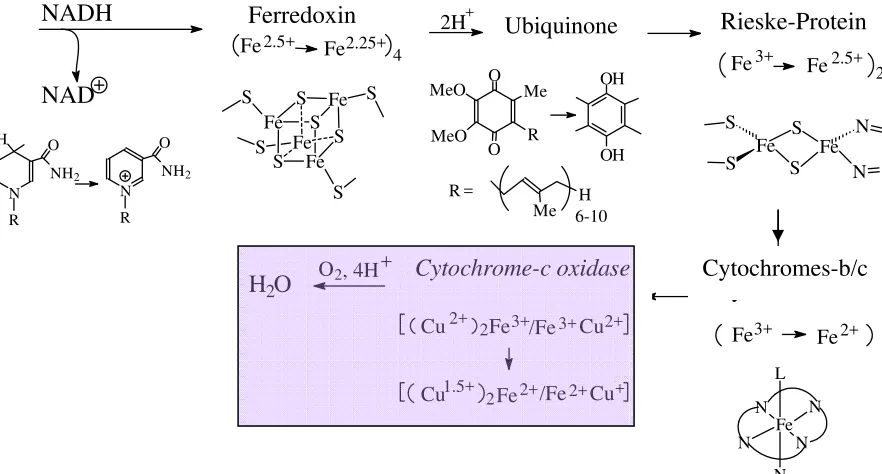
Step 1: Primary acceptor for two reduction equivalents delivered by NADH is an iron-sulphur protein belonging to the [4Fe,4S] ferredoxin family (for a systematic treatment of ferredoxins see below). Such an iron-sulphur cluster can accept and deliver just one electron per cluster. The charge is delocalised over the complete cluster system; the mean oxidation state of iron is 2.5 in the oxidised and 2.25 in the reduced form.
Step 2: Electron acceptor for the ferredoxin is a quinone (so-called ubiquinone, containing a polyisoprene side-chain in position 2), a two-electron acceptor which becomes reduced to the hydroquinone.
Step 3: Another iron-sulphur protein, the Rieske protein (or Rieske centre) then takes over. Rieske proteins are two-centre iron proteins with one of the irons carrying two His (the other one is coordinated to 2 Cys- and two bridging S2-). In the oxidised form, both iron ions are in
H3C C
H
OH
CO2- + NAD+ H3C C
O
CO2- + NADH + H+
L a c t a t P y r u v a t
the +III state, in the reduced form, the ferric iron coordinated to four sulphur functions turns to the ferrous state.
Figure 10. Reaction cascade in the mitochondrial respiratory chain (shortened).
Step 4: The reduction equivalents are now transferred to cytochrome-b (Cyt-b) and further to cytochrome-c (Cyt-c). In these haeme-type electron transporters (for details see below), iron switches between the ferric and ferrous state.
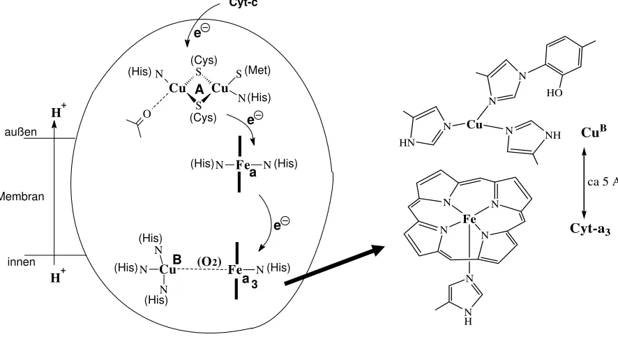
Step 5: The reduced (FeII) form of Cyt-c is re-oxidised by cytochrome-c oxidase, an enzyme that contains 5 redox active centres: 2 haeme type FeII/III (Cyt-a and Cyt-a3), a dinuclear cupper
centre{CuII2/Cu1.52} = CuAand a mononuclear CuI/II = CuB centre. In addition, there are two
structural metal centres (Zn2+ und Mg2+).
Step 6: Cytochrome-c oxidase (Cyt-c-Ox) can take up 4 electrons from 4 Cyt-c. These
electrons are employed for the reduction of O2 to H2O. 8 protons are handled in this process: 4
of the protons are needed to form water; 4 additional protons are translocated across the membrane (from the intra- to the extra-mitochondrial space); i.e. Cyt-c-Ox also works as a proton pump. Activation and reduction of the oxygen (via peroxide) occurs between the adjacent Cyt-a3 and CuB centres For the organisation of Cyt-c-Ox see Fig. 13.
4Cyt-c(Fe2+) + [Cyt-c-Ox]ox→ 4Cyt-c(Fe3+) + [Cyt-c-Ox]red
[Cyt-c-Ox]red + O2 + 8H+in→ [Cyt-c-Ox]ox + 2H2O + 4H+ex The iron-sulphur proteins
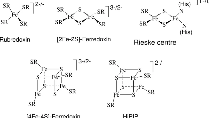
The more important (in the sense that they are more generally used) members of this family are collated in Fig. 11. (1) Rubredoxins contain one iron centre tetrahedrally
coordinated to four cysteinates. (2) [2Fe,2S] ferredoxines, with two iron centres, constitute two edge-bridged FeS4 tetrahedra. The bridging sulphur functions are inorganic sulphide S2-, the
remaining ligands are cysteinate. (3) [4Fe,4S] ferredoxins have a cubane structure. The four trebly bridging functions are again sulphide, also termed labile sulphur because they can be
H2O O2, 4H+
/Fe
2Fe Cu
Cu /Fe 2 2+ 3+ 3+ 2+ + 2+ 2+
1.5+ Fe Cu
Cu Cytochrom-c-Oxidase N N Fe N N N L 2+ 3+ Fe Fe Cytochrom-b/c N Fe S S Fe S S N 2 2.5+ 3+ Fe Fe Rieske-Protein 2H+ OH OH O O MeO MeO Me R Ubichinon Fe S Fe S Fe S S Fe S S S S 4 Fe2.25+ 2.5+ Fe Ferredoxin N R NH2 O N R NH2 O H NAD NADH
R = H
Me 6-10
Ubiquinone
converted to volatile H2S with acids. The mean oxidation state in the reduced form is 2.25, in
the oxidised form 2.5, the redox potential is typically around -200 mV. (4) HiPIPs (High Potential Iron Proteins) are identical to the [4Fe,4S] ferredoxins in as far as the core structure is concerned. However, the mean oxidation state in the reduced form is 2.5, in the oxidised from 2.75, and the redox potential is typically around +300 mV. Along with these “classical” iron-sulphur clusters, others are know, in which one iron centre is missing ([3Fe,4S] ferredoxins), or where two [4Fe,4S] ferredoxins form double-cubanes, or where a fifth ligand (Ser or His) is coordinated to one of the iron centres. The Rieske proteins have already been mentioned above; the angle N-Fe-N is ca. 90°, i.e. there is strong distortion from tetrahedral symmetry for this specific iron.
Figure 11. The iron centres of the classical (and more frequently used) iron-sulphur proteins. SR = cysteinate(1-).
Tutorial: Oxidation and reduction
An oxidation corresponds to a removal of electrons (increase of the oxidation number), reduction correspondingly to a transfer of electrons to a substrate (decrease of the oxidation number). Oxidation and reduction are coupled; an example is the oxidation of ferrous to ferric iron, coupled with the reduction of oxygen to water:
2Fe2+ + ½O2 + 2H+ → 2Fe3+ + H2O
In principal, all redox reactions are equilibrium reactions. The direction is determined by the redox potentials of the two pairs of underlying electron transfer processes. Standard redox potentials E0 are tabulated; standard conditions are: 298 K, 105 Pa, c = 1 mol/l:
Fe3+ + e- ' Fe2+;
E0 = +0.771 V
½O2 + 2e- +2H+ ' H2O; E0 = +1.229 V
Recalculation of the potential for real concentrations, Ec, is achieved with the Nernst equation: Ec = E0 + (0.059/n)log(cOx/cRed)
where n is the number of transferred electrons; cOx and cRed the concentrations of the oxidised
and reduced forms, respectively. In particular, the pH dependence has to be taken into account: At pH 7, (c(H+) = 10-7), Ec for the pair H2/H+ is -0.414 V (E0 = 0), for H2O/O2 +0.815 V.
HiPIP [4Fe-4S]-Ferredoxin
Rieske-Zentrum [2Fe-2S]-Ferredoxin
Rubredoxin
2-/-Fe
S Fe
S Fe
S Fe
S SR
SR
SR
SR
3-/2-Fe
S Fe
S Fe
S Fe
S SR
SR
SR
SR
(His) (His)
Fe S
S Fe SR
SR
N
N
3-/2-
2-/-Fe S
S Fe SR
SR
SR
SR Fe
SR
SR SR
SR
Rieske centre
innen Membran
außen
e e e
H+ H+
(O2)
(His) (His)
(His)
(His) (His) (His)
B
Cu Fe N
N N
N a3
a Fe N N
(His) (His)
(Cys) (Cys)
(Met) Cu
S
Cu
S S
N N
O
A
Cyt-c Cytochromes and cytochrome-c oxidase
Cyt-b und Cyt-c, and the cytochromes-a and -a3 of the cytochrome-c oxidase contain haeme
type iron centres. They are distinct by their substitution pattern at the porphyrin ring and the axial ligands; see Fig. 12. They transfer electrons, moving between the iron oxidation states +II and +III. Cytochromes may also attain other than simple electron transfer functions. An
example is cytochrome P450, an oxygenase in which, during turn-over, iron passes through the
oxidation state +IV (and, perhaps, +V).
L2
L1
Fe N
N N
N
CH3
CH3
H3C
HO2(CH2)2
HO2(CH2)2
R3
R2
R1
Cyt-a: R1 = vinyl, R2 = C17H34OH, R3 = formyl
L1 = L2 = His
Cyt-b: R1 = R2 = vinyl, R3 = methyl L1 and L2 free or His
Cyt-c: R1 = R2 = CH(CH3)-S-CH2-C(O)NH, R3 = CH3
L1 = His, L2 = Met
Cyt P450: R1 = R2 = vinyl, R3 = methyl
L1 = Cys, L2 = H2O (resting form) or free (active form) Mb and Hb: R1 = R2 = vinyl, R3 = methyl
L1 = His, L2 = free or O2
Figure 12 . The active centres of selected haeme-type proteins. For Cyt-P450, see the chapter on
oxigenases.
N N
N N Fe
N N H
N N
HN N
NH N Cu
HO
CuB
Cyt-a3
ca 5 A
5. Photosynthesis
Photosynthesis (assimilation) and respiration (dissimilation) are complementary processes. Photosynthesis results in reductive carbon fixation and production of oxygen, energy driven by light energy, hν.
hν
CO2 + 2H2O* → {CH2O} + O2* + H2O
Carbon dioxide is reduced, in a 4e- reduction, to {CH2O} (symbolising a carbohydrate such as
1/6 of glucose). Reducing agent is water, which is oxidised to O2. Instead of resorting to light
as an energy source, chemical energy (energy liberated in the course of a chemical reaction) can be employed, and sources for carbon other than CO2, e.g. CO or acetate, can be used.
Depending on the energy and the carbon source, one distinguishes the following categories: Light energy: phototrophic
Chemical energy: chemotrophic CO2 as C-source: autotrophic
Other C-sources: heterotrophic
Green plants, cyanobacteria and other photosynthetically active bacteria, and protozoa containing chlorophyll produce bio-mass photo-autotrophically. A 100 year old beech tree produces about 1000 l of O2 and 12 kg of carbohydrates per day (this corresponds to 100 ml of
O2 and 1.2 g of glucose per 1 m2of foliage). The major part of bio-mass is, however, produced
by chemotrophic microorganisms. Examples for chemical processes supplying energy are: Fe2+→ Fe3+ + e
-H2→ 2H+ + 2e
-HS- + 4H
2O → SO42- + 9H+ + 8e
-Mn2+ + 3H
2O → MnO(OH)2 +4H+ + 2e
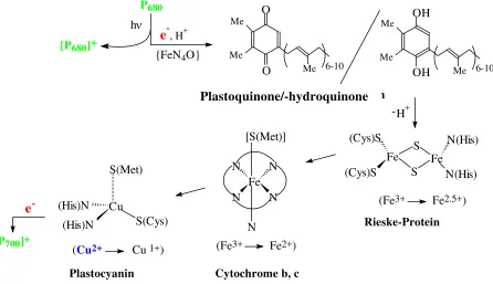
-In the photosynthetic process carried out by plants, one distinguishes between the light reaction and the light-independent (or dark) reaction on the one hand and, within the light reaction, between photosystems I and II (PSI and PSII, also referred to as light harvesting complexes LHC) on the other hand:
Light reaction:
PSII: P680 + hν→ [P680]+ +
e
- (via phaeophytin, a ”chlorophyll“ depleted of Mg2+:2[P680]+ + H2O→ 2P680 + ½O2 + 2H+ (catalysed by water oxidase)
e
- transfer chain from PSII to PSI (Fig. 14) PSI: P700 + hν→ [P700]+ +e
-[P700]+ +
e
-→ P700NADP+ +
2e
- + 2H+→NADPH + H+ (catalysed by [2Fe,2S])Dark reaction: 2(NADPH + H+) + CO2→{CH2O} + 2 NADP+ + H2O (energy driven by ATP) The photosystems are collectives of pigment molecules (ca. 200), mainly chlorophyll-a and -b, carotinoids, anthocyanes and xantophylls. These pigments act as collectors over the complete spectrum of the (visible) sun light. The energy thus collected is transferred to the reaction centres, which represent specific molecules of chlorophyll-a, termed P680 in PSII, and P700 in
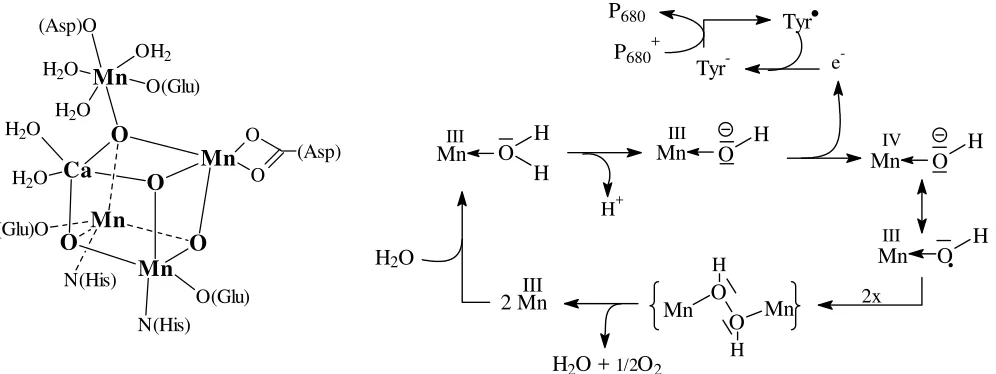
The water oxidase (oxygen evolving centre, OEC), which catalyses the oxidation of water via P680, contains 5 metal centres at its active site. Four of these metal ions (3 Mn3+/4+ and one
Ca2+) form, together with 4 O2-, a cubane-like cluster (Fig. 16). In Fig. 16, the (assumed) catalytic process is also shown. During turn-over, the four manganese centres change between the oxidation states +IV to +III step by step in the 4-electron oxidation of 2 molecules of water.
Figure 14. Simplified representation of the electron transport chain between PSII and PSI.
Figure 15: Chlorophylls ( H i s)
N H H 2 9 C 2 0
O O = C
H 2 C H 2 C H 3 C
H 3 C
N N
N N O M g O H H O N H O H Mg
R
H 2 C = C HC = O O C H 3
C H 2 CH3
C H 3
R
= CH3: Chl. a = CHO: Chl. bgesättigt im
Bakteriochlorophyll Dimerisierung über
H-Brücken in den Reaktionszentren der Photosysteme
Dimerisierung über H-Brücken in den Reaktionszentren der Photosysteme
Dimerisation via H-bridges in the reaction centres of PSI and PSII
Dimerisation via H-bridges in the reaction centres of PSI and PSII Saturated in bacterial chlorophyll Me Me Me O O 6-10 Me Me Me OH OH 6-10 H+ -Fe S S Fe (Cys)S (Cys)S N(His) N(His)
(Fe3+ Fe2.5+) Fe N N N N N [S(Met)]
(Fe3+ Fe2+) Cu
S(Cys) (His)N
(His)N
S(Met)
(Cu2+ Cu 1+)
Plastochinon/Hydrochinon
Rieske-Protein
Cytochrome b, c Plastocyanin
e
-[P700]+
P680
e-, H+ {FeN4O}
[P680]+
hν
Plastoquinone/-hydroquinone
Tyr
-Tyr
MnIII O H
MnIII O. H Mn O H
H III
H2O + 1/2O2
Mn OO Mn
H
H H+
e
-MnIV O H
2x
2 MnIII H2O
P680+ P680
Figure 16. Organisation of the metal cluster in the oxygen evolving centre (left), and the presumed catalytic process of water oxidation (right). Tyr• = tyrosyl radical, Tyr- = tyrosinate.
Plastocyanin, at the end of the electron transfer chain between PSII and PSI belongs to the category of ’blue copper proteins’, or type I Cu proteins. In these Cu proteins, Cu1+/2+ is coordinated in a flat trigonal-bipyramidal fashion by two Cys- and one His, and – in the axial position at a rather long distance of 2.9 Å – methionine. The intense blue colour of the oxidised (Cu2+, d9) form comes about by a ligand-to-metal charge transfer (LMCT), i.e. transfer of electron density from non-bonding orbitals of the coordinated Cys-S into the 3d ‘hole’ of Cu2+. While charge transfer within the d-d system of a metal ion is parity (Laport)
forbidden, and the corresponding absorption bands hence are weak in intensity (see, e.g., [Cu(H2O)6]2+), LMCT transitions are allowed and thus very intense.
Ca
O
Mn O O
Mn
O Mn
Mn O(Glu)
(Asp)O
OH2
H2O H2O
H2O
H2O
O
O
(Asp)
O(Glu) (Glu)O
N(His)
N(His)
Disgression: Systematics of copper proteins
Type I (Blue Cu-proteins): trigonal coordination geometry; ligands: 2 Cys(1-), 1 His, 1 weakly bonded Met. Strong LMCT at 600 nm; small EPR-spectroscopic hyperfine coupling constant (A = 5⋅10-4 cm-1). Function: e- transfer; Example: Plastocyanin in the e- transfer chain
PSII→PSI.
Type II: Tetragonal coordination geometry; ligands: His, Tyr(1-), H2O, no Cys. “Normal”
optical behaviour (d-d transitions); normal EPR patterns (A = 18⋅10-4 cm-1). Function: Redox
reactions; Examples: Galactoseoxidase (RCH2OH → RCHO + 2H+ + 2e-),
CuZn-superoxidedismutase (2O2- + 2H+→ O2 + H2O2).
Type III: Contain 2 cooperating Cu centres; trigonal coordination geometry; ligands: 3 His per Cu. Intensely blue in the oxidised form (O22-→Cu2+ LMCT); no EPR signal
(antiferromagnetically coupled). Function: Transport and transfer (to a substrate) of oxygen; examples: haemocyanin, Fig. 8; tyrosinase (Tyr + ½O2 + 2e-→ DOPA).
Ceruloplasmin, important for the absorption of iron, is a Cu protein containing 7 Cu centres representing types I, II and III. Nitritereductase contains type II (substrate activation) and type I Cu centres (e- transfer)
6. Hydrogenases, oxigenases, oxidoreductases, peroxidases and dismutases
Overview
Hydrogenases (often associated with the cofactors NADH or FADH2)
H2 ' 2H+ + 2e- more correct: H2 ' H+ + H- (followed by: H-→ H+ + 2e-)
May be coupled with the transfer/abstraction of hydrogen to/from a substrate (hydrogenation/dehydrogenation):
substrateH2 ' substrate + 2H+ + 2e-
Oxidoreductases generally catalyse oxidations (electron abstraction) and/or reductions (electron delivery), such as the iron-sulphur proteins or the cytochromes.
Some oxidoreductases use oxygen for the dehydrogenation of a substrate (oxidases) or water for the hydrogenation of a substrate (reductases):
SubstratH2 + ½O2 ' Substrat + H2O
Oxigenases transfer/insert, usually starting from oxygen O2, oxo groups (O2-) to/into a
substrate:
substrate + O2 ' substrateoxide/-hydroxide
often coupled to: ½O2 + 2H+ + 2e-→ H2O
The reverse process, i.e. the removal of O2- from a substrate, is catalysed by deoxygenases. Substrates can be organic in nature (RH → ROH; (CH3)2S → (CH3)2S=O), or inorganic (NO3 -→ NO2-).
Peroxidasen employ H2O2 for oxygenation:
substrate + H2O2→ substrateoxide/-hydroxide + H2O
Dismutases disproportionate oxygen species with the oxygen in the oxidation states –I (peroxide) and -1/2 (superoxide):
Catalases: H2O2→ H2O + ½O2
Sub steps: H2O2→ O2 + 2H+ + 2e- (oxidation)
H2O2 + 2H+ + 2e- → 2H2O (reduction)
Superoxidedismutases: 2O2- + 2H+→ H2O2 + O2
Sub steps: O2-→ O2 + e- (oxidation)
O2- + e- + 2H+→ H2O2 (reduction) Iron-only hydrogenase
This enzyme catalyses the charge separation in the H2 molecule via polarisation between
the NH function of the bridging
aminedithiolate and one of the iron centres, and finally the bond cleavage to form a hydridic Fe-H- and a protic R2NH2+
intermediate. Electrons from the H- are then transferred off via a [4Fe,4S] ferredoxins in direct contact with the hydrogenase.
Fe
Fe
C
O C
C C
C
C S
S S
N
O N
O O
HN
[4Fe,4S]
Hδ+
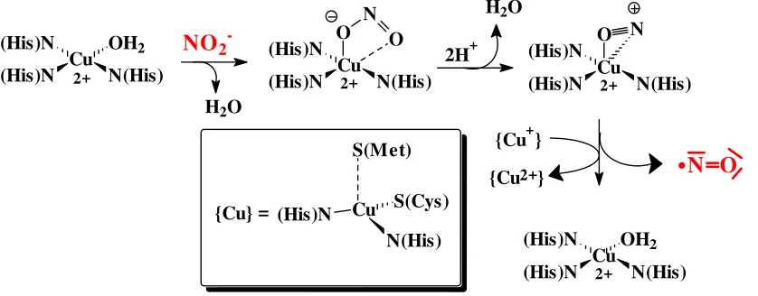
-Nitritereductase
This enzyme catalyses the deoxigenation of nitrite NO2- to nitrogenmonoxide NO via a
one-electron reduction, one of the focal steps in dinitrification (see ch. 7). The enzyme is built up of three identical subunits. Each of these subunits contains a catalytic type-II Cu centre (for the activation of nitrite) and a type-I Cu centre for electron transfer (reduction of nitrite) [see inset on p. 20 for the classification of copper enzymes). Reaction steps (cf. Fig. 17):
(1) Exchange of water for nitrite/nitrite activation (at {Cu-II}) (2) Formation of nitrosyl: NO2- + 2H+→ NO+ + H2O
(3) NO+ + e-→ NO (by {Cu-I})
(4) Reestablishment of the starting situation by exchange of NO for H2O
Cu
N(His)
OH2
(His)N (His)N
2+
NO2
-H2O
Cu
N(His) O
(His)N
(His)N O
N
2+
{Cu+}
{Cu2+}
2H+
H2O
Cu
N(His) (His)N
(His)N O
N
2+
Cu
N(His) (His)N
(His)N OH2
2+
{Cu} = Cu
N(His)
(His)N S(Cys)
S(Met)
N O
Figure 17. The course of reaction catalysed by nitritereductase. Oxigenases
Cytochrome P450 is an oxygenase belonging to the haeme-type proteins. Axial ligands are a cysteinate and – in the resting state – water. In the active state, the position occupied by water is free. Cyt-P450 detoxifies hydrophobic substrates (such as benzene) in the liver by conversion
to hydrophilic compounds (such as phenol) which are than secreted. The overall reaction can be formulated as shown:
RH + O2 + 2H+ + 2e-→ ROH + H2O
The substrate RH is bonded by hydrophobic interaction into the protein pocket close to the active centre of the active form of the enzyme. The course of reaction is illustrated in Fig. 18. In the first step, FeIII is reduced to FeII, followed by oxidative addition of O2 (FeII + O2→
FeIII-O2-), i.e. O2 is reduced to superoxide, and further – by an external e- source – to peroxide:
FeIII-O
2- + e-→ FeIII-O2-. In the succeeding step, FeIII transfers 2 electrons to the peroxo ligand.
RH + {O=FeV} → R• + {HO-FeIV}→ R-OH + FeIII
Tyrosinase and catecholoxidase: These two closely related enzymes contain type III copper centres (see inset on p. 20) and thus resemble the haemocyanins (ch. 3). The homology of the amino acid sequence is, however, restricted to the direct surroundings of the copper centres. Activation of oxygen by tyrosinase and catecholoxidase compares to that of haemocyanin, except that is irreversible:
2CuI + O
2→ CuII(O22-)CuII
One of the histidine ligands on one of the Cu centres in tyrosinase can be weakened to allow for attachment of the substrate tyrosine. Tyrosinase catalyses the oxygenation of tyrosine to dopa (o- dihydroxyphenylalanine; precursor for the neurotransmitter dopamine, and for adrenaline), catecholoxidase further oxidises dopa to the respective quinone (Fig. 18). These reactions are followed by further dehydrogenation to form indolquinone and finally melanin. Melanin, a very complex compound, is the dark pigment formed as freshly broken fruits (like apples or bananas) or vegetable (like potatoes) are exposed to air. Melanin is also the dark pigment responsible for the suntan, or present in the brown beauty patches and in melanomas.
OC N
O O
- 4[H] Indolchinon
- 2[H]
Dopa Tyrosin
O
O OC
NH
+ H2O
NH
OC OH
OH + O2 + 2H+ + 2e
-NH
OC OH
Melanin
Figure 18. Reactions which are catalysed by tyrosinase/catecholase.
N
N Fe N N
OH2
S(Cys)
N
N Fe N N
S(Cys)
III III
RH
RH N
N Fe N N
S(Cys) RH
e- II
N
N Fe N N
S(Cys) OO RH
O2
III N
N Fe N N
S(Cys) OO RH
III
e -N
N Fe N N
S(Cys) O RH
2H+ H2O
V ROH
H2O
Oxigenases/deoxygenases containing the molybdopterin cofactor, Fig. 19. O P X HN N N N O
H2N O O S S Mo O H H O P O O O OH O Cyt/Gua VI HN N N N H2N
O O X S S Mo O VI
+ Substrat
IV O X S S Mo + Substrat-O Pterin Molybdopterin
Figure 19. Oxidised form of the molybdopterin cofactor (upper right) of the sulphite reductase family. X usually is Cys-. Molybdopterin transfers oxido groups to a substrate (shown) or off a
substrate.
An example is the (dissimilatory) nitrate reductase: NO3- + 2e- + 2H+ → NO2- + H2O)
Mo O Mo O O N O O Mo O O
IV + NO3 NO2 + VI
+ FADH2
Vanadate-dependent haloperoxidases from marine algae catalyse the oxidation of halide (Hal-) to a Hal+ species such as hypohalous acid. Oxidant is hydrogen peroxide. The Hal+
species halogenates organic substrates non-enzymatically. For the mechanism, see Fig. 20. Hal- + H2O2 + H+→ HalOH + H2O
HalOH + RH → RHal + H2O
Figure 20. Active centre of vanadate-dependent haloperoxidase (left), and mechanism of the enzymatic formation of hypobromous acid from bromide (right).
N NH N NH H OH O O O V H2O
H2O
HO CH2 CH HOBr Br -V O O O H H+ V O O O H2O
+ H2O2 OH
O V
Cu,Zn-Superoxidedismutase contains a catalytically active type-I copper centre and a structural zinc centre, linked by bridging His-; Fig. 21. The enzyme catalyses the disproportionation (dismutation) of superoxide to hydrogenperoxide and oxygen.
Figure 21. Active centre of Cu,Zn-superoxidedismutase
Overall reaction: 2O2- + 2H+→ H2O2 + O2 Reaction sequence:
(1) CuII(H2O) + O2-→ CuII(O2-) + H2O (exchange of water for hyperoxide)
(2) CuII(O2-) + O2-→ CuI(O2-) + O2 (oxidation of external O2- to O2)
(3) CuI(O2-) + H+→ CuII(HO2-) (internal reduction of O2- to peroxide at the Cu centre)
(4) CuII(HO2-) + H+ + H2O→ CuII(H2O) + H2O2 (separation of hydrogenperoxide)
7. Nitrogen fixation
Nitrogen fixation is the biogenic and non-biogenic transformation of elemental N2 into
nitrogen compounds, affording to overcome the bonding energy between the two trebly bonded nitrogen atoms (949 kJ/mol).The biogenic fixation, carried out by free living nitrogen-fixing bacteria (Azotobacter) and cyanobacteria (“blue-green algae”, Anabaena), some archaea, and
by symbiotic bacteria associated with leguminous plants (Rhizobium), leads to ammonium
ions. Biogenic fixation accounts for about 60% of the overall nitrogen supply. Non-biogenic non-anthropogenic fixation, which can occur by electric discharge (lightning) in the
troposphere, and by cosmic radiation in the stratosphere [N2→ 2N; N + O2→ NO + O →
NOx], accounts for 10%. For the overall conversions cf. Fig. 22. The remaining 30% of
world-wide N2 fixation go back to the Haber-Bosch process and combustion of fossil fuels (natural
gas, coal, crude oil) and products produced from crude oil (petrol, gasoline, diesel). Comparison between the Haber-Bosch process and the biogenic N2 fixation:
Haber-Bosch biogenic
N2 + 3H2 ' 2NH3 N2 + 10H+ + 8e-→ 2NH4+ + H2
(energy driven by: 16ATP + 16H2O → 16ADP + 16Pi)
Temperature: 500 °C Temperature: ca. 20 °C Pressure: 200-450 bar Pressure: 1 bar
Catalyst: Fe (+ Al2O3 + K2O + …) Catalyst: Nitrogenase (Fe/Mo- or Fe/V-S cluster)
Yield: 17% Yield: 75%
Annual production ca. 108 t Annual production ca. 108 t II
+ (Arg)
NH2
-Cu O H H
(His)N (His)N
N(His) N N Zn
N2
N it roge n c yc le
Nitrification NH3
Biogenic nitrogen fixation
{C-N} Assimilation
Degra-dation
Nitrite NO N2O
Denitri-fication
Ammonification
Nitrate Non-biogenic nitrogen fixation
Nitrite
Figure 22: The nitrogen cycle. Processes involving the –III oxidation state of nitrogen are in red. For nitrite and nitrate reductases see the previous chapter
In Fig. 23, the organisation of the metal centres of nitrogenase is depicted. The electrons necessary for the reduction of dinitrogen are delivered by an iron protein containing a cubane-like [4Fe,4S] ferredoxin. Primary e- acceptor is the P cluster of the FeMoco, the
iron-molybdenum-cofactor. Two ATP (activated by Mg2+) are hydrolysed per electron transferred. The FeMoco contains two P and two M clusters, arranged in such a way that the complete
cofactor attains C2 symmetry. The P cluster is a double cubane containing the Fe8S7 core. The
reduction equivalents are then further transported to the M cluster, a Fe7MoS9 core, which is
responsible for the final activation and reduction of N2. The cage formed by the metal centres
of the M cluster contains electron density which can be interpreted in terms of a nitrogen atom.
The M cluster is connected to the protein matrix by just one Cys and a His, the latter
coordinated to Mo. The coordination environment of Mo is supplemented by the vicinal hydroxide and carboxylate of homocitrate. In which way activation and reduction of N2 takes
place is unknown. In the case of insufficient molybdenum supply, or at low temperatures, a vanadium-nitrogenase is activated (which is more efficient at lower temperatures than the Mo version). An iron-only nitrogenases is also known.
-Cluster M
M
S
(Cys)S
Fe Fe
Fe S
S Fe
S CH2CO2
-Gln CH2CH2CO2
-O C O-C N(His) Fe
Fe
Fe S
S Mo
S S
S
O
N
Tutorial: Nitrogen
Abundance: Atmosphere (in the form of N2, 78.1 Vol-%; 4·1015 t); hydrosphere (N2 dissolved in
water, 1012 t); in minerals (saltpetre NaNO
3) and rocks (2·1017 t); in organic form in the biomass of
soil-bound microorganisms (3·1011 t), plants and animals (1010 t).
The bond in dinitrogen is a triple bond; the bond energy amounts to 949 kJ/mol, i.e. N2 is
particularly inert.
Hydrogen compounds: NH3 (ammonia; synthesis from H2 and N2 according to the Haber-Bosch
process) and ammonium ions (NH4+), N2H4 (hydrazine), HN3 and salts derived thereof (azides, e.g.
NaN3, commonly used as fungicide and bactericide in bio-assays). Nitrides, e.g. Na3N, formally
derive from ammonia. Ammonia is an efficient complexing agent, e.g. for Ag+: AgCl dissolved in aqueous NH3 forms soluble [Ag(NH3)2]+, which gradually converts to silvernitride Ag3N (highly
explosive). The ammonium ion is a Brønstedt acid; aqueous solutions of ammonium salts consequently are acidic.
Oxygen compounds: N2O (dinitrogen monoxide, “laughing gas“), NO (nitrogenmonoxide;
synthesis by combustion of ammonia according to the Ostwald process), NO2 (nitrogendioxide, in
equilibrium with N2O4. NO2 reacts with water to form nitrous acid HNO2 + nitric acid HNO3),
N2O5 (dinitrogen pentoxide). Salts derived from HNO3 are termed nitrates, those derived from
HNO2 nitrites.
Use: Fertilisers (ammonium compounds, nitrates), explosives (nitrate; gun powder is a mixture of saltpetre, charcoal and flower of sulphur). HNO3 is used for nitrosylations in organic synthesis.
Organic nitrogen compounds: Amines (NH2R, R = phenyl: aniline; NHR2; NR3), heterocyclic
nitrogen compounds (for a selection see below), amides (1a) and peptide (1b), hydroxamic acids (2), aminoacids (3), nitro compounds (4), nitrosamines (5), diazo compounds (6).
(6) N N R
R' (5)
(4)
R N NO
H R NO2
(3)
(2)
(1b) (1a)
CH2 C R
O
N R
OH
CH C R
O
NH CH R' CH2 C
R
O
NH2
bzw. H3N CH CO2 R
H2N CH CO2H R
Pyridin Piperidin Adenin Pyrrol Imidazol NH
N NH
N
N
N
NH NH2
NH NH
N
8. Nitrogenmonoxide
N O N O
NO forms in the troposphere by electric discharges, and under stratospheric conditions under the influence of cosmic rays and high-energy UV. Further oxidation to NO2 readily
occurs:
N2→ 2 N; N + O2→ NO + O
2NO + O2→ 2NO2
With additional oxygen and moisture, nitric acid is formed, one of the constituents of ”acid rain“:
2NO2 + H2O + ½O2→ 2HNO3
Industrially, NO is obtained by passing a mixture of ammonia and oxygen through a platinum net (contact time 10-3 s, temperature 1000 °C), which is further processed to form nitric acid (Ostwald process):
2NH3 + 2½O2→ 2NO + 3H2O
2NO + 1½O2 + H2O → 2HNO3
A large amount of HNO3 goes into the production of ammonium nitrate for artificial fertiliser.
NO is also contained in the exhaust gases of automobiles (along with water and CO2 as the
main components, and fuel constituents), as well as in industrial and domestic exhaust, and rapidly is oxidised to NO2. Under the influence of UV, i.e. on sunny days, NO2 is split into NO
and oxygen atoms, which oxidise alkanes to alkylhydroperoxides, and form ozone with molecular oxygen (“summer smog”):
NO2 + hν→ NO + O
O + O2→ O3
O + C2H6 + NO2
·
→ C2H5O2H + NO·
In the stratosphere, NO catalyses ozone depletion (see also the tutorial ‘oxygen’ on p. 11): NO + O3→ NO2 + O2
NO2 + O → NO + O2
__________________
O3 + O → 2 O2 (kinetically hindered without catalyst)
In organisms, NO is an important multi-functional messenger and
neuro-transmitter, targeting, inter alia, metal centres in haeme-type proteins, and cyclic guanosine-monophosphatase (cGMPase).
Biosynthesis of NO is achieved by oxidation of one of the NH2 groups of
thus participates – as a killer agent for infectious germs – in the functioning of the immune system; (3) eNOS, in the endothelial tissue cells, where it controls the tonicity of the vascular muscles and thus the blood pressure. The NO-induced relaxation of the vascular muscles is also the basis for the medication of hypertension and angina pectoris with compounds which set free NO under physiological conditions, such as amylnitrite, (C5H11NO2); nitroglycerin
(glyceroltrinitrate CH2(ONO2)-CH(ONO2)-CH2(ONO2)) and nitroprussid sodium (disodium
pentacyanido-nitrosylferrate Na2[Fe(NO)(CN)5]).
NO is also used by the glow worm (lightning bug, firefly) to switch on its glow organs. This luminescence can be traced back to the oxidation, by O2, of luciferyl-AMP (AMP =
adenosine-monophosphate) via peroxoluciferin to oxoluciferin; cf. Fig. 24. To start this oxygen consuming formation of peroxoluciferin, the glow worm triggers NO synthesis. The NO is used to block mitochondrial cytochromes (by coordination of NO to Fe) and thus the consumption of oxygen in respiration. The O2 thus becomes available for triggering
luminescence. Other organisms capable of bioluminescence also employ this mechanism. An example is Nocticula scintillans, a dinoflagellate responsible for marine phosphorescence.
.
Figure 24. NO induced luminescence in the glow worm.
Larger amounts of NO are toxic, because NO binds to the iron and copper centres of enzymes depending on these metals. Haemoglobin binds NO ten-thousand fold more effective than O2. On coordination, NO is reduced to NO-, which is isoelectronic with O2 and binds, as
O2, in the bent end-on mode. Additional NO toxicity arises from the fact that NO nitrosylates
amines, via the intermediate formation of nitrous acid, to form carcinogenic nitrosamines: NO + H2O → HNO2 + e- + H+
R2NH + HNO2→ R2N-NO + H2O
(Nitrous acid is also formed, under physiological conditions, by reduction of nitrate, present in e.g. leafy vegetables.)
9. The biochemistry of zinc
2.5 g of zinc per 70 kg body weight makes Zn the second-to-most abundant transition metal of biological importance. Contrasting iron, copper, manganese and molybdenum, zinc is not redox active (valence electron configuration d10). In zinc proteins, Zn2+ takes over either a
+ CO2 + Licht
HOAMP
Oxiluceferin
S
N O
HO
N S
S N
C OOH HO
N
S O
AMP
(Luciferase)
O2
Luciferyl-AMP
HO P O
P O
O O OH O
ATP(Mg2+)
S
N S
N
HO COOH
NO
L u c i r i n
Peroxoluciferyl-AMP
+ CO2+ light NO
catalytic or a structural function; see the classification below. The daily requirement for zinc is 3-25 mg, a demand which, since zinc is ubiquitous, is commonly satisfied by our nutrients. Diseases due to zinc deficiency encompass disturbance of growth, arthritis-related health problems, break-down of the immune system and loss of taste. They are usually a consequence of impaired zinc absorption rather than of undersupply. In the blood stream, zinc is transported by albumin and transferrin. Zinc storage is achieved by thioneins (vide infra).
Ointments containing zinc (in the form of ZnO, Zn(OH)2, zinclactate) have already
been employed in the Middle Age and are still employed today in wound healing. The
essentiality of zinc for life had been discovered in 1869 by Raulin (inhibition of the growth of the mould Aspergillus niger caused by undersupply of zinc). In 1940, the first zinc-dependent
enzyme, carboanhydrase, was isolated by Keilin and Mann, followed by the discovery, in 1954, of the second enzyme, pancreatic bovine carboxypeptidase-A. In 1985, the role of zinc in genetic transcription (“zinc fingers”) became established, and in 1995, the Zn-based Ada repair
protein (demethylation of DNA) was characterised. A role of Zn2+ in synaptic transmission was found in 2006.
Classification of zinc proteins according to function: 1. Enzymatic:
- Ligases and synthases (C-C-bond formation, e.g. aldolases)
- Hydrolases: Here, Zn2+ is coordinated by three amino acid side-chains of the protein matrix (His, Cys and/or Asp) plus water (inactive, resting form) or a hydroxy group (active form); see the scheme below. Examples: carboxypeptidase-A (an exopeptidase acting at the C-terminus of the peptide), thermolysin (a thermophilic exopeptidase acting at the N-terminus).
Interlude: Inorganic chemistry of zinc
Important minerals: ZnS (zinc blende, wurtzite, sphalerite), ZnCO3 (zinc spar, calamine).
Earth’s outer sphere (17 km crust + hydro- + atmosphere) contains 0.007 % (by weight) of zinc.
Metallic zinc is obtained by firing of ZnS (→ ZnO + SO2) followed by reduction of ZnO with
coal, or by electrolytic reduction of aqueous zinc sulphate. Applications include alloys (such as brass, a Cu-Zn alloy), galvanisation (of iron), torch batteries (Lechlanché element).
The redox potential is E0 = -0.763 V, i.e. Zn is oxidised by H+. In air, Zn is, however, stable due to passivation [formation of compact layers consisting of ZnO, Zn(OH)2 and
Zn(OH)(HCO3)]. In aqueous media, Zn2+ exists in the form of the Brønstedt acid
[Zn(H2O)6]2+; Zn2+ itself is a Lewis acid (and this is determinant for its enzymatic actions).
Zinc hydroxide is amphoteric: Zn(OH)2 + 2H+ → Zn2+ + 2H2O; Zn(OH)2 + 2OH- →
[Zn(OH)4]2-. With halogenated alkanes RX, zinc forms reagents of composition RZnX, which
transform to ZnX2 and ZnR2 on heating. RZnX and ZnR2 are alkylating agents.
Zn2+ forms complexes mainly of coordination numbers 4 ([Zn(SR)4]2-, tetrahedral;
[Zn(Gly)2], square planar), 5 ([Zn(acac)2H2O] [acac = acetylacetonate(1-), square-pyramidal]
R C O
Z R'
+ H2O R C O
Z OH
+ HZR' Z = O: esterases
Z = NH: peptidases
Z = phosphate: phosphatases
Zn O H
2+
H+ supporting proton acid
electrophilic attack
nucleophilic attack protein
- Others: Carboanhydrase (CO2 + OH- ' HCO3-; ch. 3 and Fig. 7)
Alcoholdehydrogenase (RCH2OH → RCHO + 2H+ + 2e-; see below)
Structural: Stabilisation of the tertiary structure of protein dom