Associations between BMI, serum uric acid, serum glucose, and
blood pressure with urinary tract stone opacity
Keywords: blood pressure, BMI, serum glucose, urinary stone, uric acid, urolithiasis
pISSN: 0853-1773 • eISSN: 2252-8083 • http://dx.doi.org/10.13181/mji.v24i2.1068 • Med J Indones. 2015;24:103-8 • Received 17 Sep 2014 • Accepted 2 Apr 2015
Correspondence author: Ikhlas A. Bramono, [email protected]
Copyright @ 2015 Authors. This is an open access article distributed under the terms of the Creative Commons Attribution-NonCommercial-ShareAlike 4.0 International License (http://creativecommons.org/licenses/by-nc-sa/4.0/), which permits unrestricted non-commercial use, distribution, and reproduction in any medium, provided the original author and source are properly cited.
Ikhlas A. Bramono, Nur Rasyid, Ponco Birowo
Department of Urology, Cipto Mangunkusumo Hospital/Faculty of Medicine, Universitas Indonesia, Jakarta, Indonesia
C l i n i c a l Re s e a rc h
ABSTRAK
Latar belakang: Urolitiasis didefinisikan sebagai pembentukan
batu pada ginjal, ureter, atau kandung kemih. Beberapa penelitan menunjukkan bahwa abnormalitas metabolik merupakan hal yang umum pada pasien urolitiasis. Tujuan penelitian ini untuk melihat hubungan antara indeks massa tubuh (IMT), asam urat serum, glukosa serum, dan tekanan darah dengan opasitas batu pada pasien batu saluran kemih.
Metode: Penelitian ini dilakukan secara retrospektif dengan
melihat rekam medis pasien dengan batu saluran kemih yang menjalani prosedur ESWL pada Januari 2008 – Desember 2013 di Departemen Urologi Cipto Mangunkusumo. Data yang yang diambil adalah IMT, kadar asam urat serum, glukosa serum, tekanan darah, dan opasitas batu saluran kemih. Asosiasi antara IMT, kadar asam urat serum, glukosa serum, dan tekanan darah, dengan opasitas batu dianalisis menggunakan uji chi-square.
Hasil: Terdapat 2.889 pasien yang menjalani prosedur ESWL
pada Januari 2008 – Desember 2013. Analisis dilakukan terhadap 242 pasien yang memiliki rekam medis lengkap. Rerata usia adalah ± 12,78 (48,02 tahun). Rasio laki-laki terhadap perempuan adalah 2,27:1. Rerata IMT adalah ± 3,78 (29,91 kg/m2). IMT tinggi didapatkan pada 66,52% pasien.
Proporsi batu radioopak adalah 77,69% (188 pasien). Dua puluh dua pasien (9,1 %) memiliki tekanan darah normal. Pasien dengan kadar serum asam urat tinggi sebanyak 34,30 % (83 pasien). Secara statistik didapatkan hubungan yang bermakna antara kadar serum glukosa sewaktu dengan opasitas batu (p < 0,05).
Kesimpulan: Terdapat hubungan antara kadar serum
glukosa sewaktu dengan opasitas batu pada pasien urolitiasis. Pasien hiperglikemia cenderung memiliki batu radiolusen. Sementara pasien normoglikemia cenderung memiliki batu radioopak.
ABSTRACT
Background: Urolithiasis refers to formation of stone in the kidney, ureter, or bladder. Several studies showed metabolic abnormalities were common in urolithiasis patients. The aim of this study was to describe the association between body-mass-index (BMI), serum uric acid, serum glucose, and blood pressure toward stone opacity in urinary tract stone patients.
Methods: This study was done retrospectively by reviewing registry data of urinary tract stone patients that had undergone ESWL on January 2008 – December 2013 in Department of Urology Cipto Mangunkusumo Hospital. Data concerning body mass index, serum uric acid, serum glucose, blood pressure, and urinary tract stone opacity were recorded. Associations between body mass index, serum uric acid, serum glucose and blood pressure with urinary tract stone opacity were analyzed using chi-square test.
Results: There were 2,889 patients who underwent ESWL on January 2008 – December 2013. We analyzed 242 subjects with complete data. Mean age was ± 12.78 (48.02 years). Male-to-female ratio was 2.27:1. Mean BMI was ± 3.78 (29.91 kg/m2). High risk BMIs were found in 161 patients (66.52%). The proportion of radioopaque stone was 77.69% (188 patients). Twenty two patients (9.1%) had normal blood pressure. Patients with high serum uric acid were 34.30% (83 patients). We found a significant association between random serum glucose level and stone opacity (p < 0.05).
Urolithiasis refers to formation of stone in the kidney, ureter, or bladder. It is the third most common cause of urinary tract disease. The prevalence of urolithiasis is reported between 2 and 20% globally, with peak incidence in the second and third decade of life.1,2 The incidence
of urolithiasis increased along with the increasing life expectancy.3
There are several factors that may be related to the development of urinary stones, such as age, gender, race, genetic, climate, dietary intake, and metabolic changes.1,2 Metabolic abnormalities
were found in more than 90% of stone patients.1
Hypercalciuria and hypocitraturia are the most prevalent condition that cause stone disease, although there are also other causes such as hyperuricosuria, hypokalemia, hyperuricemia, hypophosphatemia, and low urine volume.1,2,4,5
In large epidemiological studies, obesity and type 2 diabetes have been associated with increased risk of nephrolithiasis.6 Insulin
resistance reduces the production and transport of ammonia, resulting in alterations of urine acidification and low urine pH.7 The changes in
urinary constituents, including lower urinary pH, decreased citrate excretion, and uric acid and calcium excretion also has been associated with glucose intolerance, elevated blood pressure, and dyslipidemia. These conditions lead to increased risks of uric acid and calcium stone formation.6,8,9
Urolithiasis as renal manifestations in gout patients usually is due to low urine pH.10 Several
studies have shown association between recurrent uric acid kidney stones and metabolic syndrome. Kidney stones patients with type 2 diabetes have higher prevalence of uric acid kidney stones than other patients with kidney stones. Increased soluble undissociated uric acid will lead to uric acid stone genesis.11
Increased body mass index (BMI) not only associated with kidney stone formation but also associated with stone size. Mechanisms of obesity to affect urolithiasis genesis are still being investigated. A study by Amaro, et al.1 has shown
that metabolic syndrome (including obesity and high blood pressure) patients were at risk of certain stone formation (uric acid and calcium oxalate stones). Significant contribution of high BMI to kidney stone formation were through its
influence in lowering urine pH. Obesity not only induces acidic urine, but also increases excretion of uric acid, calcium, and oxalate.12 Maalouf, et
al.12 have suggested visceral obesity as a factor
to disrupt insulin resistance. Insulin resistance will lead to low urine pH. Inadequate ammonium excretion and/or production occurred, if there was reduced insulin bioactivity in the kidney proximal tubule.11
Knowing about the association between metabolic abnormalities and type of stone formation is really important, because metabolic factors can be modified to prevent the risk of certain urinary stones. It also can be used to select proper medical and dietary therapies to prevent recurrent stone formation.2 The aim of the study was to describe
theassociations between BMI, uric acid serum, glucose serum, and blood pressure with stone opacity in urinary tract stone patients.
METHODS
We collect data from patients who underwent extra-corporeal shock-wave lithotripsy (ESWL) procedure from January 2008 to December 2013. Inclusion criteria were urinary tract stone disease patients who came to Department of Urology, Cipto Mangunkusumo Hospital to undergo ESWL procedure. Exclusion criteria were patients whose medical record data were not complete. Medical records that contained complete data, i.e. baseline demographic data (age, sex) and physical characteristics (bodyweight, height, and calculated BMI), systolic and diastolic blood pressure, stone size (length and width), uric acid serum level, and random glucose serum level were used. Confidentiality of subject identity was guaranteed.
Blood pressure (BP) (mmHg), which was measured according to the Eighth Joint National Committee (JNC 8) guidelines13 was noted. BMI
that was calculated as weight in kilograms divided by square of height in meters, and radiographic data that showed opacity and size of urinary tract stone were taken from medical record.
WHO expert consultation stated BMI cut off points for determining overweight and obesity in Asian populations: <23 kg/m2 as normal and
>23 kg/m2 as increased risk.14 According to
Radioopaque stone (n = 188)
Radiolucent stone (n = 54) Age (years) 48.21 ± 12.30 46.86 ± 12.58 Height (cm) 162.47 ± 7.44 163.69 ± 7.94 Weight (kg) 65.96 ± 11.88 65.92 ± 12.69 BMI (kg/m2) 24.91 ± 3.71 24.53 ± 3.87 Systolic BP
(mmHg)
125.81 ± 12.41 129.86 ± 14.09
Diastolic BP (mmHg)
81.44 ± 6.87 82.96 ± 7.70
Stone length (mm)
12.34 ± 8.60 9.00 ± 5.20
Stone width (mm)
9.36 ± 6.56 8.06 ± 4.11
Uric acid serum (mg/dL)
6.81 ± 7.40 5.98 ± 1.75
Random glucose serum (mg/dL)
111.54 ± 36.67 127.98 ± 5.15
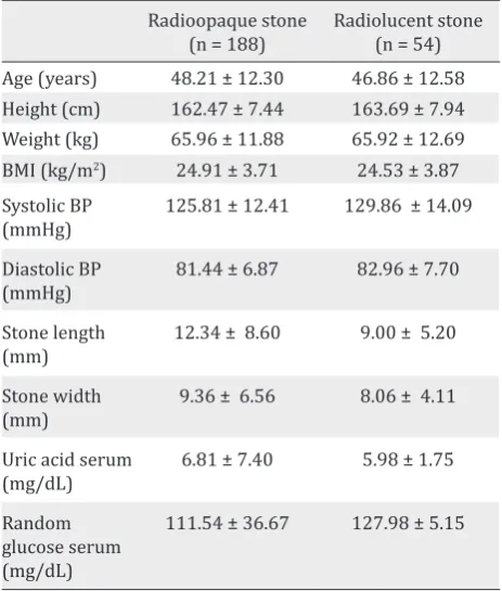
Table 1. Clinical characteristics of patients according to urinary stone opacity.
Radio-opaque stone Radiolucent stone
OR (95%CI) p OR (95%CI) p
High BMI 0.992 (0.552 – 1.885) 0.981 0.971 (0.503 – 1.873) 0.929
High uric acid 1.871 (0.938 – 3.732) 0.073 0.612 (0.305 – 1.229) 0.165
High random glucose 0.219 (0.064 – 0.747) 0.009 3.444 (1.006 – 11.792) 0.038
Hypertension 1.026 (0.360 – 2.923) 0.961 1.190 (0.384 – 3.687) 0.763
Table 2. Risk estimation of stone opacity in urinary tract stone patients
mmHg and diastolic blood pressure <80 mmHg was defined normal.13 American College of
Rheumatology Guidelines 2012 for Management of Gout defined hyperuricemia as serum uric acid greater than 6.8 mg/dL.10 American Diabetes
Association described hyperglycemia as casual or random plasma glucose ≥200 mg/dL. Casual or random is defined as any time of day without regard to time since last meal.15
Statistical analyses were carried out using SPSS version 16.0 (SPSS, Chicago, IL, USA). A p-value was calculated by using chi-square test. Chi-square analysis was also used to determine the odds ratio (OR) and 95% confidence interval (CI) of associations between BMI, serum uric acid, serum glucose and blood pressure, with urinary tract stone opacity. A p-value of less than 0.05 was considered statistically significant. Parameters that were considered statistically significant would undergo probability analysis of radiolucent or radioopaque stone formation.
RESULTS
There were 2,889 patients who underwent ESWL procedure from January 2008 through December 2013. We retrospectively analyzed 242 medical records with complete data. The patients mean age was ± 12.78 (48.02 years). Radioopaque stone was found in 77.69% patients and radiolucent stone was found in 22.31% patients. The male-to-female ratio was 2.27:1. Mean BMI was ± 3.78 (29.91 Kg/m2). High risk BMI were found
in 161 patients (66.52%). The proportion of radioopaque stones were 77.69% (188 patients). Twenty two patients (9.1%) had normal blood pressure. Patients with high serum uric acid were 34.30 % (83 patients).
Table 1 shows the clinical characteristics of patients. Radioopaque stone patients that
had high risk hypertension were 171 patients (90.96%), high risk BMI were 125 patients (66.49%), high uric acid serum level were 70 patients (37.23%), and high random glucose serum level were 5 patients (2.66%). Radiolucent stone patients that had high risk hypertension were 49 patients (90.54%), high risk BMI were 36 patients (66.67%), high uric acid serum level were 13 patients (24.07%), and high random glucose serum level were 6 patients (11.11%).
Statistically there was a significant difference between random glucose serum level with stone opacity (p<0.05). Patients with high level of random glucose serum tended to have radiolucent stone (OR = 3.444; 95% CI = 1.006– 11.792; p = 0.038; Table 2). BMI, uric acid serum, and blood pressure showed no assocation with
*statistically significant
stone opacity. The probability of patient with high random glucose serum towards radioopaque and radiolucent stone formation was 17.97 % and 77.45 % respectively.
DISCUSSION
This study was to describe the associations between metabolic profile with stone opacity in urinary tract stone patients. Examples of radio-opaque stones are calcium oxalate dihydrate, calcium oxalate monohydrate and calcium phosphates. Examples of poor radio-opacity stone are magnesium ammonium phosphate, apatite, and cystine, while examples of radiolucent stone are uric acid ammonium urate xanthine and 2,8-dihydroxyadenine.16
Our study showed that males had a two times higher incidence of stone formation compared to females, indicating that urolithiasis formation might be influenced by sex hormones. A study by Naghii, et al18 has concluded that there were
an association between high plasma androgen concentration and incidence of renal calculi. They proposed gonadal steroids role in male idiopathic urolithiasis formation.17 Some animal studies had
shown that testosterone enhanced excretion of urinary oxalate and increases evolution of calcium oxalate stone.18 Moreover, glycolic acid oxidase
(GAO) level in the liver is induced by testosterone. Higher testosterone serum level will lead to increased GAO synthesis.19 Higher GAO hepatic
level will cause hyperoxaluria condition, resulting in raised formation of calcium oxalate.19,20 This
mechanism increases the likelihood of urinary stone genesis by reducing expression of renal osteopontin and promoting excretion of urinary oxalate.21
Shakhssalim, et al23 have proposed the likelihood
of testosterone influence in renal calculi formation. The results of their study, although stated that there was no statistical difference between male active renal calcium stone formers and control groups, testosterone serum level influenced higher excretion of urinary uric acid and urinary oxalate.22 On the other hand, low
estrogen serum level in post menopausal women counterfeit the hormonal status in men. Urinary calcium level and calcium oxalate saturation are higher in menopausal women compared to premenopausal women. A study by Kato, et al23
had shown that lower citrate and higher calcium excretion were found in menopausal women. This condition will lead to calcium stone formation. Estradiol has protective effect in premenopausal women compared with menopausal women in reducing urolithiasis development.21
A study by Cho, et al2 showed that metabolic
syndrome was associated with a significantly increased risk of uric acid calculi development, especially in those with impaired fasting glucose. The opacity of uric acid stone will be radiolucent. In this study, random glucose serum level showed statistically significant association with opacity of stone formation. Therefore, the result of our study was well in line with the earlier study by Cho, et al2 moreover, a study by Letendre, et al24 stated
that uric acid stone and hyperuricemia usually associated with high glucose serum level.23
Metabolic syndrome is associated with several systemic disorders. Hyperglycemia that happens in diabetes patients can disrupt urinary chemistry that exerts its effect on stone formation.23 Insulin
resistance, which is happened in patients with type 2 diabetes mellitus, reduces amonia production. Decrease amount of ammonia would result in urine pH and acidity. Insulin resistance can be influenced not only by hyperglicemia but also by hyperuricemia condition.7
In this study, the probability of patient with high random glucose serum towards radioopaque and radiolucent stone formation was 17.97% and 77.45% respectively. People with impaired glucose metabolism often have radiolucent stone. Impaired glucose metabolism can disrupt another metabolism. Sometimes people with imbalance glucose metabolism will have blood pressure problem and also uric acid metabolism disorder.7
Although several studies showed that BMI, hypertension, and uric acid serum were associated with composition of stone formation2,24,26 our study
showed no statistically significant associations.
Hypertension can be induced by uric acid by decreased nitric oxide. While urate reaches the vascular smooth muscle, it will promote cellular proliferation, followed by renal microvascular disease progression. Further, reduced number of nitric oxide will lead to renin-angiotensin system (RAS) activation that will lead to proliferation of smooth muscle cells and formation of various inflammatory mediators. Decrease amount of nitric oxide and RAS activation will procede to endothelial dysfunction. This condition will cause vasoconstriction in kidneys.27 Several studies
proposed theory that in the beginning uric acid will initiate hypertension. As time goes by, uric acid serum will have a role in salt-sensitive hypertensive condition rather than direct effect in vascular dysfunction.28
Strohmaier, et al7 have concluded that uric acid
serum level significantly correlate with uric acid stone formation. Moreover, Skolarikos, et al29
have proposed that patients with radiolucent stones (uric acid and ammonium urate) may have high level of uric acid serum. However, there is weak evidence in association between radiolucent stones and hyperuricemia.31 A study
by Jeong, et al33 concluded that increased uric
acid excretions were not only associated with uric acid stone formation but also calcium oxalate stone formation. Calcium oxalate stone is formed by salting-out mechanism in a hyperuricosuria condition. Lower urine pH will cause reduced formation of calcium phospate crystals, which happen in the rising formation of calcium oxalate stones.30
Several metabolic parameters were not analyzed in our study due to lack of data. Clinicians often provide care to urolithiasis patients with numerous co-morbidities, but not all of metabolic parameters were checked. European Association of Urology (EAU) Guidelines 2014 on Urolithiasis did not include metabolic syndrome parameters work-up (HDL cholesterol & triglycerides).16 EAU
Guidelines 2014 on Urolithiasis suggest several ways to evaluate and treat urolithiasis patient. Each stone type has its specific work-up to prevent stone recurrence.29
Our study showed that there was no significant association between hyperuricemia with radiolucent stone formation. We assumed that a bigger sample size should be obtained to get a more significant result. A further study with longer time needs to be done to confirm the results of our study.
In conclusion, there is an association between random glucose serum level with stone opacity in urinary tract stone patients. Patients with high level of random glucose serum tend to have radiolucent stone. Whereas, patients with normal level of random glucose serum tend to have radioopaque stone.
Acknowledgment
We would like to thank University of Indonesia Research Grant 2013-2014 who have given financial support for this study.
Conflict of Interest:
The authors affirm no conflict of interest of this study.
REFERENCES
1. Amaro CR, Goldberg J, Damasio PC, Leitao VA, Turney B, Padovani CR, et al. An update on metabolic assessment in patients with urinary lithiasis. World J Urol. 2015. 33(1):125-9.
2. Cho ST, Jung SI, Myung SC, Kim TH. Correlation of metabolic syndrome with urinary stone composition.
Int J Urol. 2013;20(2):208-13.
3. Ahmad I, Pansota MS, Tariq M, Tabassum SA. Frequency of metabolic abnormalities in urinary
stones patients. Park J Med Sci. 2013;29(6):1363-6.
4. Freitas Junior CH, Mazzucchi E, Danilovic A, Brito AH, Srougi M. Metabolic assessment of elderly men with
urolithiasis. Clinics (Sao Paulo). 2012;67(5):457-61.
5. Pascual E, Perdiguero M. Gout, diuretics and the kidney.
Ann Rheum Dis. 2006;65(8):981-2.
6. Naseri M, Varasteh AR, Alamdaran SA. Metabolic factors associated with urinary calculi in children. Iran
J Kidney Dis. 2010;4(1):32-8.
7. Strohmaier WL, Wrobel BM, Schubert G. Overweight,
insulin resistance and blood pressure (parameters of the metabolic syndrome) in uric acid urolithiasis. Urol
Res. 2012;40(2):171-5.
8. Fernandez A, Fuller A, Al-Bareeq R, Nott L, Razvi H. A
comparison of the metabolic profiles of diabetic and
non-diabetic uric acid stone formers. Can Urol Assoc J.
2013;7(3-4):E190-2.
9. Maalouf N. Approach to the Adult Kidney Stone Former.
Clin Rev Bone Miner Metab. 2012;10(1):38-49.
guidelines for management of gout. Part 1: systematic nonpharmacologic and pharmacologic therapeutic approaches to hyperuricemia. Arthritis care & research.
2012;64(10):1431-46.
11. Maalouf NM, Sakhaee K, Parks JH, Coe FL, Adams-Huet B, Pak CY. Association of urinary pH with body weight
in nephrolithiasis. Kidnet Int. 2004;65(4):1422-5.
12. Mosli HA, Mosli HH, Kamal WK. Kidney stone composition in overweight and obese patients: a
preliminary report. Res Rep Urol. 2013;5:11-5. 13. James PA, Oparil S, Carter BL, Cushman WC,
Dennison-Himmelfarb C, Handler J, et al. 2014 evidence-based guideline for the management of high blood pressure in adults: report from the panel members appointed to the Eighth Joint National Committee (JNC 8). JAMA.
2014;311(5):507-20.
14. WHO Consultation Expert. Appropriate body-mass
index for Asian populations and its implications for policy and intervention strategies. Lancet.
2004;363(9403):157-63.
15. American Diabetes Association. Diagnosis and
classification of diabetes mellitus. Diabetes Care. 2008;33(Suppl1):S55-60.
16. Uroweb.org [Internet]. Europe: European Association
of Urology [update 2014; cited 2014 Dec 19. Available
from: http://uroweb.org/wp-content/uploads/22-Urolithiasis_LR.pdf.
17. Naghii MR, Babaei M, Hedayati M. Androgens involvement in the pathogenesis of renal stones
formation. PloS One. 2014;9(4):e93790.
18. Yagisawa T, Ito F, Osaka Y, Amano H, Kobayashi C, Toma H. The influence of sex hormones on renal osteopontin
expression and urinary constituents in experimental
urolithiasis. J Urol. 2001;166(3):1078-82.
19. Soundararajan P, Mahesh R, Ramesh T, Begum VH. Effect of Aerva lanata on calcium oxalate urolithiasis in
rats. Indian J Exp Biol. 2006;44(12):981-6.
20. Fan J, Chandhoke PS, Grampsas SA. Role of sex hormones in experimental calcium oxalate nephrolithiasis. J Am
Soc Nephrol. 1999;10 Suppl 14:S376-80.
21. Kato Y, Yamaguchi S, Kakizaki H, Yachiku S. Influence of
estrus status on urinary chemical parameters related
to urolithiasis. Urol Res. 2005;33(6):476-80.
22. Shakhssalim N, Gilani KR, Parvin M, Torbati PM, Kashi AH, Azadvari M, et al. An assessment of parathyroid
hormone, calcitonin, 1,25 (OH)2 vitamin D3, estradiol
and testosterone in men with active calcium stone disease and evaluation of its biochemical risk factors.
Urol Res. 2011;39(1):1-7.
23. Kang HW, Seo SP, Kim WT, Kim YJ, Yun SJ, Lee SC, et al. Hypertriglyceridemia is associated with increased risk for stone recurrence in patients with urolithiasis.
Urology. 2014;84(4):766-71.
24. Tang W, Fu Q, Zhang Q, Sun M, Gao Y, Liu X, et al. The association between serum uric acid and residual beta -cell function in type 2 diabetes. J Diabetes Res.
2014;2014:709691.
25. Miyake T, Kumagi T, Furukawa S, Hirooka M, Kawasaki K, Koizumi M, et al. Hyperuricemia is a risk factor for the onset of impaired fasting glucose in men with a high plasma glucose level: a community-based study.
PloS One. 2014;9(9):e107882.
26. Bhole V, Choi JW, Kim SW, de Vera M, Choi H. Serum uric acid levels and the risk of type 2 diabetes: a prospective
study. Am J Med. 2010;123(10):957-61.
27. Wang J, Qin T, Chen J, Li Y, Wang L, Huang H, et al. Hyperuricemia and risk of incident hypertension: a systematic review and meta-analysis of observational
studies. PloS One. 2014;9(12):e114259.
28. Samimi A, Ramesh S, Turin TC, MacRae JM, Sarna MA, Reimer RA, et al. Serum uric acid level, blood pressure, and vascular angiotensin II responsiveness in healthy
men and women. Physiol Rep. 2014;2(12) pii: e12235.
29. Skolarikos A, Straub M, Knoll T, Sarica K, Seitz C, Petrik A, et al. Metabolic Evaluation and Recurrence Prevention for Urinary Stone Patients: EAU Guidelines.
Eur Urol. 2015;67(4):750-63.
30. Jeong JY, Doo SW, Yang WJ, Lee KW, Kim JM. Differences in Urinary Stone Composition according to Body