Brome mosaic virus defective RNAs generated during
infection of barley plants
Tri Asmira Damayanti,
1†
Hideaki Nagano,
1Kazuyuki Mise,
1Iwao Furusawa
1and Tetsuro Okuno
21Laboratory of Plant Pathology, Graduate School of Agriculture, Kyoto University, Kyoto 606-8502, Japan
2Laboratory of Plant Pathology and Biotechnology, Faculty of Agriculture, Kochi University, Nankoku, Kochi 783-8502, Japan
Brome mosaic virus (BMV) purified from systemically infected barley leaves 8 weeks post-inoculation (p.i.) contained defective RNAs (D-RNAs). The D-RNAs were detected in total and virion RNAs extracted from infected plants at 8 weeks p.i. or later, but not before, when barley plants had been inoculated with virions either containing or lacking D-RNA. The D-RNAs were derived from genomic RNA3 by double or mainly single deletions in the 3a protein ORF, and formed a heterogeneous population. By usingin vitro transcripts of D-RNA synthesized from full-length cDNA clones, the D-RNAs were shown to replicate in a helper virus-dependent manner and to be packaged into virions in barley protoplasts. Subgenomic RNA4 was produced from the D-RNA and the coat protein was also expressed. Existence of the D-RNAs together with BMV genomic RNAs in inoculated protoplasts decreased the accumulation of 3a protein but it had no apparent effect on the accumulation of BMV genomic RNA3 or the coat protein. This is the first report of naturally occurring D-RNAs generated during prolonged infection with BMV.
Introduction
Defective RNAs (D-RNAs) are deleted RNAs containing portions of the parental virus genome. They maintain cis-acting elements that contain virus replication and encapsidation signals (Rouxet al., 1991 ; Whiteet al., 1991) and accumulate only in mixed infection with non-defective helper virus, which supplies essential componentsin trans(Rouxet al., 1991). D-RNAs that interfere with the replication of the helper virus genome are referred to as defective interfering RNAs (DI-RNAs). Interference with the parental helper genome may result in an increase (Romero et al., 1993) or a decrease in symptom severity (Roux et al., 1991 ; Hillman et al., 1987 ; Burgyanet al., 1989). DI-RNAs have been found in association with animal virus infections (Perrault, 1981 ; Holland, 1990) and have proved useful in identifying sequence elements
Author for correspondence :Kazuyuki Mise.
Fax81 75 753 6131. e-mail kmise!kais.kyoto-u.ac.jp †Permanent address :Faculty of Agriculture, Department of Plant Pests and Diseases, Bogor Agriculture University, Bogor 16144, Indonesia.
The EMBL accession number of the sequence reported in this paper is X58459.
involved in virus functions such as encapsidation and rep-lication (Levis et al., 1986 ; Weiss et al., 1989 ; Schlesinger, 1988). They have also been implicated as important com-ponents in driving virus evolution (Steinhauer & Holland, 1987).
In recent years, D- and DI-RNAs have been found and well characterized in many plant viruses, including several tombus-viruses (Hillman et al., 1987 ; Burgyan et al., 1989 ; Rochon, 1991 ; Rochon & Johnson, 1991), carmoviruses (Liet al., 1989), potexviruses (White et al., 1991), broad bean mottle bromo-virus (BBMV) (Romeroet al., 1993 ; Poganyet al., 1995) and cucumber mosaic cucumovirus (CMV) (Graves & Roossinck, 1995).
Brome mosaic bromovirus (BMV) is a small, spherical plant virus that infects cereals, including barley (Lane, 1981). The genome of BMV consists of three species of messenger-sense single-stranded RNA, 1, 2 and 3 (Ahlquist, 1992). RNA1 (3±2 kb) and RNA2 (2±9 kb) encode the 1a and 2a proteins, respectively, which are required for virus RNA replication (Frenchet al., 1986 ; Kibertiset al., 1981). RNA3 encodes the 3a protein, which is required for cell-to-cell movement of the virus (Schmitz & Rao, 1996). Subgenomic RNA4, which encodes the coat protein (CP), is synthesized by the virus replicase from a promoter present in the (®)-strand of RNA3 (Miller et al., 1985).
We have found D-RNAs in purified virions of wild-type BMV and in several CP mutants after prolonged infection (8 weeks) of barley plants. The D-RNAs were derived from RNA3 by single or double deletions in the 3a protein gene. Here we report on the molecular characterization of two D-RNA clones that were derived from a CP mutant of BMV.
Methods
+ Virus strains.Strains of BMV used were ATCC66, which has been propagated in our laboratory (Mise et al., 1992), and ATCC PV-47, ATCC PV-178, ATCC PV-180 and M1 (Ahlquistet al., 1984).
+ Plasmid clones.Plasmids pBTF1, pBTF2 and pBTF3W contain the full-length cDNAs of BMV RNA1, RNA2 and RNA3, respectively (Mori et al., 1991 ; Miseet al., 1992). Progeny virus derived from infected plants inoculated within vitrotranscripts from these plasmids was referred to as KU2 strain (Naganoet al., 1997).
A plasmid pBTF3WSS5R25 was constructed as follows. pBTF3W was digested withSalI andSacI and the resulting 0±2 kbp fragment was
exchanged with the corresponding fragment of pAT3J5, a plasmid that contains the CP gene of BMV strain ATCC PV-47 (accession number X58459), to create pBTF3WSS5. The nucleotide sequences of the SalI}SacI region differed between pBTF3W and pAT3J5 at six sites [A1312G, T1314C, A1323G, C1350A, A1356G and G1374A, where the two letters refer to nucleotides of pBTF3W and pAT3J5 and numbers indicate nucleotide position (Miseet al., 1994)]. On the basis of the nucleotide differences, single or double point mutations were introduced into pBTF3W or pBTF3WSS5 by site-directed mutagenesis (Kunkelet al., 1987). Six amino acids contained in the resulting plasmids are listed in Table 1. Progeny virus derived from inoculation within vitrotranscripts of pBTF3WSS5R25 together with those from cDNA clones of BMV RNA1 and RNA2 was named R25.
+ Cloning and sequencing of D-RNA cDNA.Full-length cDNAs of BMV RNAs or D-RNA were synthesized by RT–PCR with a set of primers ; 5«primers that contained aPstI site and corresponded to the 5«
Table 1.Summary of amino acid differences among BMV RNA3 cDNA clones
Amino acid encoded at the indicated site in the CP gene†
Plasmid* 21 22 25 34 36 42
pBTF3W His Trp Arg Leu Thr Val
pBTF3WSS5 Arg Arg Gly Ile Ala Ile
pBTF3WR21 Arg Trp Arg Leu Thr Val
pBTF3WSS5H21 His Arg Gly Ile Ala Ile
pBTF3WSS5R25 Arg Arg Arg Ile Ala Ile
pBTF3WG25 His Trp Gly Leu Thr Val
pBTF3WR21G25 Arg Trp Gly Leu Thr Val
* Plasmids pBTF3WR21, pBTF3WSS5H21, pBTF3WSS5R25, pBTF3WG25 and pBTF3WR21G25 were constructed on the basis of the amino acid differences between pBTF3W and pBTF3WSS5.
†Bold letters indicate amino acid substitutions created by site-directed mutagenesis.
end of BMV RNAs 1 and 2 or RNA3, and a 3«primer that contained an EcoRI site and was complementary to the 3«end of the BMV RNAs. The RT–PCR was conducted under conditions described previously (Nagano et al., 1997). The amplified cDNA products were separated by agarose gel electrophoresis, cloned directly into a TA cloning vector (pCR II, Invitrogen) or cloned into pUC119 (Vieira & Messing, 1987) at the PstI}EcoRI sites after digestion of the cDNA with these enzymes. To ascertain the heterogeneity of D-RNA, cDNA was partially synthesized from D-RNA and amplified by using the 5« primer specific to BMV
RNA3 and a 3«primer, B6, the sequence of which is complementary to
nucleotides 1324–1338 of RNA3 (Mise et al., 1992). The amplified products were digested withPstI andAor51HI and ligated into pUC118 (Vieira & Messing, 1987) at thePstI}SmaI sites.
Nucleotide sequences of two full-length and several partially synthesized cDNA clones of D-RNA were analysed with an automated DNA sequencer (Applied Biosystems, model 373A) according to the manufacturer’s recommendations.
+ In vitrotranscription, inoculation, purification of virus and RNA extraction.All plasmids were linearized withEcoRI and used as templates forin vitrotranscription. Capped full-length transcripts were synthesizedin vitroby using T7 RNA polymerase (Moriet al., 1991).
Barley (Hordeum vulgareL. cv. Gose-shikoku) plants were grown under conditions described previously (Fujitaet al., 1996). Six-day-old seedlings were used for inoculation. Virions and virion RNA were purified from infected plants as described previously (Okuno & Furusawa, 1979).
Isolation of protoplasts of barley (cv. Hinode-hadaka) and inoculation of in vitro transcripts and virion RNAs were performed as described previously (Okuno & Furusawa, 1978 ; Kroner & Ahlquist, 1992). Total RNAs were extracted and virion fractions were obtained by PEG precipitation (Kroner & Ahlquist, 1992) from infected protoplasts at 24 h after inoculation.
+ Northern blot analysis.Total or virion RNA was denatured and separated in a 1±5 % agarose gel containing formaldehyde and MOPS and transferred to a nylon membrane (Hybond-N+, Amersham). ()- and (®)-strand RNAs were detected by using$#P-labelled SP6 transcripts fromHindIII-linearized pBSPL10 (Kaidoet al., 1995) andEcoRI-linearized pBSMI10 (Mori et al., 1993), respectively. The RNA signals were quantified with a digital radioactive imaging analyser (Fujix BAS 2000, Fuji).
+ Western blot analysis. Proteins were extracted from barley protoplasts with sample buffer (Laemmli, 1970) and separated by electrophoresis on 15 % polyacrylamide gels containing 0±1 % SDS (Laemmli, 1970). Proteins were transferred to PVDF membranes (Millipore) by using a transfer-blot SD semi-dry transfer cell (Bio-Rad) according to the manufacturer’s instructions. Accumulation of the 3a protein and the CP of BMV was analysed with anti-BMV 3a monoclonal antibody (Fujitaet al., 1998) and anti-BMV antisera (Naganoet al., 1997), respectively. The proteins were detected with alkaline phosphatase-conjugated anti-Ig secondary antibody, followed by a colour reaction with 5-bromo-4-chloro-3-indolyl phosphate in combination with nitro blue tetrazolium. Protein bands were scanned by an Epson Scan II (Seiko Epson) and protein accumulation was quantified with the Quantity One program (PDI) version 3.0.
Results
Occurrence of BMV D-RNA
Barley seedlings were inoculated with BMV RNA3 trans-cripts together with transtrans-cripts of RNAs 1 and 2 (inoculum
12 2 (a)
Weeks p.i.
1 2
3
4
23.1 9.4 6.6 4.4
2.3 2.0
0.6
P1/2 P3 P3
5′ primer
D-RNA λ R25 R25 + D-RNA
D (b)
1 2 3 4 5
P1/2
Fig. 1.(a) Agarose gel electrophoresis of RNAs extracted from virions 2 and 12 weeks p.i. RNAs were analysed on a 1 % agarose gel in TBE buffer followed by ethidium bromide staining. The additional RNA is indicated by an arrowhead (left) and positions of RNAs 1, 2, 3 and 4 are indicated on the right. (b) Electrophoresis in a 0±8 % agarose gel of RT–PCR products of
R25 lacking D-RNA (lanes 2 and 3) and R25 containing D-RNA (lanes 4 and 5). Lane 1 shows aλDNA marker digested with HindIII. Sizes of the markers are indicated on the left (kbp). P1/2 (lanes 2 and 4) and P3 (lanes 3 and 5) are primers specific to the 5«termini of RNAs 1 and 2 and RNA3, respectively. The 3«-terminal primer used is complementary to all BMV RNAs. The RT–PCR product of D-RNA is indicated on the right (D).
Table 2.Time-course analysis of generation and maintenance of D-RNA in infected barley plants
Seven-day-old barley seedlings were inoculated with virions lacking (®D) or containing (D) D-RNA. Total and virion RNAs were extracted as described in Methods. Detection of D-RNA, assessed by Northern blot analysis of both total and virion RNAs extracted from infected leaves, is scored as positive () or negative (®). Similar results were obtained by RT–PCR with 3«and 5«primers specific for BMV RNA3.
Time p.i. (weeks)
Inoculum 1 2 4 6 8 10 12
®D ® ® ® ®
D ® ® ® ®
R25) and virions were purified from systemically infected leaves at 2 and 12 weeks post-inoculation (p.i.). Analyses of virion RNA by agarose gel electrophoresis showed that an additional RNA that was not detectable in the 2 weeks p.i. virion sample was present in the 12 weeks p.i. virion sample (Fig. 1a). The additional RNA migrated between RNA3 and RNA4 and appeared to be approximately equimolar with each genomic RNA.
To test the maintenance and generation of the additional RNA, barley seedlings were inoculated with either the 2 weeks p.i. or 12 weeks p.i. virions. Total and virion RNA obtained from infected leaves at various time p.i. up to 12 weeks were analysed. An additional RNA was first detected in each inoculation by at least 8 weeks p.i., but not before (Table 2). It was also detected by Northern blot analysis with a probe
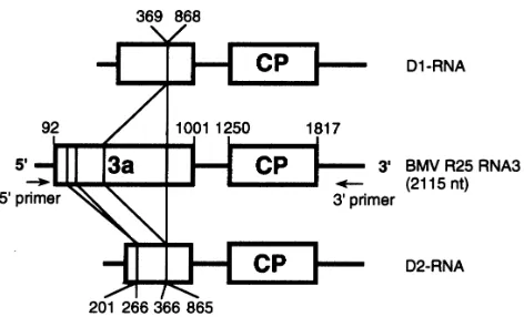
Fig. 2.Schematic representation of the BMV D-RNAs based on the nucleotide sequences of two cDNA clones and comparison with the parental RNA3. The deleted region in the 3a ORF is indicated by vertical lines. D1 RNA contains one deletion, at nt 369–868. D2 RNA contains two deletions, at nt 201–266 and 366–865. The first nucleotide of the initiation codon and the third nucleotide of the termination codon of the 3a and CP genes are shown above the RNA3 diagram.
complementary to the 3«conserved sequence of BMV RNAs. This suggested that the additional RNA was not maintained or generated in the early stages of infection but was generated naturally during infection between 6 and 8 weeks p.i. To test whether the additional RNA could generally be observed in BMV infection, several BMV strains and BMV CP mutants derived from the KU2 strain were inoculated to barley seedlings and total RNAs obtained from systemically infected leaves at 8 weeks p.i. were analysed by Northern blot analysis. Similar additional RNAs corresponding to that observed in the R25 infection were found to be associated with KU2, ATCC66, ATCC PV-47, ATCC PV-178, ATCC PV-180 and M1 strains and also with the BMV-KU2 CP mutants SS5, R21, G25, H21 and R21G25 (Table 1) (data not shown).
(a) (b)
(c) (d)
Fig. 3.(a)–(b) Northern blot analyses of progeny virus RNA in barley protoplasts inoculated with a mixture ofin vitrotranscripts containing or lacking D-RNA. Total RNA was extracted from infected protoplasts 24 h p.i., separated by electrophoresis in a 1±5 % agarose gel, transferred to a nylon membrane and detected with probes for ()-strand (a) or (®)-strand (b) BMV RNAs. Inocula : lanes 1, water (mock) ; 2, transcript of D2 RNA ; 3, transcripts of BMV RNAs 1 and 2 and D2 RNA ; 4, transcripts of BMV RNAs 1, 2 and 3 and D2 RNA ; 5, transcripts of BMV RNAs 1, 2 and 3. Identities of the RNAs are indicated on the left. (c) Western blot analysis of the accumulation of 3a and coat proteins in barley protoplasts inoculated with a mixture ofin vitro transcripts containing or lacking D-RNA. Protein was extracted from 4¬104protoplasts 24 h p.i. and was loaded onto a 15 % SDS–PAGE gel, transferred to a PVDF membrane and detected with anti-BMV 3a or anti-BMV antibody. Inocula : lane 1, water (mock) ; 2, transcripts of BMV RNAs 1 and 2, and D2 RNA ; 3, transcripts of BMV RNAs 1, 2 and 3 and D2 RNA ; 4, transcripts of BMV RNAs 1, 2 and 3. (d) Northern blot analysis of the encapsidation of D-RNA in barley protoplasts. Virions were obtained at 24 h p.i. by PEG precipitation. RNAs were extracted and analysed with a probe for ()-strand BMV RNAs. Inocula : as (a) and (b).
To determine the origin of the additional RNA, virion RNA of R25 containing the additional RNA was amplified by RT–PCR with the 3« primer common to all BMV genomic RNAs and the 5«primer specific to BMV RNAs 1 and 2 or the 5« primer specific to RNA 3 (Fig. 1b). cDNA fragments corresponding to the additional RNA and RNA3 were amplified successfully by using the 5«primer specific to BMV RNA3, while only RNA1 and RNA2 cDNAs were amplified by using the 5« primer specific to BMV RNAs 1 and 2. No cDNA fragments corresponding to the additional RNA were amplified in virion RNA obtained from the 2 weeks p.i. samples (data not shown). These results suggest that the additional RNA was a defective RNA derived from BMV RNA3. Therefore, it will be referred to as D-RNA.
Two full-length cDNA clones of the D-RNA were obtained and their nucleotide sequences were determined. These clones had sequences similar to that of R25 RNA3. However, one clone (D1) had a single large deletion (500 bp) in the 3a ORF (Fig. 2). The other clone (D2) had a similar 500 bp deletion and an additional small deletion (66 bp) (Fig. 2), both in the 3a ORF. There was no deletion in the CP ORF or the non-coding region (sequence data not shown) in these clones.
Replication and encapsidation of D-RNA in protoplasts The two full-length cDNA clones (D1 and D2) of the D-RNA were tested for their ability to replicate and to interfere with BMV RNA replication in barley protoplasts. In vitro
(a) (b)
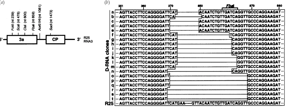
Fig. 4.(a) Schematic representation of restriction enzyme map of BMV R25 RNA3. The enzymes are identified above the RNA3 diagram with the nucleotide position of cleavage. (b) Nucleotide sequence heterogeneity of BMV D-RNA with varied deletion junction sites. RT–PCR products from the D-RNA were cloned into a plasmid vector and sequenced. Deleted regions are drawn as horizontal lines. Individual clones are indicated alphabetically on the left. Clones a–e possess anFbaI site as indicated. R25, partial nucleotide sequence of the parental virus (Miseet al., 1994).
transcripts of the D-RNAs from each clone were inoculated into barley protoplasts together with transcripts of BMV RNAs 1 and 2 or with those of BMV RNAs 1, 2 and 3. Total RNA from infected protoplasts was examined by Northern blot analyses. Fig. 3 shows the results obtained with clone D2. Similar results were obtained with clone D1 in other in-dependent experiments (data not shown). Regardless of whether RNA3 transcript was included in the inoculum, the D-RNA was detected by both probes for () (Fig. 3a) and (®) strands (Fig. 3b) of BMV RNAs. Both ()-and (®)-strand D-RNAs were also detectable when protoplasts were inoculated with BMV virion RNA containing D-RNA (data not shown). These results indicate that D-RNAs can replicate in protoplasts. When D-RNA transcripts were inoculated with transcripts of BMV genomic RNAs 1 and 2, accumulation of subgenomic RNA4 was detected (Fig. 3a). However, when D-RNA was inoculated without BMV RNAs 1 and 2, no accumulation of D-RNA was detected (Fig. 3a), indicating that the replication of D-RNA was helper-dependent.
The effects of the presence of D-RNA on the accumulation of virus RNAs and proteins were examined. The level of RNA3 accumulation relative to RNA12 was reduced to 91 %, while that of RNA4 increased to 117 % [on the basis of measurements from Fig. 3 (a) and three other experiments]. The presence of D-RNA increased the accumulation of CP to 114 % [on the basis of measurements from Fig. 3 (c) and two other experiments]. In contrast, the presence of D-RNA reduced the accumulation of protein 3a to 54 %. These results suggest that the presence of D-RNA does not interfere or interferes only slightly with the replication of RNA3 but apparently interferes with the synthesis of protein 3a in protoplasts.
Encapsidation of D-RNA was tested in barley protoplasts. The virion fraction was prepared from infected protoplasts and
packaged RNA was analysed by Northern blot analysis. The D-RNA was detected together with BMV genomic RNAs (Fig. 3d, lanes 3 and 4), indicating that D-RNA was encapsidated into virions.
Heterogeneity of D-RNA sequence
To analyse any heterogeneity that may be present in the D-RNA population, virion D-RNA containing D-D-RNA was extracted at 8 weeks p.i. and full-length cDNAs were amplified by RT–PCR as described above. The PCR products of RNA3 and D-RNA were separated and purified after low-melting-point agarose gel electrophoresis and digested with restriction enzymes FokI, ScaI, ClaI, FbaI, Aor51HI or SacI (Fig. 4a). Digested cDNAs were analysed by agarose gel electro-phoresis. Full-length cDNA of R25 RNA3 used as control was completely digested by all the enzymes tested, resulting in the expected fragments corresponding to the enzyme cutting sites (Fig. 4a). The cDNA products of D-RNA were completely digested byFokI,Aor51HI andSacI. AfterAor51HI digestion, D-RNA cDNA as well as RNA3 cDNA produced a 1±0 kbp fragment corresponding to the 3«half of RNA3, indicating that there were no deletions in that region, which includes most of the intercistronic region, the CP ORF and the 3« non-coding region. However, the cDNA of the D-RNAs was not digested byScaI or ClaI (central part of the 3a ORF), indicating that those sites did not exist in the D-RNA. Since the cDNA of the D-RNA was only partially digested byFbaI (3« region of 3a ORF), the D-RNA population was assumed to be hetero-geneous. This restriction enzyme mapping also confirms the sequencing result (Fig. 2) that a deletion occurred around the central region of 3a ORF. However, the cDNA of the D-RNA was completely digested byFokI, which has no recognition site
in the D2 sequence, suggesting that D2 RNA could be a minor species.
For further examination of D-RNA heterogeneity, we synthesized partial cDNA of the D-RNA by using the 5«
primer specific to BMV RNA3 and the 3« primer B6. We obtained 20 cDNA clones and determined their nucleotide sequences. In this experiment, all the D-RNA clones contained single deletions in the 3a ORF and no D-RNA contained two deletions, such as were found in D2 (Fig. 2). The sequence data also confirmed the results of restriction enzyme mapping, showing that both theScaI andClaI sites were absent from all the clones and that only a portion of the clones (5 of 20) contained anFbaI site. The 5« borders of the deleted region were roughly the same among the clones, though the exact junction sites varied (Fig. 4b), resulting in deletions ranging from 477 to 500 bp.
Discussion
The structure of the additional RNAs described here suggests that they are the first reported naturally occurring D-RNAs associated with BMV infection, although artificial DI-RNAs constructed from BMV RNA2 were reported previously (Marshet al., 1991). A number of features of the BMV D-RNAs distinguish them from previously characterized plant virus D} DI-RNA. First, smaller RNAs with electrophoretic mobilities similar to that of the D-RNAs of R25 were detected in several BMV strains or BMV-KU2 CP mutants. This suggests that the presence of D-RNAs is a general occurrence after prolonged BMV infection of barley plants. It also suggests that mutations in the 5«-terminal region of the CP gene did not affect the generation of D-RNA.
Second, the D-RNAs were generated by either single or double deletions, exclusively in the 3a ORF (Figs 2 and 4b). The deleted regions are nt 369–868 of D1 RNA and nt 201–266 and 366–865 of D2 RNA, suggesting that the regions retained are essential for the accumulation of BMV D-RNAin planta. The D-RNA clones with single deletions had different deletion junctions (Fig. 4b), which resulted in sizes of deletions from 477 to 500 bp. In barley protoplasts inoculated with in vitro transcripts of D-RNA with either one or two deletions, the D-RNAs were replicated and encapsidated into virions when co-inoculated with BMV genomic RNAs. When present together with the genomic RNAs, the D-RNA reduced the accumulation level of protein 3a (Fig. 3c). This could be the result of competition for ribosomes between D-RNA and wt RNA3 in the synthesis of the truncated and wt 3a proteins.
The third specific feature of the BMV D-RNAs is their generation. As previously reported, repeated passage at high m.o.i. is required for the generation of DI-RNAs in animal viruses (Holland, 1990) and D-}DI-RNA in some plant viruses (Morris & Hillman, 1989 ; Knorr et al., 1991 ; Graves & Roossinck, 1995). However, the BMV D-RNAs were generated in rather unique circumstances. Generation of
D-RNA was demonstrated by inoculation of barley seedlings with virion inocula either containing or lacking D-RNA. In both cases, the D-RNA was not detected at 1–6 weeks p.i., but was detected after prolonged infection (8 weeks p.i.) (Table 2). These features raise interesting questions ; why isn’t the D-RNA with deletions in the 3a ORF maintained even in the initially inoculated leaves, and why does the 3a ORF become a target for deletions after prolonged infection? Explanations for these observations could be : (i) since D-RNAs replicate efficiently in protoplasts (Fig. 3a) and are encapsidated into virions (Fig. 3d), the lack of cell-to-cell movement rather than replication may be responsible for the lack of D-RNA maintenance in initially inoculated leaves. Because the 3a gene has a crucial role in virus cell-to-cell movement (Schmitz & Rao, 1996), RNA3 with a truncated 3a gene may be less advantageous for further cell-to-cell movement than that with an intact 3a gene. Therefore, the D-RNA may fail to accumulate to detectable levels even in initially inoculated leaves. (ii) It is possible that there may be a unique interaction between either BMV strains or BMV CP mutants and 8-week-old barley plants. Physiological changes in old barley may interact with and}or alter the 3a gene. Alternatively, the intact 3a gene might be dispensable in old barley plants. Therefore, the dispensable region in the 3a gene might be deleted and regions that are presumed to be necessary for RNA replication and encapsidation in plants could be retained.
Previously, D- and DI-RNAs have been found to be associated with CMV (Graves & Roossinck, 1995) and BBMV (Romeroet al., 1993 ; Poganyet al., 1995), respectively. Both viruses, as well as BMV, belong to the familyBromoviridaeand their genomic organization is similar. D-RNAs of CMV and BMV originate from parental genomic RNA3, while DI-RNAs of BBMV are from RNA2. On the other hand, the generation processes are quite different. The CMV D-RNAs are produced upon serial passage of the wild-type Fny strain and are maintained by virus even after additional passage, while the BBMV DI-RNAs occur naturally in strains Tu and Mo (Romero et al., 1993) and are generatedde novoby serial passage at high m.o.i. (Pogany et al., 1995). In contrast, BMV D-RNAs are produced during prolonged infection of BMV and cannot be maintained even in the initially inoculated leaves. This might result from young-barley-mediated inhibition of D-RNA encapsidation or an increased instability of D-RNA-containing virions, as previously reported in the case of BBMV DI-RNAs in pea (Romeroet al., 1993). Further studies to find a favourable host that could support the maintenance of BMV D-RNAs are needed to investigate whether the existence of D-RNAs has any effect on the symptom development induced by the helper virus.
The mechanism of formation of BMV D-RNA is unknown. However, short regions showing sequence similarity and}or complementarity were found at the 5« and 3« junction sites (Figs 4band 5). These local complementarities could juxtapose two molecules of ()-strand RNA3, which might allow the
(a)
(b)
(c)
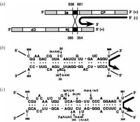
Fig. 5.Proposed model for template switching during the generation of BMV D-RNAs. Large arrows on the right between RNAs indicate the direction of (®)-strand RNA synthesis during template switching. A similar model can be conceived in which switching occurs during ()-strand
RNA synthesis. (a) Schematic representation of a heteroduplex formed between two molecules of ()-strand RNA3. Open rectangles are the 3a
protein and CP ORFs. Shaded rectangles indicate non-coding regions. Black rectangles indicate partially complementary sequences adjoining deleted regions. (b)–(c) Two types of heteroduplexes for D-RNA clones with anFbaI site (clones a–e) (b) and those without anFbaI site (clones f–t) (c). The top and bottom lines show downstream and upstream sequences around the deleted region, respectively. Each resulting D-RNA clone shown in Fig. 4 (b) contains the top sequences up to the arrow and then continues with the bottom strand sequences. Vertical arrows labelled with the name of the D-RNA clone indicate the locations of the putative recombination sites. Base pairing is shown by bold dots.
replicase to switch from one template to another (Fig. 5a), as proposed previously for recombination events among BMV RNAs (Bujarski & Dzianott, 1991 ; Bujarskiet al., 1994). Such a template-switching mechanism has also been largely accepted to be responsible for the generation of D-}DI-RNAs in animal viruses (Huang, 1977 ; Holland, 1990) and in plant viruses (Poganyet al., 1995 ; Graves & Roossinck, 1995).
We thank Paul Ahlquist for the cDNA clones of BMV M1 strain. This work was supported in part by a Grant-in-Aid (06660051) for Scientific Research and a Grant-in-Aid (09NP1501) for Creative Basic Research from the Ministry of Education, Science, Sports and Culture, Japan and a Grant-in-Aid (JSPS-RFTF96L00603) from the ‘ Research for the Future’ programme of the Japan Society for the Promotion of Science. T. A. D. was the recipient of a Sagawa Fellowship.
References
Ahlquist, P. (1992). Bromovirus RNA replication and transcription. Current Opinion in Genetics and Development2, 71–76.
Ahlquist, P., French, R., Janda, M. & Loesch-Fries, L. S. (1984). Multicomponent RNA plant virus infection derived from cloned viral cDNA. Proceedings of the National Academy of Sciences, USA 81, 7066–7070.
Bujarski, J. J. & Dzianott, A. M. (1991). Generation and analysis of nonhomologous RNA–RNA recombinants in brome mosaic virus : sequence complementarities at crossover sites. Journal of Virology 65, 4153–4159.
Bujarski, J. J., Nagy, P. D. & Flasinski, S. (1994).Molecular studies of genetic RNA–RNA recombination in brome mosaic virus.Advances in Virus Research43, 275–302.
Burgyan, J., Grieco, F. & Russo, M. (1989).A defective interfering RNA molecule in cymbidium ringspot virus infections. Journal of General Virology70, 235–239.
French, R., Janda, M. & Ahlquist, P. (1986).Bacterial gene inserted in an engineered RNA virus ; efficient expression in monocotyledonous plant cells.Science231, 1294–1297.
Fujita, Y., Mise, K., Okuno, T., Ahlquist, P. & Furusawa, I. (1996).A single codon change in a conserved motif of a bromovirus movement protein gene confers compatibility with a new host. Virology 223, 283–291.
Fujita, M., Mise, K., Kajiura, Y., Dohi, K. & Furusawa, I. (1998).Nucleic acid-binding properties and subcellular localization of the 3a protein of brome mosaic bromovirus.Journal of General Virology79, 1273–1280. Graves, M. V. & Roossinck, M. J. (1995).Characterization of defective RNAs derived from RNA 3 of the Fny strain of cucumber mosaic cucumovirus.Journal of Virology69, 4746–4751.
Hillman, B. I., Carrington, J. C. & Morris, T. J. (1987). A defective interfering RNA that contains a mosaic of a plant virus genome.Cell51, 427–433.
Holland, J. J. (1990).Defective viral genome. InVirology, 2nd edn, vol. 1, pp. 151–165. Edited by B. N. Fields & D. M. Knibe. New York : Raven Press.
Huang, A. S. (1977). Viral pathogenesis and molecular biology. Bacteriological Reviews41, 811–821.
Kaido, M., Mori, M., Mise, K., Okuno, T. & Furusawa, I. (1995). Inhibition of brome mosaic virus (BMV) amplification in protoplasts from transgenic tobacco plants expressing replicable BMV RNAs.Journal of General Virology76, 2827–2833.
Kibertis, P. A., Loesch-Fries, L. C. & Hall, T. C. (1981).Viral protein synthesis in barley protoplasts inoculated with native and fractionated brome mosaic virus RNA.Virology112, 804–808.
Knorr, D. A., Mullin, R. H., Hearne, P. Q. & Morris, T. J. (1991).De novo generation of defective interfering RNAs of tomato bushy stunt virus by high multiplicity passage.Virology181, 193–202.
Kroner, P. & Ahlquist, P. (1992).RNA-based viruses. InMolecular Plant Pathology:A Practical Approach, vol. 1, pp. 23–34. Edited by S. J. Gurr, M. J. McPherson & D. J. Bowles. Oxford : Oxford University Press. Kunkel, T. A., Roberts, J. D. & Zakour, R. A. (1987).Rapid and efficient site-specific mutagenesis without phenotypic selection. Methods in Enzymology154, 367–382.
Laemmli, U. K. (1970). Cleavage of structural proteins during the assembly of the head of bacteriophage T4.Nature227, 680–685. Lane, L. C. (1981).Bromoviruses. InHandbook of Plant Virus Infections: Comparative Diagnosis, pp. 333–376. Edited by E. Kurstak. Amsterdam : Elsevier}North-Holland.
Levis, R., Weiss, B. G., Tsiang, M., Huang, H. & Schlesinger, S. (1986). Deletion mapping of Sindbis virus DI RNAs derived from cDNAs defines the sequences essential for replication and packaging.Cell44, 137–145. Li, X. H., Heaton, L. A., Morris, T. J. & Simon, A. E. (1989).Turnip crinkle virus defective interfering RNAs intensify viral symptoms and are generatedde novo.Proceedings of the National Academy of Sciences,USA86, 9173–9177.
Marsh, L. E., Pogue, G. P., Connell, J. P. & Hall, T. C. (1991).Artificial defective interfering RNAs derived from brome mosaic virus.Journal of General Virology72, 1787–1792.
Miller, W. A., Dreher, T. W. & Hall, T. C. (1985).Synthesis of brome mosaic virus subgenomic RNAin vitroby internal initiation on (®)-sense
genomic RNA.Nature313, 68–70.
Mise, K., Tsuge, S., Nagao, K., Okuno, T. & Furusawa, I. (1992). Nucleotide sequence responsible for the synthesis of a truncated coat protein of brome mosaic virus strain ATCC66.Journal of General Virology 73, 2543–2551.
Mise, K., Mori, M., Nakayashiki, H., Koyama, T., Okuno, T. & Furusawa, I. (1994).Nucleotide sequence of a set of cDNA clones derived from brome mosaic virus ATCC66 strain and comparison with the Russian strain genome. Annals of the Phytopathological Society of Japan 60, 454–462.
Mori, M., Mise, K., Kobayashi, K., Okuno, T. & Furusawa, I. (1991). Infectivity of plasmids containing brome mosaic virus cDNA linked to the cauliflower mosaic virus 35S RNA promoter. Journal of General Virology72, 243–246.
Mori, M., Kaido, M., Okuno, T. & Furusawa, I. (1993). mRNA amplification system by viral replicase in transgenic plants.FEBS Letters 336, 171–174.
Morris, T. J. & Hillman, B. I. (1989).Defective interfering RNAs of a plant virus. In Molecular Biology of Plant–Pathogen Interactions, pp. 185–197. Edited by B. Staskawitcz, P. Ahlquist & O. Yoder. New York : Alan R. Liss.
Nagano, H., Okuno, T., Mise, K. & Furusawa, I. (1997).Deletion of the C-terminal 33 amino acids of cucumber mosaic virus movement protein enables a chimeric brome mosaic virus to move from cell to cell.Journal of Virology71, 2270–2276.
Okuno, T. & Furusawa, I. (1978). Modes of infection of barley protoplasts with brome mosaic virus. Journal of General Virology 38, 409–418.
Okuno, T. & Furusawa, I. (1979).RNA polymerase activity and protein synthesis in brome mosaic virus-infected protoplasts. Virology 99, 218–225.
Perrault, J. (1981). Origin and replication of defective interfering particles.Current Topics in Microbiology and Immunology93, 151–207.
Pogany, J., Romero, J., Huang, Q., Sgro, J.-Y., Shang, H. & Bujarski, J. J. (1995).De novo generation of defective interfering-like RNAs in broad bean mottle bromovirus.Virology212, 574–586.
Rochon, D. M. (1991).Rapidde novogeneration of defective interfering RNA by cucumber necrosis virus mutants that do not express the 20-kDa nonstructural protein.Proceedings of the National Academy of Sciences,USA 88, 11153–11157.
Rochon, D. M. & Johnston, J. C. (1991). Infectious transcripts from cloned cucumber necrosis virus cDNA : evidence for a bifunctional subgenomic mRNA.Virology181, 656–665.
Romero, J., Huang, Q., Pogany, J. & Bujarski, J. J. (1993). Charac-terization of defective interfering RNA components that increase symptom severity of broad bean mottle virus infections.Virology194, 576–584.
Roux, L., Simon, A. E. & Holland, J. J. (1991). Effects of defective interfering viruses on virus replication and pathogenesisin vitroandin vivo.Advances in Virus Research40, 181–211.
Schlesinger, S. (1988).The generation and amplification of defective interfering RNAs. In RNA Genetics, Retroviruses, Viroids and RNA Recombination, pp. 167–185. Edited by E. Domingo, J. J. Holland & P. Ahlquist. Boca Raton, FL : CRC Press.
Schmitz, I. & Rao, A. L. N. (1996).Molecular studies on bromovirus capsid protein. I. Characterization of cell-to-cell movement-defective RNA3 variants of brome mosaic virus.Virology226, 281–293. Steinhauer, D. A. & Holland, J. J. (1987). Rapid evolution of RNA viruses.Annual Review of Microbiology41, 409–433.
Vieira, J. & Messing, J. (1987).Production of single-stranded plasmid DNA.Methods in Enzymology153, 3–11.
Weiss, B., Nitschko, H., Ghattas, I., Wright, R. & Schlesinger, S. (1989). Evidence for specificity in the encapsidation of Sindbis virus RNAs. Journal of Virology63, 5310–5318.
White, K. A., Bancroft, J. B. & Mackie, G. A. (1991).Defective RNAs of clover yellow mosaic virus encode nonstructural}coat protein fusion products.Virology183, 479–486.
Received 11 March 1999 ; Accepted 17 May 1999