H e t e r o l o g ~ u s antilyriphacyte serum (i',LF) r e f e r s t o antiserum t h n t w a s m i s e d i n one species zg-:inst lymphoid c e l l I n t i g e n s of o t h e r s p e c i e s . Slthough the p r e p ? r n t i o n of ALS was a l r e a d y r e p o r t e d by Metchnikoff i n
1899
t h e use of t h i s e n t i s e r ? f o r modulation of the innune response had t o w 2 i t u n t i l t h e r o l e of lynpknid t i s s u e i n t h e i m ~ u n e r e z c t i o n w 2 s recognized. I n t h e f a t e f i f t i e s t h e experiments of I n d e r b i t e e n (1956) were t h e f i r s t t o demonstrate t h a th e t e r o l o g o u s a n t i s e r u r ~ directeif Ei,@inst lynphoict c e l l s c ~ u l d F n h i b i t delayed h y p e r s e n s i t i v i t y r e a c t i o n , S e v e r a l y e a r s
l a t e r Woodruff snd Anderson (1963) showed c l e a r l y t h a t ALS used e i t h e r a l o n e o r as a n a d j u n c t t ? t h o r o c i c duct drainage could prolong the s u r v i v a l of s k i n a l l o g r a f t i n r a t s .
S i n c e 1967 ALS h a s been t h e s u b j e c t of i n c r e a s i n g n m b e r s o f p u b l i c ~ t i o n s and reviews. I t s main use is i n t h e f i e l d of irmnunosuppression. I t hEts t h e ~ b i l i t y t o msrkedly diminish c e l l u l a r im-iune r e s p o n s e s p a r t i c u l a r l y s k i n a l l o g r a f t and h e t e r o p a f t r e j e c t i o n i n experimental animals. It a l s o czn n b o l i s h t h e inmunity evoked by a p r e v i o u s e l l o g r a f t . The g r a f t v e r s u s h o s t r e ~ c t i o n i n i r r a d i a t e d animals such
a s mice o r monkeys caused by t h e i n j e c t i o n o f a l l o g e n i c s p l e e n o r bone narrow c e l l s can be n e u t r a l i z e d by :?LS i n j e c t i o n .
3 cell functions i n the immune response. Ls a n experimental tool it i s still used to inliibit preferentially cellular immune response, while lesving humoral antibody mechanism generally intact. With regard to clinical immunosuppr~saion
ALS represents the first agent whose actions were limited
to cells of the lymphoid syxtcm and which would specifically interact with lymphocytes i n both resting and proliferative
stages. Thc immunosuppressive effects of
ALS
can be increasedby prior thymectomy, whole body irradiation, administration of corticosteroid and lymphocyte depletion by thoracic duct fistula-
ALE has been raised in horses, rabbits, pigs and rumi-
nants against lymphocytes of the dos, mouse, rat, monkey and human. The sera usually possess optimal immunosuppressive activity when injected into the lymphocyte donor species. Anti-rat and anti-mouse antisera have also Seen rcised in
chickens a n d ducks, but their spectra of activity are some- what narrower than mammalian antisera, because of their poor interaction with mammalian complement. Xany different immu- nization techniques have been tried, yielding antisera of different potency.
Non-lymphoid tissues have been used to produce
A L S
witheffective immunosuppressive action including mouse cell
cultures
(L
cells), epidermal cells o r plasma cells. Lymphoid cell a n d thymocyte cell fractions have also been used to pro-The 7 5 I& f r a c t i o n s o f ALS c o n t a i n v i r t u a l l y a l l immuno - s u p p r e s s i v e p o t e n c y , b u t t h a i n t a c t a n t i b o 5 g m o l ~ c u l c i s r e - q u i r e d f o r o p t i m a l immunosuppressive a c t i v i t y . Thz p u r e 1 9 s f r a c t i o n o f ALS s e p . ? r a t e d by i m f i u n o e l e c t r o p h o r ~ s i s h a s l i t t l e o r no immunosuppressiva a c t i v i t y , The ~ ( a b ) 2 p o r t i o n of the
a n t i b o d y molecule, o b t a i n e d by d i g e s t i o n w i t h p e p s i n from h o r s e - - a n t i - r u t l y n p h o c y t I& f z i l c d t o p r o l o n y t h e s u r v i v , z l o f s k i n a l l o g r a f t i n r a t s a s r e p o r t e d by ?Foodruff
~4
? & - ( 1 9 6 7 ) -S i n c e t h e r e p o r t o f S t s r z l g t a&, (1367) ALS has been usad c l i n i c a l l y i n humrns. A l t h o u g h A L 7 r e m a i n s t h e most p o w e r - f u l s i n g l e a g e n t f o r p r o l o n g a t i o n o f s k i n g r a f t s i n m i c e , i t s e f f e c t s i n hwu:?ns hnva b e e n f a r l e s s d r a m a t i c . I t h a s bccome c l e a r t h a t ALS alone c a n n o t be used t o o b t a i n n c o n t i n u o u s i m m u n o s u p p r e s s i o n , but r a t h e r must be a p p l i e d i n c o m b i n a t i o n w i t h c o n v e n t i o n a l immunosuppressive a : y c n t s used i n c l i n i c a l
t r a n s p l a n t a t i o n s . The m a j o r c l i n i c a l u s c o f ALS a t p r e s e n t i s a s nn . a d j u n c t t o u s u < ? l islmunosupprcssive 6 r u . z ~ s u c h a s a z a t h i - o p r i n e a n d p r c d n i s o n e i n t h o management o f p a t i o n t s w i t h k i d n c y o r l i v e r a l l o s r , a f t , where t h o A L S a c t s on t h e c e l l u l a r i r m u n i t y a n d a z a t h i o p r i n c on t h e humoral ivmuuli t y I n c a r d i s c t r a n s p l a n -
A n o t h e r i n p o r t a n t use o f a n t i l y l l p h o c y t e g l o b u l i n ( A L G ) i s i n p a t i e n t s w i t h fymphoS1NsZ;ic anemia t : ~ allo-f: t h e t a k e of HLA n o n i d e n t i c a l bone marrow. ALS h a s a l s o been a d d e d t o t h e
x i s t
of n g e n t s which may be of v a l u e i n t h e ! 3 u t o i m u n e d i s o r d e r s o r o t h c r d i s e n c e s w i t h irniune fc2ntures. I n t h e p a s t4 L G h a s a l s o been used i n t h e t r e a t m e n t o r l y m p h o p r o l i f e r a t i v e d i s o r d e r s l i k e l y m p h a t i c 1eukr:min.
I n V e t e r i n a r y Medicinr. t h e usi! o f ALS i s m o s t l y f o r e x p e r i m e n t a l p u r p o s o s , Ths e f f e c t s of A L S i n m i c e , r a t s , dogs and primatez. hava been s t u d i e d i n t e n s i v e l y * I t i n known t h a t t h e chimpznze,r; s h ~ r e s organ a n t i g e n s w i t h humans. ?$any human l u u c o c y t e a n t i g e n s a r e p r a s e n t on t h e l c u c o c y t e s of chimpan-, z e c s . S e v e r a l p r i m a t e s p e c i e s ( c h i n p n n z c e ) and r h c s u s monkegs have been used f o r t h z q u a l i t y a n d s s f a t y c o n t r o l of a n t i -
human lymphocyte scrum, A n t i - d o s lymphocyte s e r u m h a s been used i n t r a n s p l a n t a t i o n s t u d i e s . Howcvor, t h e c l i n i c a l a p p l i c a t i o n of
a n i m a l A L B i s v e r y l i n i t a d .
So f a r t h e e f f s c t s o f a n t i Lymphocyt serum i n c a t t l e h a s n o t been p u b l i s h e d .
O r i g i n a l l y t h e p u r p o s e o f t h e p r o d u c t i o n o f h o r s e ~ n t i - - b o v i n e lymphocyt serum (ABLc;) w a s f o r t h e t r a n s m i s s i o n o f ju- v e n i l e bovine l e u k o s e , which i s a d i s e a s e e s p e c i a l l y a f f e c t i n 2 c a l v e s
.
2.1. P r o d u c t i o n . o f ~ n t i l y r ? p h o c y t o s c r u r ? "
FTethods o f r a i s i n g A L S havc b e c n d e s c r i b e d by m a n y i n v e s t i q n t o r s u s i n ? lymphoid c e l l s suspended i n a nedima o r e m u l s i f i e d i n complc t e F r o u n d . * s a d j u v a n t
.
M ' : n y immu- n i z a t i o n t o c h n i q u e o h a v e b e e n used y i e l d i n g ALS o f h i g h i m r ~ u n o s u p p r c s s i v e p o t cncy. T ~ L most cornrfion 2 r e t h e tvuo o r t h r c o n u b c u t a n e o u s o r i n t r a v e n o u s i n j e c t i o n s q i v e n 3-t t w o weeks ~ p n r t f o l l o w e d by n b o o s t s r i m m u n i z z t i o n f o u r weeks t h e r e e f t e r . M n n y d i f f e r e n t h o s t s p e c i e s h a v e b c e n used t o r a i s c o f f c c t i v e ALS, The most used s p z c i e s a r e t h e r a b b i t a n d t h e h o r s s . P i g s and rurciinrnts h a v ea l s o b e e n used b u t t h e y proiluca = n t i c c ? r e x of l o w e r q u - ? l i t g .
n
~ n % i s e m h a v e boon r a i s c C a g a i n s t lymphoid t i s s u e s o f human, monkey, d o g , r a t , b o v i n e a n d n o u s e -
The p r o d u c t i o n o f a n t i - m t B L R i n r z b b i t s wes r e p o r t e d by Woodruff nnc? Anderson ( 1 9 6 3 ) . T h e y i n j e c t e d i n t r a p e r i t o n c a l l y 200.0
x
l o 6
r a t t h o r a c i c d u c t lympho- c y t e s t h r e e t i m e s a t weekly i n t e n r " 1 s . T h c r l b b i t s \-?ere b l e d t e n d:?ys a f t e r t h e l a a t i n j e c t i o n and b o o s t e r i n t r a - p e r i t o n c z d i n j z c t i o n o f 1-2x
lo6
l y m p h o c y t e s were given t e n d a y s b e f o r e e a c h s u b s e q u e n t b l e e d i n g .two i n t r a v e n o u s i n j e c t i o n s of 4-10 x
l o 8
lymphoid c e l l s i n r a b b i t s a t f o u r t c a n d ~ y s c g s r t . The r a b b i t n TA?ere t h e n b l e d7
d a y s a f t e r t h e s e c o n d i n j a c t i o n and t h e s e r ? were c o l l c c t c d and h c n t i n ~ c t i v a t e d ( 5 6 O ~ f o r 30 m i n u t e s ) , S e i t z f i l t e r e d a n d s t o r e d a t -20°C u n t i l u s e d . Gray e_f:21,
( 1 9 6 6 ) and blonaco~ - 5
%&. ( 1 9 6 6 ) r e p o r t e d t h e i r methoci o f r a i s i r i f : A I - T i n r ~ b b i t - a s f o l l o w s , Each r a b b i t r e c e i v e d a t o t s 1 o f 100.0 x 10' lymph n o d e c e l l s e m u l s i f i e d i n c o m p l e t e P r e u n d S s a d j u v a n t . The e ~ . l u l -s i o n was e q u a l l y d i v i d e d and i n j e c t e d i n t o e a c h orr" t h e f o u r f o o t p a d s . The b o o s t e r i n j u c t i o n a o f s p l i n t ? c e l l s u s p e n s i o n of 100.0
x
lo6
c e l l s w i t h o u t a d j u v n n t weri: g i v e n i n t r n v c n o u s l y on t h r e e s u c c e s s i v e d a y s f o u r weeks l a t e r . Thc r a h b i t a wGrc b l e d f o u r d a y s a f t e r t h e l a s t i n j e c t i o n . The blood was l e f t t o c l o t a t room t e m p e r a t u r e a n d s e r m was c ~ l l e c t a d t h e n e x tday, h e a t i n z c t i v a t e d and s t o r e d n t - 2 0 ' ~ .
lymphoid c a l l s on 4 t o 6 o c c a s i o n s a t 2 t o 4 weeks i n t e m l o f o r t h e p r o d u c t i o n o f anti-hu.nan A L S . Thc. i ~ o r s e s were b l e d one week a f t e r t h e l a s t i n j z c t r o n . They r e p o r t e d t h n t s e r a produced i n t h i s manner prolang,d s k i n . g r a f t s u r v i v a l i n zlon- k e y s . Lucko @ a2. (1968) imlunizud h o r s e s by,
3
s u b c u t z n e o u s i n j e c t i o n s o f 1.0x
10' p i g lymph node c e l l s i n m e d i a 1 9 9at
7 d r y s . Blood was c o f l e c $ c d one wcek a f t o r t h e l a s t i n j e c t i o n . Twenty f o u r h o u r s t h e r e a f t e r t h e h o r s e s werc g i v e n a n o t h e r i n j e c t i o n of 0.44x
10' l y n p h noiic c e l l s and b l e d 7 dcys l a t e r . The blood wes r l l o w e d t o s t a n d f o r 2 h o u r s zt room t e n p e r Q t u r a and t h e n k e p t o v e r n i g h t n t ~ O C . The s a r m w a s s e p a r r t e d , a&sorbed w i t h one t h i r d of i t s v o l w l e w i t h p i g e r y t h r o c y t e s end s t o r i l i z c d by S c i t z f i l t r a t i o n and s t o r e d a t - 2 0 O ~ u n t i l used. They observed t h a t s e r a produced i n t h i s n s n n c r were e f f e c t i v e and p r o l o n g e d s k i n g r a f t s u r v i v a l in p i g s . C l r o l l e _ t E C . (1976) immunized 2 h o r s e s by two s u b c u t a n e o u s i n j e c t i o n s of 2.0 x 1 0 6 b o v i n e m i l k l e u c o c y t e s i n c o r ~ 7 l e t e P r e u n d ' s a d j u v ? n t f o l l o n e d by intravenous injections o f leucocytcs s u s p e n s i o c i n s a l i n e e v e r y wsek t h e r e a f t e r u n t i l 5 1 wcek. Blood was c o f l o c t e d e v e r y week. The h o r s e a n t i b o v i n e l e u c o c y t e serum when i n j c c t e Z i n t o
c a t t l a caused n e u t r o p e n i a ~ i t h i n 1 h o u r ,
a n i m a l s were given two i n j e c t i o n s ~t t w o week i n t o r v a l s . They observed t h a t t h e nethod developed f o r t h e groduction of imnu- nosuppressive scrurl i n t h e p i g n f l i n s t m o u s e lymphocytes proved
s z t i s f a c t o r y f o r t h e p r e p l r a t i o n of a n t i - r s t , 2nti--dog 2nd a n t i - c h i c k e n dLS. B o v i n < ~ , sheep and gont ALS nnd* a g a i n s t mouse thyinocytcs a l s o had s t r o n g irmunosuppressivo n c t i v i t y .
I n t h e bovine, production o f im-tunosuppressive ~ c t i v i t y seemed age dependent. They noted t h a t poor immunosuppressive a c t i v i t y was found i n c n l v c s which were l e s s than 2 months o l d .
Th.3 p r o d u c t i o n of anti-nouac, anti-hunan, anti-dog 2nd anti-guinea pi.? ALS i n g o a t s has b e e n r c p o r t e d by Roll-d
a1. (1971). These nnimnls reccivud f o u r i n t r a v e n o u s i n j e c t i o n s
-.-
of
5.0
x
lo7cells
p e r kg body weight . q t weelrly interv.?.ls and were k i l l e d and bled out one week z f t e r the l e s t 5nj::ction. They found a c o r r e l n t i o n bet~voen t h e2.q
vLKr2 membrcne i m u n o - f l u o r e s c e n c e t i t e r of t h e s e r a s m i n s t nouse thymocytes t o the i n vivo i m u n o s u ~ p r e s s i v e a c t i v i t y 0s judge6 by profon,+?tion--
---
of t a i l s k i n h o r ~ o g r a f t s u r v i v a l ,
S h e i l g&
9 2 .
(1971) communicated t h e p r o d u c t i o n of 8 n t i - human lymphocyte s c r m i n 3 g o e t s . Eech goc:t rec-eivad 5.0 x 10⁹was i r ~ ~ u n o s u p p r e s s i v i -
i n no.nkeys w i t h s k i n xi?nogc.ft.
E f f o c t i v c anti-mouso 6LS i n c h i c k e n s
-3-1ducks was pro-
d u c d bg j o ~ s t c
~ t ,
21.
( l c 6 R ) .
Thcyfound t h a t i n v i t g o
.-.-
.-
t h e
s e r a wera s t r o n g l y c y t o t o x i c t o roouse l y n ~ h o c y t e s
i _ n .t h a p r e
s e n c e o f duck o r r a b b i t c o s p l c n e n t b u t i n j c c t i o n i n t o 'nice
b r o u s h t nbout o n l y z i n o r t r z n s i c n t l p s h o p e n i r : .
Many h y p o t h e s e s have been ~ r o p o s e d
on t h e node of a c t i o n
o f
ALS.
Nost o f t h c s z hypothesi-s have b e s n t r i e d o u t a n 3
d i s c a r d e d and o r ~ y
t h e h y p t h c s i s proposcd
byLance
(1968)
vias a c c e p t n b l c . Lance :€nun? t h R t tho T l o b u l i n f r a c t i o n of
BLS
w 3 s a c t i v e i n i;munosugpressiun.
H e
r e p o r t e d t h a t t h e
mti-
lynphocjtte . z l o b u l i n a c h i e v e 3
i t s
irmuno-suppressive a c t i o n by
c a u s i n g s o l c c t i r r i ~
d e p l e t i o n of c a l l s
~f
t h o r e c i r c u l a t i n g
p o o l
of
l y n p h o c y t e s . T h i s
ic<tiss u p p o r t e d by t h e f i n Z i n g s o f
s e v e r s 1 i n v e s t i g 3 t o r s li'?rrs
h n r i ~ n
and F r c n k o l (1968) who
s t u d i e d t h e d i s t r i b u t i o n
o f
ALG
i n r a t s
;andn i c a by ir.ununo-
f l u o r e s c e n c e nnd 1311 labelin:.
The r e s u l t s s u g g e s t e d t h e t
t h o
ilLGp e n o t m . t e d t h e t h y n u s , s p l e e n and l y r ~ p h
nodes
ts
a
l i m i t e d e x t e n d . These f i n d i n g s e x p i a i n e d t h e s e l e c t i v e i m u - -
n o s u p p r e s s i v e a c t i o n of ALG. I n a n o t h e r e x p e r i n e n t
Demen
snd. F r e n k e l
(1968)
found t h c t t h e thymus w e i y b t d e c r e a s e d i n
c o n j u n c t i o n w i t h
2g e r s i s t c n t lymphopenia. Thcy conclu3eci
c y t e s i n t o t h e c i r c u l a t i o n . Lymphopoiesis i n t h e t h p u s and s p l e e n w.28 n o t i n h i b i t e d by
ALE and
plosrm c y t o s i s was n o t i c e d i n t h o s p l e e n . These f i n d i n g s supported t h e i d e z t h a t ALS 'in.rt2nI.y a c t e d on t h e p e r i p h e r a l blood lynphocytes '2nd t h eimfnunc f u n c t i o n of t h e s e c e l l s w a s suppressed.
The nechqnisn of a c t i o n of t h e a c t i v e component of 2hLS
w n s s t u d i e d i n mice by Lnncc (1963a) who used an a n t i b o d y e l u a t e propnrcd by s b s o r p t i o n and s u b s c q u ~ n t e l u t i o n f r o n thymocyto mambrnne. The r e s u l t i n g -%tibody o l u a t e was l a b e l l e d w i t h 1 2 5 ~ and i n j e c t e d i n t o mice which were k i l l ~ d a t i n t e r - v e l s and t h e i r t i s s u e s examined by radiography t o dcterrrtine t h e a n t i b o d y a c t i v i t y . Thi. r e s u l t indicritec? t h a t t h e l n b e l l e d a n t i b o d i e s were e l i m i n ? t e d from t h e r e c i p i e n t s extreinely r a - ' p i d l y . The mechanism of t h i s r n ; 7 i d c l e a r a n c e nppenr-.e t o
depend upan t h e a b s o r p t i o n o f nntibody molccule on tha lynpho-- c y t e s u r f a c e a n d t h e subsequent c l e a r i n g and degr2.dation o f t h e antibody-lymphocyte c ~ ~ ~ ~ l e x e s by t h e r z t i c u l o e n d o t h e l i n l system. D i s t r i b u t i o n s t u d i e s confirmad t h a t t h e n s j o r s i t e of t h e a n t i b o d y lymphocyte i n t e r a c t i o n was i n tho ~ e r i p h e r a l blood w i t h r e l a t i v e l y l i t t l e p e n e t r a t i o n of a n t i b o d y w i t h i n lynphoid organs. Rediographic s t u d i e s showed t h a t t h e p c t t e r n of l o c a l i z a t i o n w i t h i n lymphoid ant! o t h e r orrT2ns was confined t o r a t h e r s p e c i f i c a r e a s , These o b s e r v z t i o n s were b e l i e v e d t o o f f e r s t r o n g s u p p o r t f o r t h e n o t i o n t h a t A L S achioved its
p o p u l a t i o n of r e c i r c u l a t i n g lymphocytes.
Fron t h e i r s t u d i e s on t h e ncch.misn n f a c t i o n o f A L S E v e r e t t
2:
g .
(1970) demonstrated thz!t 25x3 S e l e c t i v e l yd e s t r o y e d l o n z l i v e d s m a l l lymphocytes, a p o p u l a t i o n c o n t a i n i n g t h e immunocompctent c e l l s . This f i n d i n g s u p p o r t s the suggested machanisra of ~ c t i o n of ALS- by Ii?nc:; (1368)
.
2,.3.
The e f f e c t s of A L S& g x & % g -
The e f f e c t s of A L S
kt?_
x t t r g
have been d e s c r i b s d by s e v e r a l i n v e s t i g a t o r s . Gray @g.
(1966) r e p o r t e d t h a t in- c u b a t i o n of lyqphocytes w i t h ALS caused a g g l u t i n a t i o n of t h e s e c e l l s . They observed t h a t i n c u b a t i o n of 1 s - q h o c y t e si n undecomplanented r a b b i t anti-n3use lymphocyte serum (m71%~S)
k i l l e d approximately 50% of raouse s p l e e n o r lynpll: node c e l l s . H e a t i n g t o 5 6 O ~ f o r 30 minutes rcmovcd t h e c y t o t o x i c e f f e c t
c y t e s and destroyed 1.ymphocytes i n t h e presence o f conplume$- Greaves
et
el. (1963) found n c o r r e l l + i o n Setween the s u p p r e s s i o n of s k i n ~ r S t r ~ j e c t i o n and n ' y - r , , l u t i n z t i n g , cytc- t o x i c , n i t o g e n i c a n d opsonizing p r o p e r t i e s of A L ? , They o b e r -ved t h a t t h e o p s o n i z a t i o n t i t e r Lmve t h e b e s t c o r r e l a t i o n with i m u n o s u p p r e s s i o n -
Zach nnP Antqine (1968) ane Bach
(1970)
r c i ~ o r t ~ dt h s t
I
l y n p h o c y t e s , a f t e r i n c u b a t i o n with ALS a t 3T°C f o r 90 minutes, l o s t t h e i r c a p a c i t y t o f o n ~ srontoneous r o s e t t e with 5R3C. They observed t h a t the i a h i b i t i o n could take p l a c a i n comple- n e n t f r e e ~ n c d i u n , b u t the s e n s i t i v i t y was nuch i n c r e a s e d by c o a p l e n c n t . Bach (1970) found a p o s i t i v e c o r r e l a t i o n be tween r o s e t t e i n h i b i t i o n t i t e r of .JLS and im~unosuppresoive a c t i v i t y , b u t no good c o r r e l a t i o n observed between c y t o t o x i ~ i t y t i t e r and r o s e t t e i n h i b i t i o n t i t e r , R o s e t t e i n h i b i t i o n w-'s obt?ined a t nontoxic c o n c e n t r a t i o n of ALS. Paraskevas
e?
3L.
(1972) s t u d i e d *he r e a c t i o n of ?LS with lymph -rtesxitit2
u s i n g t h e technique of r e v e r s e i m ~ u n o - c y t o t o x i c adherence which de- t e c t e d surf-tce ,yammaglobulin through the f o r n a t i o n of r o s e t t e s . They observed t h - t ALS blocked the d e t e c t i o n of qmrmglobulin i n 30-
40:k o f mouse s p l e e n lymphocytes. This was shown by i n - h i b i t i o n of r o s e t t e f o r m l t i o n - The 79 o r 19s f r n c t i o n s used s e p a r a t e l y o r combined, o l s o blocked the s u r f a c e gam?aglobulin.observed t h a t t h e r e was a c o r r e l a t i o n between t h e membrane immunofluorescence t i t e l * and mean g r a f t s u r v i v a l time.
2,4. The e f f e c t s o f ALS
in
k v g
2.4.1. E f f e c t s on t h e c i r c u l a t i n g white blood c e l l s .
G e n e r a l l y a f t e r ALS i n j e c t i o n t n e r e i s a slirght depres- s i o n i n t h e l e u c o c y t e c o u n t s , b u t t h e a b s o l u t e n u ~ b e r of lymphocytes i s s i g n i f i c a n t l y reduced. G r a y , e
a&.
(1966), B r e n t9 2
a&,
(1967) and T u r s ia&.
(1969) working withmice r e p o r t e d t h a t a s i n g l e i n t r a p e r i t o n e a l h j e c t i o n of MMLS c ~ u s e d a profound drop i n t h e a b s o l u t e number of lymphocytes i n n i c e . The t o t a l white blood c e l l counts were s l i g h t l y d e p r e s s e d , but t h i s w8s e n t i r e l y d u e t o t h e disrpnenrance o f lymphocytes. However, t h e a b s o l u t e numbers of polymorphonuclear l e u c o c y t e s and o t h e r c e l l s were i n c r e a s e d . They observed t h a t t e n days a f t e r the i n j e c t i o n o r RAMLS t h e t o t a l white c e l l c o u n t s were normal, but t h e d i f f e r e n t i a l c u n t s a : ~ o w e d t h i s t o be s t i l l due t o a n i n c r e a s e i n polymorphonucle?r l e u c o c y t e s and o t h e r non-lymphocyte l e u c o c y t e s . The t o t a l lyrlphocyte
counts o n l y reached 60% o f t h e p r e i n j e c t i o n l e v e l . Two t o
th-e waeks a f t e r t h e i n j e c t i o n of mMLS 'Pursi
a _ l ,
(1969) n o t e d t h a t t h e lymphocyte count g r a d u a l l y r e t u r n e d t o normal.The same change i n t h e blood p i c t u r e was a l s o observed i n r a t s by Agnew (1968) and Curry and Z i f f (1966).
p h e r a l blood lymphocytes o f dogs and humans t r e a t e d w i t h ALS-
B e y n o t e d t h a t lymphopenia was produced e f t e r ALS a b i n i s t r a -
t i o n . The d i m i n u t i o n of t h e a b s o l u t e number- o f lymphocytes
/
however, was l e s s s t r i k i n g b e c a u s e of t h e i n c r e a s e
i n
t h e
t o t a l w h i t e c e l l c o u n t s . Thzre
was
o f t e n a n i n c r e a s e d a u m b e r
o f inmature g r a n u l o c y t e s . When dogs were i n j e c t & w i t h unab-
sol-ped ALS t o r e d c e l l s , a c u t e anemia developed.
BIonaco
5%.
(1467) r e p o r t e d t h a t h m a n p a t i e n t s i n j e c -
t e d w i t h r e b b i t anti-human lymphocyte serum (RAHLS) developed
a
profound, b u t t r a n s i e n t p e r i p h e r a l lymphopenia. The a b s o l u t e
number o f lymphocytes r e a c h e d 1 0
-
20%
of t h e p r e - i n j e c t i o n
l e v e l and p e r s i s t e d f o r 2
-
4
days a f t e r t h e l a s t i n j e c t i o n .
They observed
an
i n v a r i a b l e t r a n s i e n t g r a n u l o c y t o s i s , b u t no
f a l l
in
h a e n n t o c r i t and p l a t e l e t counts. No change i n lympho-
c y t e
c o u n t s
w a s
observed i n p a t i e n t s w i t h c h r o n i c l y m p h a t i c
leukemia when t r e a t e d w i t h RilFILS and t h e y presumed t h a t t h i s
was due t o t h e r e l a t i v e l y s n a l l d o s e s o f Y S L S a s compared
w i t h t h e l a r g e number o f p e r i p h e r s l aad t i s s u e lyr-~phocytes.
H a y
e t
(1974)
working w i t h s h e e p r e p o r t e d t h a t r a b b i t
a n t i - s h e e p lymphocyte serum (RASLS) and r a b b i t a n t i - s h e e p
lymphocyte g l o b u l i n (FZiSLG) g i v e n l o c a l l y e i t h e r by subcuta-
n e o u s i n j e c t i o n o r by endolymphatic i n f u s i o n , b o t h p r e p a r a -
t i o n s a l m o s t c o m p l e t e l y e l i m i n a t e d r e c i r c u l a t i n g lymphocytes
from t h e e f f e r e n t lymph of t h e r e g i o n a l node. When g i v e n
in-
l e v e l of lymphocytes i n t h e blood was reduced t o about t e n p e r c e n t of t h e p r e t r e a t m e n t v a l u ~ w i t h i n n p e r i o d of 4 t o 5 days. This lymphopenia p e r s i s t e d f o r s e v c r a l weeks without
any f u r t h e r i n j e c t i o n of
ALS.
A s t h e l e v e l of lymphocytes i n t h e blooii y e l l , t h e r e was a concomitant f a l l i n t h e con- c e n t r a t i o n of l y m ~ h o c y t e s i n t h e iym&and i n the output of lymphocytes from t h e p e r i p h e r a l lymph nodes. They a l s o obser-. ved t h a t a d m i n i s t r n t i o n ofRRSLS
o r R A S L G , e i t h e r l o c a l l y o r i n t r a v e n o u s l y , had no a p p a r e n t e f f e c t on t h e production of b l a s t c e l l s o r antibody-forming c e l l s w i t h i n t h e p o p l i t e a l lymph node i n response t b c h a l l e n g e with Salmonella a n t i g e n s . These c e l l s appearedi n
t h e lymph i n normal numbers. They observed t h a t even when more t h a n 99 p e r c e n t of t h e circu- l a t i n g lymphocytes was e l i m i n a t e d from t h e lymph, c e l l s w i t h i n t h e lymph node coyld r e a c t t o a n t i g e n and g i v e r i s e t o essen- t i a l l y normal n w b e r s of b l a s t c e l l s and a n t i b o d y farming c e l l s . The a&n5$istration o f ALS had no effr;,.ct on t h e t r a f f i c of polymorphonuclear c e l l s o u t of the node. Becnuse of t h e r e s t r i c t e d a c t i o n of A L S oq r e c i r c u l a t i n g lymphocytes, i t can be usedin
&v*;o
o b t a i n l a r g e numbers of antigen-stimu- l a t e d c e l l s , antibody-fbrming c e l l s , o r polymorphonuclear l e u k o c y t e s uncontamim3 t e d w i t h s m a l l lymphocytes.E l l i o t a l . 41978) r e p o r t e d t h a t r h e s u s monkeys in-
c r e a s e i n e r y t h r o c y t e s , packed c e l l v o f m e s and haemoglobin and by t h e i n c r e a s e i n r e t i c u l o c y t e s . This e f f e c t , however, was abrogated by aSsorbing t h e serum with human e r y t h r o c y t e s .
T h e y n o t e d t h a t 2 4
-
48 hours f o l l o w i n g BHTG i n f u s i o n the t o t a l leukocyte counCs were decreased. Ifowever, t h e a b s o l u t e n e u t r o p h i l count was e l e v a t e d .2.4.2. E f f e c t of A L S on the t i s s u e h i s t o l o g y .
d o s e s , a r e a s of d e p l e t i o n i n s m a l l lymphocytes developed i n t h e s p l e e n , These were s h a r p l y r e s t r i c t e d t o t h e p e r i a r t e r i a l s h e e t s of lymphoid follicles, A c o l l e c t i o n o f immunoblasts u s u a l l y p e r s i s t e d a s a r i n of c e l l s j u s t surrouncling t h e
c e n t r a l f o l l i c u l a r a r t e r i o l e s . Except f o r t h e d e c r e a s e i n w e i g h t , they found t h a t alltiserum a d m i n i s t r a t i o n d i d n o t a f f e c t t h e thymic a r c h i t e c t u r e and o e l l u l a r p o p u l a t i o n , even when t h e lymph nodes and s p l e e n had been s e v e r e l y d e p l e t e d 3f lymphocytes. They a l s o noted t h a t m u l t i p l e i n j e c t i o n s u s u a l l y induced a h y p e r p l a s i a of e r y t h r o i d elements i n t h e bone marrow. A f t e r 3 months of weekly t r e a t m e n t of ALS o r normal r a b b i t serum most animals developed h i s t o l o g i c evidence o f n e p h r i t i s w i t h f i b r i n o i d clccumulations wi-thin the g l o m e r u l i and t h i e c - n i n g o f Bouvnnan's capsule s i m i l a r t o l e s i o n s seen i n serum s i c k n e s n e p h r i t i s . Denman and Frenkel (1968) found t h a t a f t e r ALS a d m i n i s t r a t i o n lymph nodes were uniformly enlarged and l o s s of a r c h i t e c t u r e , lymphocyte d e p l e t i o n and plasmacytosis were e v i d e n t . The p e r c e n t c g c s of b l a s t c e l l s and plasma cells were comprisingly h i g h i n t h e lpiphopenic r a t s . I n t h e s p l e e n t h e r e was s e v e r e d e p l e t i o n of lymphocytes and i n t h e thymus t h e r e was r e d u c t i o n of c o r t i c a l t h i c k n e s s accgnpan$ed by decrease i n we%ght.
Iwasaki
22
(1967) found lymphoid h y p e r p l a s i a i n dogs t r e a t e d w i t h anti-dog lymphocyte plasma, serum g l o b u l i n ,contained numerous pyroninophilic cells similzr in structure to Choee encorntered in the splenic fnllicles. The number of small lymphocytes surrounding the lymph folliclcs were greet-
ly
reduced. The medulla coqtained varying numbers of smaller,deeply pyroninophi1Fc cells and some plasma cells. 'They re-
ported %hat in the spleen there was follicular hyperplasia
and the follicles were b i g ~ e $ and more numerous than in
un-
treated dogs, The follicular centers were crowded with large and medium sized cells with lightly pyroninophilio cytoplasm and large pale nuclei with prominent nuuleoli and many of these cells were in mitosis. Same reticular cells and macro-- phages contained ingested nuclear debris. In the kidney of dogs treated with anti-dog lymphocyte sera they observed
periodic aaid Shiff
(PAS)
positive thickening of the glome-rular capillary basement membrane.
Monaco
et
a&-
(1966) reported thatmice
continuouslytreated with
RAMLS
became severely wastes, showed progres-sive weight lose, hunching of the back, severe plopecia and
a variable degree of diarrhea. All mice die? 42 t o
56
dayspostgrafting. Postmortem examination of these animals showed profound atrophy of the lymphoid organs particularly the lymph nodes. Areas of coagulation necrosis in the liver and spleen were common,
Gray
(1966)
reporfed that consistent changes1 9
FLAILS. The thymus was d e p l e t e d of lymphoid c e l l s b u t e p i t h e l i a l elements were p r e s e r v e d , The P e y e r ' s p a t c h e s showed decreased number of lymphocytes which were found t o be lsse t i ~ & t l y packet? and r e p l a c e d by h i s t i o c y t e s a n d macrophagcsr The most pronounceit changes were a p p a r e n t i n t h e lymph noc'es and t o a l e s s e r e x t e n t i n t h e s p l e e n . One week s f t e r R!'FLS a d m i n i s t r a - t i o n t h e r e was a marked diminution i n t h e number and s i z e of germinal c a n t e r s i n t h e nodes and t o a l e s s e r e x t e n t i n t h e s p l e e n . A f t e r one week of t r e a t m e n t many nodes were seve- r e l y d e p l e t e d o f lymphoid c e l l o and evidence of c e l l dec-th was p r e s e n t i n t h e form o f c e l l d e b r i s and pyknotic n u c l e i - They observed t h a t two weeks a f t e r t e r m i n a t i o n o f serum t r e a t - ment t h e lymph node and s p l e e n showed evidence of r e c o v e r y w i t h r e p o p u l a t i o n of lymphoif? c e l l s and reappearznce of ger- minal c e n t e r s .
E l l i o t
g .
(1978) r e p o r t e d t h a t i n monkeys i n f u s e dw i t h anti-human lymphocyte serum t h e lym3h nodes became s s a l l e r and t h e c o r t i c a l a r e a w n s l e s s d e n s e l y populnted* The lymph nodes and s p l e e n were o f t e n d e p l e t e 6 o f s m a l l lymphocytes i n
t h e s o - c a l l e d thymus dependent a r e a s . They observed a rzither c o n s i s t e n t f i n d i n g of t h r o m b o p h l e b i t i s i n t h e femoral and saphenous v e i n s .
2.4-3.
fChe e f f e c t of A L S on g r a f t s u r v i v a l ,r e p o r t e d t h a t mice given d a i l y RAMLS f o r one week p r i o r t o g r a f t i n g showed a s i g n i f i c a n t p r o l o n g a t i o n of g r a f t s u r v i v a l between 2 1 and 2 4 days. Tk-y observed thr t h e rc: j e c t i o n oorresponded t o t h e time when r e p o p u l a t i o n of t k , l p ~ ~ h nodes had occurred. When t h e mice were i n j e c t e d d a i l y with RBMLS f o r
7
days p r i o r t o g r a f t i n g and then continuously f i v e time a week a f t e r g r a f t i n g t h e s k i n g r a f t showed no s i g n of r e j e c t - i o n a t any time. A f t e r 5 woeko of continuous t r e a t m e n t ,however, t h e animals became s e v e r e l y wasted a n d e v e n t u a l l y a l l died 42 t o 56 days p o s t g r a f t i n g w i t h a l l s k i n g r a f t re- maining i n p e r f e c t c o n d i t i o n . They a l s o found that Rf"MLS s i g n i f i c a n t l y i n h i b i t e d second-set a l l o g r a f t r e j e c t i o n r e s - ponse. Levey a n d Meciawar (1966, 1966a) comununicnted t h a t t h e a d m i n i s t r a t i o n of RAMLS was most e f f e c t i v e on t h e second a d
f i f t h day a f t e r g r a f t i n g , I n t h i s manner t h e y obtained a n average g r a f t s u r v i v a l p r o l o n g a t i o n of 40 days. They a l s o found t h a t RAIYILS abrogated second s e t g r a f t response and
concluded t h a t t h i s p r o p e r t y d i s t i n g u i s h e i A L Z film a l l irununo- s u p p r e s s i v e agents. Under continuous ALS t r e z t m e n t Levey and Medawar (1968) and Lance and nliedawar (1969) observed t h a t mice
could a c c e p t s k i n h e t e r o g r a f t from donors of v e r y d i s t a n t g e n e t i c a l r e l a t i o n s h i p e.g, guinea p i g , r a b b i t o r human.
t h a t oombination of lymph ctrainage f r o m d a y -5 .to day 0 and d a i l y RARLS t r e a t m e n t a f t e r g r a f t i n g f o r
14
days r e s u l t e d i n an average g r a f t s u r v i v a l t i n e o f35
days, Anderson21.
(1967) observed t h a t t h e F ( a b 9 ) 2
and
Fab* fragments of h o r s e a n t i - r a t fgG h a d no e f f e c t on t h e homograft s u r v i v a l time. Besides they found t h a t r a b b i t a n t i - r a tI@
~ T V E ? mean sur-v i v a l time of 28
-
35
days and h o r s e a n t i - r a tI@
o n l y 20.8 days, The g r a f t s u r v i v a l time of c o n t r q l r a t s a n l r a t e r e - c e i v i n g normal h o r s e and r n b b i t serum was 8 days,Malek
g q
a l - (1969) performed s k i n a l l o g r s f t i n g i n15
dogs followed by daily t r e a t m e n t with horse anti-dog lympho- c y t e serum (HADLS) f o r 4 weeks o r until g r a f t r e j e c t i o n . The dogs were divided i n t o t h r e e p;roups, c o n ~ i s t i n ~ y of dogs r e - c e i v i n g HADLS i n t r a v e n o u s l y , dogs r e c e i v i n g HADLS subcutaneous- l y Fn the right f o r e l e g anif subcutaneously i n t h e l e f t h i n d l e g . %!key found t h a t i n t h e group of dogs r e c e i v i n g t h e HADLS on t h e g r a f t e d l e g , t h e g r a f t showed no evi6encc o f r e j e c t i o n throughout t h e 4 week o b s e r v a t i o n period. I n dogs r e c e i v i n gHXDLS i n t r a v e n o u s l y and subcutaneously f a r f r o m the g r a f t , however, the s k i n g r a f t s were r e j e c t e d w i t h i n 8
-
1 8 days.S k i n g r a f t s u r v i v a l of 1 8
-
20 days w a s observed i n monkeys t r e a t e d subcutaneously w i t h anti-human lymphocyteserum
by B a k e ret
s.
(1568), L-mce a n d Medawar (1970) and Balner (1972). Lance and Medawar (1969) found a l o n g e s t pro-days p r o l o n g a t i o n i n homografts.
Sheil.
52
a l . (1971), N a j a r i a n and Simmons (1971), N a j a r i a net
e.
(1976) and Thomasa&.
(1977) r e p o r t e d t h a t antihuman lymphocyte g l o b u l i n i n j e c t ei? i n t o p a t i - n t c with r e n a l transpl?.nt gave a succesf u l g r a ft
p r 0 1 o n ~ ~ ~ t i o n . There was a much h i g h e r s u r v i v a l of r e n a l t r a n s p l a n t in p a t i e n t s r e c e i v i n g A L Gas
compared t o p a t i e n t s without.2.4-4. E f f e c t of A L S on t h e humors1 a n t i b o d y response.
I t h a s been shown t h a t ALS i n h i b i t s t h e humoral a n t i b a d y response i n mice and r a t s . Monaco
g g
51.
(1965b, 19661, Denman e ta&.
(1966) and Barth&&
a2. (1968) r e p o r t e d t h a t RFlMLS--
depressed t h e primary immune response t o SRBC in mica. They observed t h a t with t h e i n c r e a s e i n number of i n j - c t i c n s of
RAMLS a more r e l e v a n t d e p r e s s i o n o f t h e humoral a n t i b o d y response was achieved. Lance (1970) f o u n d t h a t t h e depression of the primary immune resp2nse was e s p o c i ? l l y e f f e c t i v e i f ALS was given p r i o r t o t h e a n t i g e n . He observed t h a t t h e magni-
tude o f t h e d e p r e s s i o n variec? d i r e c t l y w i t h RAILS dose and i n - v e r s e l y with t h e a n t i g e n dose.
albumin
( 8 s ' )
and pneumococcus type I1 polysaccharideinoculation in nice injecteO with RnMLG. -phey fn3unl that
the suppression varic4 from one nousc strnin to the other.
Monaco
&
(1966)
and Lance(1970)
observed thatALS administr2tion harT lesser effect on the secondnry
i m u n e response*
On
the other hand Monaco&
PA.
(1067)
found th=t human patients injcctcd with RAHLS formed de-
m o n s t r ~ b f e antibody to rabbit gammaglobulin. One patient
showed serum sickness 2
-
3
weeks after nntiser? injectionwith high antirabbit globulin titer at that time.
2.5.
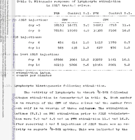
Effect of ALS on the responsiveness of lymphocytes tophytohaemagglutinin (PHA)
.
The responsiveness of lymphoid cells from k L S treated
mice was reported by Tursi
~ 1 ,
(1963). T h c g ctbserved that thymus, lymph node ant! peripheral blood lymphocytestaken from mice treated with FLG showec7 r -:re~tly diminis-
had response to
PHI:
2
v&tcg.
Recovery of the circulating lymphocyte level preceded recovery of respansiveness toFHA, The responsiveness to PHI: could be prevented by re-
injection of ALG o r thymectomy. They observeP t h a t after
PRA respon=%veness had recovereci to the v3lue of epproxi-
metely 207: of the normal, graft rejection also bcgnn to
by a s s e s s i n g t h e
PHn
s e n s i t i v i t y of t h e p e r i p h e r n l white blood c e l l s *2.6- The e f f e c t of ALS on the cfelayed h y p e r s e n s i t i v i t y r e a c t i o n . I n t e r h i t z e n (1956) and Waksnsn
&
21.
(1961) r e p o r t e d t h a t BCG s e n s i t i z e d guinea p i g s r e c e i v i n g ALS showed a m?rked d e p r e s s i o n o r a b o l i t i o n of t h e t u b e r c u l i n r e a c t i o n . lTFaksman ~ l _ . showed t h a t t h e s u p p r e s s i v e e f f e c t s of a s i n g l e dose of ALS on t u b e r c u l i n r e a c t i o n was c l e a r l y r e - l a t e d t o the t i n e a t which t h e ALS w n s a d n i n i s t r ? t e r l . They noteft t h a t t h e n f f c c t c d t u b e r c u l i n r e a c t i o n w c s ccused by ? diminution i n t h e ancmnt of c e l l u l a r i n f i l t r q t i o n i n t h e s k i n . They a l s o observed t h a t t h e d e l ~ y e d r c ? c t i o n t o p u r i f i e d d i p h t h e r i a tox3id w n s reduced o r completelysuppressed i n guinen p i g s trentec? w i t h
ALS.
The s u p p r e s s i v e e f f e c t w q s observed i n guinea p i g s t h a t had lymphocyte counts bclow 4 0 0 0 b 3 . Hurg-n p t i e n t s i n c t e ? with R:HLSshowed no delayed h y p e r s e n s i t i v i t y s k i n rc.ncti?r t o t u b e r - c u l i n a s r e p o r t e d by Plonxco
22
s.
(1967).2.7. The e f f e c t of ILS on t h e B and 92 lymphocytes.
The e f f e c t 2f ALS on the B .and T c e l l s was reporte?. by B i s h o p
~2
s.
( 1 9 7 5 ) * C o s i m igt
a?.
(1976), Thomas g t21.
caused a prof3und f a l l in t h e T lymphocytes t o below 10% of t h e p r e t r e a t m e n t l e v e l . Thomas
9
5%.
( 1 ~ 7 7 ) f o u n d t h a t t h i s e f f e c t p e r s i s t e d f o r more than one week a , t e r LFJTG a d m i n i s t r a - t i o n .AH%%
a d m i n i s t r s t i o n was a s s o c i a t c 6 with r i s e s i n s u r f a c e irmnunoglobin-positive ( B ) c e l l s t o 7 2 t o 93s cnc? E Z C r e c e a t o r p o s i t i v e c e l l s t p 36 t o 687% of t h e mononuclear c e l l s , i n d i c e t - i n g t h e s e l e c t i v e e f f e c t of bHTG on t h e c i r c u l z t i n g T c e l l s . Thomas&
92,
(1978) sugzested t h l t t h e immunosuppressive pstency of r a b b i t AHTG i n t h e primate s k i n ~ m f t a s s a y w a sr e l a t e d l a r g e l y t o i t s m t i - T c e l l a c t i v i t y , Bishop
~2
a&.
(1975) proposed t h a t t h e monitoring o f c i r c u l a t i n g r o s e t t i r t g( T ) c e l l s may be 2 u s e f u l c l i n i c a l g u i d e t o t h e degree of T
26
3.
MIATERIALS AND METHODS3.1. P r e p a r a t i o n of a n t i - b o v i n e lymphocyte SeFLml.
'Pha a n t i - b o v i n e lymphooyte serum (ABLS) used i n t h e s e e x p e r i m e n t s was produced by
Dr
J . G . K r e e f t e n b e r g and F.L e e r l l n g from t h e N a t i o n a l I n s t i t u t e of P u b l i c S e Q l t h , B i l t - - hoven, t h e N e t h e r l a n d s and was r a i s e d i n f o u r h o r s e s a c c o r d i n =
t o t h e method d e s c r i b e d by Monaco
t
gl.
(1966) w i t h some modifications. Lymphocytes were d e r i v e d from a thymus g l a n dof a c a l f . The thymus g l a n d o b t a i n e d from a l o c ? l s l a u g h t e r house was c o l l e c t e d i n c o l d H a n k s * .
ESS
medium. O n a r r i v a l i n t h e l a b o r a t o r y t h e t i s s u e s were c l e a n e d , d e f a t t e d and homo- genized i n Hanks* BSS medium w i t h a l o o s e f i t t i n g P o t t e r t u b e . The c e l l s u s p e n s i o n was f i l t e r e d through a double l a y e r of s t e r i l e gauze t o removed c o a r s e thymus p a r t i c l e s , washed t w i c e and t h e n resuspended f n Hanks* BSS medium. The concen- t r a t i o n was a d j u s t e d t o 2.5x
lo8
lymphocytes p e r m l . When n o t d i r e c t l y used t h e c e l l s u s p e n s i o n w a s s t o r e d i n l i q u i d n i t r o g e n . The c e l l s were f r o z e n i n 10% h o r s e s e w and 10% dimethyl s u l p h o x i d e .ctult c l i n i c a l l y nor%rrl h o r s e s mere useir f o r i m m i
-
*tian* F i g u r e 1 d i s c r i b e s t h e method o f immunization. E - c h h o r s e w a s first i n j e c t e d s u b c u t a n e o u s l y w i t h 3.0
x
10' thymicC e l l s i n complete F r e u n d ' s a d j u v a n t . The b o o s t e r immunization
R a i s i n g
ABLS
i n
.*
=9
r--3,0x10
CFA
s , c ,
t---
1 . 5 ~ 1 0 ~
P l a s m a p h e r e r e s
I I I I I I I
I I l l 1 1 1t I I I J 1 l l l l l i
t l l ( t I i I ' l t L 1 ' ~ I I ( I
0
10
20
3
040
Day
CFA
= c o m p l e t e F r e u n d ' s a d j u v a n t
F i g ,
1.
The m o d i f i e d Monaco's method f o r r a i s i n g
A B L Si n a h o r s e .
9
T h e
h o r s e r e c e i v e d s u b c u t a n e o u s l y
3 . 0 ~ 1 0t h y m o c y t e s i n
CFA
on
d a y0, f o l l o w e d
bytwo i n t r a v e n o u s i n j e c t i o n s
o f9
1 . 5 ~ 1 0
t h y m o c y t e s i n Hanks'
B S Smedium
on day
2 3and
2 4 .days, Between day
10
and
24
a f t e r t h e l a s t i n j e c t i o n t h e h o r s e w a s s u b j e c t e d t o a ~ e r i e s of 6>'plasmaphereses. A f t e rc o l l e c t i o n t h e serum was s t o r e d i n - 2 0 O ~ u n t i l used. T h e
serum was used wit;hout i n a c t i v a t i o n o r a b s o r p t i o n w i t h bovine e r y t h r o c y t e s .
3.2, Determination of c y t o t o x i o and haemolysin t i t e r s -
The c y t o t o x i o t i t e r o f t h e ABLS was determined by t r y p a n blue dye e x c l u s i o n t e s t i n the presence of r a b b i t complement i n a m i c r o t i t e r p l a t e . F i f t y 4 of two f o l d ABLC d i l u t i o n s were i n c u b a t e d with
5 0 4
of bovine p e r i p h e r a l blood lympho- c y t e suspension i n Hanks.' BSS medium c o n t a i n i n g 6.0x
10 6 c e l l s p e r r n l and 5 0 ~ 1 of f r e s h r a b b i t seruQ (complef~ent)d i l u i a d 1:10 i n s a l i n e . A f t e r one hour i n c u b a t i o n a t 3 7 O ~ t h e percentage of dead c e l l s was estimated by the a d d i t i o n o f one drop of 0.1% t r y p a n blue. The t i t e r was t h e r e c i p r o c a l of t h e h i g h e s t d i l u t i o n which k i l l e d a t l e a s t 507 of t h e bovine
lymphocytes.
.-
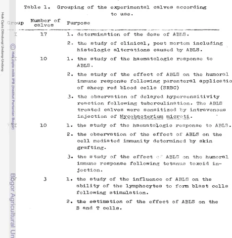
3 . The experimental c a l v e s .Table 1. Grouping of the experimental calves according to use.
Group
N
~
~
~
~
Purposes
O
f
-.-
---
---
----
A - - ----
.---
I
17
I.
determination of the dose of A B I S ,2 - the study o f clinical, post mortem including
histologic alterations caused by ABLS.
1. the study of the haematologic response to ABLS
.
2. the study of the effect of A B L S on the hulloral immune response following parenteral applicatiofi
of sheep red blood cells (SRBC)
3 . the observation of delayed hypersensitivity
reaction following tuberculination. The ABLS
treated calves were sensitized F y intravenous
injection of
M~coba~m~~~
mncco
qz.
1. the study of the hacnato&ogic response to
ABLS.
2 - the observation of the effect ot. ABLS on the
cell mediated immunity determined by skin grafting
.
3.
the study of the effect , > ' ' A B L S an tho humoral immune response following tetanus toxoid in- jection.1. the study of the influence of A R L S on the ability of the lymphocytes to form blast cells following stimulation.
[image:40.516.32.502.53.537.2]-3.1. Group I
3 - 3-1a1. D e t e r m i n a t i o n of t h e d o s e s of ABLS.
F o r t h i s purpose
17
c a l v e s were u s e d , These c a l v e s were d i v i d e d i n t o 5 subgroups .as shown i n t a b l e 2 . T h e ABLS was i n j e c t e d s u b c u t a n e o u s l y a t v a r i o u s q u a n t i t i e s . Subgroups 4 and 5 c o m p r i s i n g t h r e e c a l v e s were g i v e n normal h o r s e serum a s c o n t r o l .From t h i s e x p e r i m e n t i t was found t h a t I nl ABLS p e r kg
body w e i g h t was s u f f i c i e n t t o d e p r e s s t h e p e r i p h e r a l blood lymphocyte p o p u l a t i o n , whereas t h e e r y t h r o c y t e , haemoglobin and thrombocyte c o u n t s remained normal and t h e animals showed o n l y s l i g h t c l i n i c a l s i g n s ,
Table 2. Nunber of e x p e r i m e n t a l c a l v e s i n groul3 I ,
dose and r o u t e of a p p l i c a t i o n o f a n t i - b o v i n e lymphocyte serum f o r t h e e s t i m a t i o n o f t h e e f f e c t i v e dose,
Sub- Number o f ~ o s e / k g body g r o UP c a l v e s w e i g h t
---.--
---
---.---.---1. 4 7.50-10 m l ABLS
2. 3 3.00- 6 r n l ABLS
3
7 0.75- 1 rnl ABLS4 1 5 m i normal h o r s e serum
5. 2 3 E l 1 tt
NurnSzr and node Days o f o f i n j e c t i o n s i n t e r v a l s
.---- --
-
3 Subcutaneous 4 and 2
2 tr 1
[image:41.516.56.500.315.490.2]3.3.1.2- The c l i n i c a l and post-mortem o b s e r v a t i o n s .
The c a l v e s were observed d a i l y f o r c l i n i c a l symptoms and temperatures were taken twice a day. The animals t h a t succum- bed were n e c r o p s i e d and a s t u d y of t h e g r o s s and h i s t o l o g i c a l t e r a t i o n s were made. For h i s t o l o g i c a l e x m i n a t i o n s p i e c e s of organs i n c l u d i n g lymph node, thymus g l a n d , l u n g , h e a r t , l i v e r , s p l e e n , kidney, i n t e s t i n e , bone narrow a n d b r a i n were c o l l e c t e d and f i x e d i n 10% b u f f e r e d f o r m a l i n , T h s r e a f t e r the t i s s u e s were processed a c c o r d i n g t o s t a n d a r d h i s t o l o g i c a l techniques. S e c t i o n s of 5 microns were s t a i n e d with r o u t i n e haematoxylin e o s i n . Other s t a i n i n 3 methods such a s van G i e s o n and P . A . S . were a p p l i e d when n e c e s s s r y .
<.3-2- Group 11.
T h i s group c o n s i s t e d of 7 ABLS t r e a t e d c a l v e s a n d 3 c o n t r o l s . They r e c e i v e d t e n subcutaneous i n j e c t i o n s of AELS a t one day i n t e r v a l s a t a dose of 1 m l p::r kg bociy weight. The i n j e c t i o n s were givftn a t t h e l e f t and r m i g h t s i d e s o f t h e neck. The c o n t r o l c a l v e s were l e f t u n t r e a t e d ,
3.3.2-1. iGwma%aiogical examinations.
P r i o r t o t h e i n i t i a t i o n of the experiments, haematolo- g i c a l examinations were performed twice a week f o r t w o
e x a m i n a t i o n s were performed a t 6 h o u r s , 2 4 h o u r s and t h e n a t one d a y i n t e r v a l s u n t i l s e v e n t i m e s follov,i;.ng t h e l a s t in-. j e c t i o n .
Blood was obtaincld by j u g u l a r veni-punckure and c o l l e c t e d i n a t u b e c o n t a i n i n g a s o l u t i o n of sodium ethylene-diamine- t e t r a - a c e t i c a c i d (EDTA) t o p r e v e n t c o a g u l a t i o n t a a f i n a l c o n c e n t r a t i o n of 1
mg
EDTA p e r r n l blood.F o r c e l l u l a r c o u n t s and hstemoglobin d e t e r m i n a t i o n s 2 0 , ~ l EDTA b l o o d was d i l u t e d i n 1 0 n l I s o t o n I1 s o l u t i o n ( C o u l t e r E l e c t r o n i c s , Gmbh, 4150 K r e f e l d , G a j l i n g s f n d 5 3 , 61. Germany)
a n d 1 0 0 4 of t h i s l t 5 0 0 d i l u t e d blood was suspended i n 1 0 m l I s o t o n I1 s o l u t i o n (1:50.000) f o r e r y t h r o c y t e c o u n t s and
h a e n a t o c r i t d e t e r m i n a t i o n s . F o r l e u c o c y t e c o u n t s 3 d r o p s o f Z a p o g l o b i n e s o l u t i o n ( C o u l t e r E l e c t r o n i s L t d , Coldharbour Lane, Harpenden, H e r t s , England) were added t o t h e r e m a i n i n g
s u s p e n s i o n i n o r d e r t o l y s e t h e e r y t h r o c y t e s ,
The thrornbocyte c o u n t s were deternin:;;d by u s i n g t h e C d k e r P l a t e l e t K i t (Coldharbour Lane, Harpenden, H e r t z , England). Approximately 0 . 1 ml of EDTA blood was a s p i r a t e d i n t o a s p e c i a l p l a s t i c s e d i m e n t a t i o n t u b e and allowed t o s e t t l e f o r 2
-
3
h o u r s , Thc t u b e was t h e n c u t a t t h e j u n c t i o n o f the r e d c z l l s and t h e plasma. The plasma r i c h thrombocytes was sucked i n t o a3,3,&
p i p e t and d i l u t e d i n 1 0 m l I s o t o n I1s o l u t i o n ,
The l e u c o c y t e s , e r y t h r o c y t e s a n d thrombocytes were
L t d , C o l d h a r b o u r L a n e , H a r p e n d e n , H e r t s , ~ n g l a n d ) , w h i c h w a s p r o v i d e d w i t h a h a e m o g l o b i n m e t e r a n d h a e c a t o c r i t m e t e r . T h e l a t t e r wets c o n n e c t e d w i t h t h e C o u z t e r C o w l : e r . ' " l z e n t h e e r y -
t h r o c y t e s w e r e c o u n t e d t h e h a e m n t o c r i t n e t e r d e t e r m i n e d t h e h a e m a t o c r i t p e r c e n t a g e z t t t h e s a m e t i m e .
l ' h ~ h a e m o g l o b i n w a s d e t e r m i n e d w i t h the h n o r n o g l o b i n
m e t e r u s i n g t h e s a m e b l o o d d i l u t i o n a s f o r t h e l e u c o c y t c c o u n t c o n t a i n i n g Z a p - o g l o b i n e s o l u t i o n .
T h e l e u c o c y t e s w e r e d i f f e r e n t i a t e d i n b l o o d snears s t a i n e d a f t e r M a y G r e t n w a l d and G i e m s a (6. M e r c k , D a r m s t a d t ) f o r r e s p e c t i v e l y 5 and
1 5
m i n u t e s . D i f f e r e n t i a t i o n w a s c a r r i e d n u t w i t h a C a r l Z c i s s l i g h t m i c r o s c o p e u s i n g o i l i m m e r s i o n a t 600 x r n a m i f i c a t i o n .3.3.2.2. T h e s t u d y of t h e e f f e c t o f A B L S on t h e h u m o r a l i m m u n e r e s p o n s e f o l l o w i n g p a r e n t e r a l a p p l i c a t i o n o f sheep r e d b l o o d c e l l s (SRBC)
.
3.3.2.2-1. P r e p a r a t i o n , d o s e a n d a p p l i c a t i o n of S R B C -
c s l v c s were i n j e c t e d i n t r a v e n o u s l y w i t h 1 m l (75.0 x 1 0 6 c e l l s ) SRBC suspension.
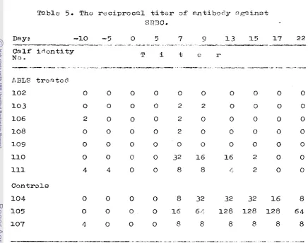
3.3.2.2.2, A g g l u t i n a t i o n t e s t f o r t h e d e t e r m i n a t i o n of a n t i b o d y t o S R R C .
The a n t i b o d y t i t e r a f = a i n s t SRBC was determined by t h e d i r e c t haemagglutination t e s t . Norrnal sheep blood i n Alsevere
j l : l j was c e n t r i f u g e d a t 340 g f o r 1 0 minutes and t h e p e l l e t
w i l s washed 3 times i n s a l i n e . A f t e r washing t h e haernoglobin
t i t e r was determined and adjuster2 t o 16.0 ng 5 with s a l i n e ?nd t h i s c o n c e n t r a t i o n was considered a s a 100% SRBC suspen- s- sn. From t h i s suspension a
15
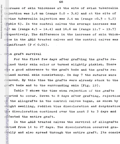
SRBC suspension was prepared i n a b u f f e r c o n s i s t i n g of 80% n u t r i e n t g e l a t i n b u f f e r (400 mg n u t r i e n t g e l a t i n d i s s o l v e d i n 1 l i t e r d i s t i l l e d w a t e r ) , 207' v e r o n a l b u f f e r and 0 . 5 5 i n a c t i v a t e d bovine f o c t u s serum which w s p r e v i o u s l y absorped w i t h S R B C . Two f o l d d i l u t x o n s of the t e s t s e r a were made i n t h i s b u f f e r i n m i c r ? t i t e r p l a t e s , To 5 0 ~ 1 of t h e serum d i l u t i o n s 5 0 4 o f t h e SRBC suspension3.3.2.3.
Observation of delayed hypersensitivity reaction by tuberculination.F o r testing the influence of ABLS on this type of
delayed hypersensitivity reaction all ABLS treated and control
c llves were injected intravenously with 1 ml of a &lfc@c-~~e-ilr-~
g & c r G &
suspension containing 3.0x
la5 viable cells units perm l at
24
hours after the administration of the first ABLS.The classical hypersensitivity skin test was performed
4
weeks after the administration of X¶~~~a_wte_r_~~-m_&c~q't&.
This was done by injecting intrademally all ABLS treated and
control calves with 0.1 ml of bovine and avian tuberculin.
E?ch calf thus received 5 0 0 0
TU
bovine tuberculinPPD
and2000 TIJ nvian tuberculin PPD on the right side of the neck. Bzfore the tuberculin was administered the thickness of the
skin was measured with a cutimeter. The skin reaction was
determined
72
hours after the tuberculin injection by mea-suring the increase in the thickness of the skin a t the site
of injection,
3.3.3.1.
The study of haematologic response to ABLS (see group I1 c.)3.3-3.2.1,
Treatfnent of t h e c a l v e s before s k i n g r a f t i n g .Animals of group I11 c o n s i s t e d o f 1 0 c a l v e s were divided i n t o 6 A A L S t r e a t s d c z l v e s and 4 c o n t r o l s . The ABLS t r e a t e d
c a l v e s r e c e i v e d t o n i n j e c t i o n s o f 1 m l ABLS p e r kg b o d y weight a t one d a y i n t e r v a l s . The c o n t r o l c a l v e s were i n j e c t e d with norm71 h o r s e s c r m a t t h e same dose cnd a l s o a t one day i n t e r - v a l s f o u r times. T h e a d m i n i s t r a t i o n of ABLS and normal h o r s e s z r u m w ~ s t a r t e d s t w o days b e f o r e g r a f t i n g (day -2) on t h e
d a y of g r a f t i n g (d* 0 ) and every two days t h e r e a f t e r . Table 3 shows the donor-recipient p a i r s o f c a l v e s i n skin g r e f t i r , _
T n b l e 3. Donor-recipient pairs of c a l v e s i n s k i n g r n f t i n g .
R e c i p i e n t G r a f t donor
C a l f i n d e n t i t y N o . Calf i d e n t i t y No.
--
.- .-------
---
-
A?3L_.? TR_EaE_E!
1 5 6 158
158 156
161 162
162 1 6 1
1 2 3 250
2 50 1 2 3
csn_trol_
157 159
1 5 9 157
207 208
3-3.3.2.2, Skin g r a f t i n g
3.3.3.2.2-a. P r e p a r a t i o n o f t h c c a l v e s ,
Twenty f o u r hours b e f o r e s k i n g r a f t i n g foo-I was with- h e l d f r o m a l l c a l v e s . The donor s k i n s i d e and the g r a f t i n g bod s i d e were c l e a n l y shaven aqd washed with w8ter c o n t a i n i n g cytopogen ( ~ i s t - ~ r o c n d e s N V , D e l f t , the ~ e t h e r l ~ n d s ) and
d h s i n f e c t e d with 70% e t h a n o l . The c a l v e s were s e d a t e d by in- j e c t i o n of 1 ml 2% Rompun s o l u t i o n (Bayer) in&ramuscularly. General a n a e t h e s i a was c a r r i e d o u t with N m b u t a l s o l u t i o n
(Abbott Lab) a t a dose of 0.2 ml p e r kg b o d y weight, admi- n i s t e r e d i n t w o divided doses. The f i r s t dose wns i n j e c t e d b e f o r e t h e removal of t h e g r a f t and the second was given p r i o r t o t h e p r e p a r a t i o n of t h e g r a f t bed and i t s fixation.
3.3-3.2.2.b. P r e p a r a t i o n of t h e g r a f t s ,
White s k i n was chosen f o r t h e g r a f t s t o f a c i l i t a t e
e v a l u a t i o n and comparisor~ with t h e s k i n
.
arrotm3ing the g r a f t beds. G r a f t s were rsmoved f r o m t h e l a t e r a l s i d e of t h e hind l e g below t h e s t i f l e j o i n t . The g r a f t was t h e s l z e of a m i -2
croscope s l i d e (2.5
x
6 cm ) , which was used a s a template ( f i g u r e 1 1 ) . A f u l l t h i c k n e s s s k i n g r a f t of t h i s s i z e was removed by i n c i s i o n and b l u n t d i s s e c t i o n . A f t e r removal. t h e2
a p e t r i d i s h c o n t a i n i n g a s m a l l volume o f Hanks* B S S s o l u t i o n and a n t i b i o t i c s , The g r a f t s were cleaned by removing t h e
a d h e r i n g f a t and cormective t i s s u e s with a c i s s o r s o r s c r a p i n g w i t h a s c a l p e l .
The donor's wound was s u t u r e d w i t h N o . 1 s i l k s u t u r e a n d t h e s u r g i c a l wound was smeared with a t h i n l a y e r of S o c a t y l p a s t e (Ciba Geigy, Bazel, S w i t h z e r l a n d ) , The wound was covered w i t h a s t e r i l e gauze and f i n a l l y with p l a s t e r tape (Figs.
12-13).
3.3.3.2 -2. c. P r e p a r a t i o n of the g r a f t beds.
40
(Figs. 15-1 6 ) a
3.3.3.2.2.d. Treatment of t h e gr~fts.
Following fixaCion the g r a f t s were covered with Neba- c e t i n powder (H, Lunback & Co A
/S
,
Copenhagen),
a p i e c e of p a r a f f i n impregnated gauze, a picce of d r y gauze and then with Hmsopor p l a s t e r , and t h e r e a f t e r f i x c d with p l a s t e r t a p e . F i n a l l y t h e backs a f t h e c a l v e s were covered with a white p i e c e of c l o t h , which w ~ s h e l d i n p l a c e by two e l a s t i c bandage8 around t h e w a i s t and c h e s t ( ~ i s s - 17-18). The g r a f t s were i n s p e c t e d every two days and a t t h e s a m e time t h e p a r e f - fin-impregnated and dry gauze were changed. To kcep theg r a f t o moist they were smesred with ur'lder cream ( I C I Phnrmrl.- c e u t i c a l s D i v i s i o n ) . The s u t u r e s were removed on d a y 7 a f t e r t h e opcrotion.
Thc parameters used f o r the e v a l u a t i o n of g r a f t s u r v i v a l were t h e g e n e r a l macroscopic appearance, ? : L i a b i l i t y * t h s
occurencc of haemorrhugos and d e s i c c r i t i o n , decret,se o f g r a f t s i z e and h a i r growth. N o microscopic e v a l u a t i o n was p e r f o m e d .
3.3.3.3. The study of t h e e f f e c t of ABLS on t h e h u ~ o r a l immune response f o l l o w i n g t e t a n u s toxoid i n j e c t i o n .
3-3.3.3.1. O r i g i n and dose o f t e t a n u s toxoid.
4 1
H e a l t h i n B i l t h o v e n , t h e N e t h e r l a n d s , Each ABLS and c o n t r o l c a l v e s were i n j e c t e d i n t r a m u s c u l a r l y w i t h 3 m l t e t a n u s t o x o i d s u s p e n s i o n 48 h o u r s a f t c r t h e f i r s t a d m i n i s t r a t i o n of ABLS.
3.3.3.3.2. D e t e r m i n a t i o n of t h e a n t i b o d y t i t e r against t e t a n u s t o x o i d i n blood serum.
The serum from e x p e r i r x e n t a l anirnnls was collected t w i c e s week. These s e r a were t i t r a t e d by