Young and Ali.
http://mji.ui.ac.id
178 Med J Indones
Endovascular treatment of traumatic carotid cavernous istula with
trapping technique
Benny Young, Ahmad Faizal M. Ali
Department of Radiology, School of Medicine, University Malaysia Sarawak (UNIMAS), Hospital Umum Sarawak, Kuching, Malaysia
Abstrak
Penatalaksanaan istula arteri karotis kavernosa secara endovaskular konvensional melibatkan penempatan material secara langsung, baik itu koil, balon atau keduanya. Tujuan akhir adalah tertutupnya istula dan terjaganya arteri karotis. Tetapi dalam beberapa kasus dengan laserasi arteri karotis yang parah, tatalaksana endovaskular yang memungkinkan adalah dengan menutup pengisian istula dari sirkulasi otak. Metode ini dikenal dengan nama ‘trapping technique’ yang melibatkan metode penutupan arteri karotis, dikerjakan pada kasus ini dengan hasil yang baik. (Med J Indones. 2013;22:178-82. doi: 10.13181/mji.v22i3.588)
Abstract
Conventional endovascular treatment for carotid cavernous istula (CCF) involves a direct delivery of either coils, detachable balloon or both to the istula with end point of CCF resolution and carotid artery preservation. But in few cases with severe laceration of carotid artery, the feasible endovascular technique applicable is by blocking the illing of istula from cerebral circulation. This method known as trapping technique which implicates carotid artery occlusion, was performed in our present case with good result. (Med J Indones. 2013;22:178-82. doi: 10.13181/mji.v22i3.588)
Keywords: Carotid cavernous istula (CCF), carotid occlusion, trapping technique
Correspondence email to: [email protected]
Trauma is known as the most common cause of direct carotid cavernous istula (CCF). A carotid artery injured by bony fragment has a through communication to the cavernous sinus with inherent fast low via the istula. It was grouped as type A in Barrow classiication; commonly and simply known as CCF. It is a progressive medical problem where spontaneous resolution rarely occur. If left untreated, CCF can lead the patient to devastating conditions. Arterialization of the cavernous and cerebral venous low may cause loss of eye vision, cerebral infarction or hemorrhage associated with venous hypertension,
and sinus or venous aneurysm.1,2
Endovascular treatment for CCF has widely replaced surgical treatment. Direct delivered coils or balloons to the istula either using trans-arterial or transvenous approach has become the standard conventional endovascular treatment. An ideal goal of endovascular treatment is achieving preservation of the main carotid artery with complete occlusion of the CCF.3 The success of these methods relies on a clear delineation of the istula’s track and the accessibility of the intended vessels. Several maneuvers such as Mehringer-Hieshima maneuver [gentle ipsilateral internal carotid (IC) injection during manual compression of the ipsilateral carotid artery] and Heuber maneuver (ipsilateral carotid compression during vertebral artery injection) are theoretically useful
to aid outlining on the site of carotid’s tear.4
The other factor that inluences success of endovascular treatment is the size of carotid tear. Oversize tear
associates with a risk of coils or balloon migration to the carotid artery. Subsequently, a large tear is accounted for total diversion low into cavernous sinus with ensuing poor outline of the long course of cerebral carotid artery. Hence, this circumstance precludes interventionists from placing a non-detachable balloon as a safety mean to avoid coil or balloon migration during their deployment. Due to these aforementioned dificulties, a trapping technique
is considered as an alternative endovascular method for
CCF with large tear. The principal of trapping technique is obliteration of istula’s illing from the carotid artery
and retrograde intracranial perfusion.5,6 We report one
case of CCF with inherent dificulty that warranted endovascular trapping technique with description on
the technical consideration and suggestions to achieve success ful treatment.
CASE REPORTS
A 46-year old male was admitted to our hospital for a close head injury following a high speed motor collision. He suffered from multiple facial and skull base fractures. Two days after, we noted that he had a loss of right eye’s vision, proptosis and chemosis with decreased level of consciousness. A post traumatic direct CCF was suspected and a diagnostic angiogram was performed.
Technique
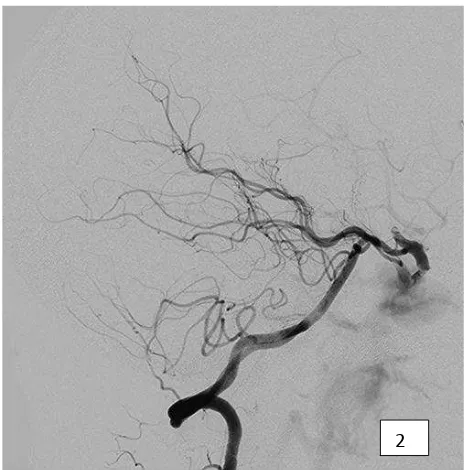
F vertebral angiogram catheter was used for cerebral arteriogram. Right internal carotid artery (ICA) arteriogram showed direct type of right CCF secondary to severe laceration of carotid artery. CCF demonstrated a total diversion low to cavernous sinus and its tributaries with almost absence of right intracranial carotid low. The right ophthalmic artery was totally not opaciied nor received any collateral; which accounted for the loss of patient’s right eye vision. The involved and opaciied venous drainage were the right ophthalmic vein, bilateral pterygopalatine plexus, retroclival plexus, and the contralateral cavernous sinus (Figure. 1). The right vertebral arteriogram demonstrated a retrograde illing of the CCF through the right posterior communicating artery (PCOM) (Figure 2).
We estimated that it was not possible to navigate the
microcatheter across the cavernous segment of carotid
artery and advance it into the istula due to the fact that a delineation of long segment carotid artery was not present. The non-opaciied inferior petrous sinus or a good size of ophthalmic vein had deterred us from the
option of transvenous approach.
Therefore, we chose a method to close the istula by trapping its low but with a consequence of occluding the carotid artery. This procedure comprised a closing of the proximal carotid artery with a detachable balloon and blocking the retrograde low of the istula from any of the collateral in the circle of Willis. The latter was achieved by occluding the pre-clinoid cavernous part of carotid via right PCOM.
Figure 1.
Figure 2.
A) Pre-treatment diagnostic angiogram via right carotid artery from lateral view, B) AP view demonstrated a direct right CCF with torrential low to cavernous sinus and nearly absent of distal intracranial low. The subsequent venous drainage to the superior ophthalmic vein, retroclival plexus, and pterygo-palatine plexus were seen
Lateral view of right vertebra arteriogram showed right CCF recruited a retrograde perfusion through the right posterior communicating artery. It demon-strated clearly that the distal and proximal portion of the cavernous segment of right carotid artery was not communicating, consistent with severe laceration
Prior to the occlusion procedure, the patient was heparinized by giving a bolus of intravenous 3,000 units of heparin. A 6 F Fagomax introducer catheter was advanced carefully into the proximal right ICA. A 0.019-in over the wire Excelcisor SL 10 microcatheter
Young and Ali.
180 Med J Indones
Figure 3.
Figure 4.
Post delivery of 2 GDC microcoils (arrow head) to the pre-clinoid carotid artery. Angiogram via micro-catheter in the PCOM showed minimal illing to the istula at the posterior aspect (arrow)
The short portion of pre-clinoid carotid artery was totally occluded after delivery of 50% histoacryl glue into the pre-existing packed GDC coils. A subsequent closing of the anterograde illing of the istula was performed by proximal carotid occlusion with detach -able balloon
(Boston Scientiic, Fremont, CA) with a 0.014-in hydrophilic guidewire (Synchro 14; Boston Scientiic) were run across coaxial within the guider. A continuous lushing system using a pressurized bag illed with 1,000 units of heparin in 1 L of normal saline was connected to the introducer catheter via the Tuohy-Borst adaptor. The irst step of our treatment was to microcatheterize the right PCOM from the right vertebral artery. Following it, we advanced our microcatheter into the short part of the pre-clinoid part of the carotid artery and deploying two soft platinum GDC coils, size 4 mm x 12 cm and 5 mm x 20 cm (Helix Axium ev3, Irvine CA) each. A control
run post coiling revealed a persistent small opening to
the istula at the posterior aspect (Figure 3). In order to have a complete closing of this retrograde illing, a 3 mls of 50% mixed Histoacryl liquid glue: Lipiodol solution (Lipiodol; Guerbet, Genova, Italy; Histoacryl, B Braun, Melsungen, Germany) was delivered into the pre-existing coils pack. Post embolization with combination of coils and adhesive histoacryl liquid showed a complete closing of the retrograde perfusion to the istula with preservation of the PCOM (Figure 4).
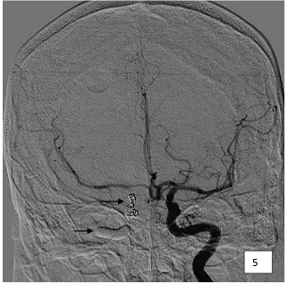
The next step was to occlude the carotid artery just proximal to the site of istula by using a detachable balloon. A number 9 latex gold valve balloon (Nycomed, France) was prepared and mounted on 2.7 F MABDE microcatheter. Through the 6 F Fagomax introducer catheter, the balloon was loated past the cervical ICA and navigated into the proximal cavernous segment of carotid artery. After the balloon was correctly placed into this area, it was fully inlated with 1 mL of Visipague (GE Health Care, US) contrast media and detached. A control run via the introducer catheter showed a complete occlusion of the proximal carotid artery. A left carotid angiogram showed the right intracranial perfusion was well maintained via the collateral perfusion at circle of Willis (Figure 5).
One day after endovascular treatment, the patient showed improvement of his right eye’s proptosis and chemosis even though his right eye’s vision was not able to be salvaged. Follow up after 3 months showed that the patient remained free from right CCF symptoms with absence of any cerebral ischemic due to carotid occlusion.
DISCUSSION
Direct CCF is considered as a progressive neurosurgical case which needs an early diagnostic ruling with
prompt management. The indications for urgent
treatment of CCF include the following :1) progressive visual loss, 2) epitaxis, 3) sphenoid sinus aneurysm, 4) comatose patients in whom intracranial lesions have been excluded, 5) re-routing of the venous drainage
3
4
5
Figure 5. Final angiogram via left carotid artery post posterior coiling and glue (ar-row) and balloon occlusion at the proximal right carotid artery (arrow head) showed trapping of right istula’s low. The right intracranial low was main -tained via collateral from the left carotid circulation
Most of the reported success rate of conventional
endovascular treatment of direct CCF using detachable balloon or platinum coils are about 75%-88% with preservation of the main carotid artery.4-6For the rest
of the cases, the occlusion of the main carotid artery is contrived to treat the istula. Cases which are treated by occlusion of the main artery inherently have large tear in which the low of carotid artery is profoundly terminated
in the cavernous sinus.
The occlusion of carotid artery as treatment of choice
has a relevance in our present case. The technical
dificulties conceived from the lacerated artery were addressed. The associated massive low due to large tear into istula and cavernous sinus created a poor delineation of the istula’s oriice and its track. Hence, the consequence was a blind track, making it dificult to navigate any microcatheter system to the istula and deploying the embolized materials. Furthermore, the placement of any coils or detachable balloons within the istula carried a high risk of migration to the carotid artery from the large artery rent to the istula. In addition, the predominant redirection of the CCF to pterygoid plexus had also precluded our option to approach the
istula via inferior petrous or inferior ophthalmic veins for transvenous delivered coil embolization.
A case series of trapping technique for CCF consisted of 4 patients was reported by Coley et al. In three cases, a retrograde low trapping of CCF was performed via anterior communicating artery access with one case required PCOM for the access. In all these cases, closing of the distal cavernous carotid artery was successfully achieved by packing of platinum coils. Similar with our reported case, the occlusion of the proximal carotid artery in this case series was performed using a detachable balloon.8
An alternative method for treating severe direct CCF
is placing a stent dedicated for intracranial carotid
Young and Ali.
182 Med J Indones
Carotid artery occlusion method is not a risk free procedure. Brain ischemic is a known early complication of post carotid occlusion which is secondary to abrupt changes of the brain auto-regulation. A prior balloon test occlusion (BTO) for assessing the availability of collateral circulation while judging the patient’s tolerance to carotid occlusion treatment by assessing patient’s neurocognitive during BTO should be done to reduce the risk of post occlusion brain ischemic. However, some patient with critical clinical condition can be exempted from BTO.
11-13 Hypotension is known as a predisposing factor for
this cerebrovascular event post carotid occlusion. A close monitoring with comprehensive intensive care is mandatory to minimize this pertaining complication after
carotid occlusion procedure.
In conclusion, trapping technique with endovascular approach is feasible for treating a high low carotid cavernous istula in severely injured carotid artery. In the availability of stent designed for intracranial use and a good experience from the operators, a method of stent placement for carotid reconstruction and excluding istula’s low can be considered. Hypotension is a known predisposing factor for brain ischemic complication post carotid occlusion, for which preventive measurement must be carried out accordingly.
Acknowledgments
The author thanks Dr. Lau Jia Him of Radiology Department, General Hospital Kuala Lumpur for his contribution on technical assistance in performing the
endovascular procedure.
REFERENCE
1. Krings T, Geibprasert S, Brugge KG. Case Based Interventional Neuroradiology. New York: Thieme; 2011.
2. Halbach VV, Higashida RT, Hieshima GB, et al. Interventional neuroradiology. AJR Am J Roentgenol. 1989;153(3):467-76.
3. Sarbienko FA. Balloon catheterization and occlusion of major cerebral vessels. J. Neurosurg. 1974;41(2):125-45. 4. Halbach VV, Hieshima GB, Higashida RT, Reicher M.
Carotid cavernous istulae: indications for urgent treatment. AJR Am J Roentgenol. 1987;149(3):587-93.
5. Morris PP. Balloon reconstructive technique for the treatment of a carotid cavernous istula. AJNR Am J Neuroradiol. 1999;20(6):1107-9.
6. Moris PP. Carotid Cavernous Fistulas. In: Moriss PP, editors. Interventional and Endovascular Therapy of the Nervous System: A Practical Guide. New York: Springer-Verlag; 2002. p. 177-90.
7. Cho JH, Jung Cy, Sheen SH, Kwon BJ, Han MH. Traumatic carotid cavernous istula caused by intradural aneurysm rupture: A case report. Neurointervention. 2006;1:39-43.
8. Coley SC, Pandya H, Hodgson TJ, Jeffree MA, Deasy NP. Endovascular trapping of traumatic carotid cavernous istulae. AJNR Am J Neuroradiol. 2003;24:1785-8. 9. Gomez F, Escobar W, Gomez AM, Gomez JF, Anaya CA.
Treatment of carotid cavernous istulas using covered stents: midterm results in seven patients. AJNR Am J Neuroradiol. 2007;28(9):1762-8.