The benefit of pulmonary rehabilitation against quality of life alteration
and functional capacity of chronic obstructive pulmonary disease (COPD)
patient assessed using St G
eorge’s respiratory questionnaire
(SGRQ) and
6 minutes walking distance test (6MWD)
Wiwien Heru Wiyono*, Joko Riyadi*, Faisal Yunus* Anita Ratnawati, Sabarina Prasetyo
Abstrak
Pasien penderita penyakit paru obstruktif kronik ( PPOK ) tampaknya mendapatkan manfaat dari program rehabilitasi paru. Penelitian ini mengkaji manfaat program rehabilitasi paru pada pasien rawat jalan yang menderita PPOK, dengan menggunakan St George Respiratory Questionnaire (SGRQ) dan six min walking distance test (6MWD), yang mengukur kualiti hidup kesehatan dan toleransi latihan fungsional sebagai hasil pengukuran utama. Penelitian ini merupakan penelitian prospektif, terbuka, acak dengan kelompok kontrol paralel yang diberikan program rehabilitasi pasien rawat jalan pada 56 pasien penderita PPOK (52 orang laki-laki dan 4 orang perempuan). Kelompok aktif (n= 27) diberikan program edukasi dan latihan selama 6 minggu. Kelompok kontrol (n= 29) diperiksa secara rutin sebagai pasien medis rawat jalan. SGRQ dan 6MWD dilakukan pada saat awal penelitian dan setelah 6 minggu. Didapatkan hasil SGRQ dan 6MWD sebelum dan sesudah terapi. Berdasarkan statistik, SGRQ menurun dan skor 6MWD meningkat secara signifikan pada kelompok aktif dibandingkan kelompok kontrol. Disimpulkan bahwa program selama 6 minggu pada pasien rawat jalan ini secara signifikan telah meningkatkan kualiti hidup dan kapasitas fungsional pasien PPOK derajat ringan hingga sedang. (Med J Indones 2006; 15:165-72)
Abstract
Patients with chronic obstructive pulmonary disease (COPD) have been shown to be benefit from pulmonary rehabilitation programs. We assessed an entirely outpatient-based program of pulmonary rehabilitation in patients with COPD, using the St George’s Respiratory Questionnaire (SGRQ) and six minutes walking distance test (6MWD) (which measures health-related quality of life and functional exercise tolerance) as the primary outcome measure. We undertook a randomized, opened, prospective, parallel-group controlled study of outpatient rehabilitation program in 56 patients with COPD (52 men and 4 women). The active group (n=27) took part in a 6-weeks program of education and exercise. The control group (n=29) were reviewed routinely as medical outpatients. The SGRQ and 6MWD were administered at study entry and after 6 weeks. Outcome with SGRQ and 6MWD before and after therapy was performed. Decrease score SGRQ and increase 6MWD in both groups of study, it was analyzed by statistic study and in active group the decrease of SGRQ and the increase of 6MWD was statistically significant. In conclusion 6-weeks outpatient-based program significantly improved quality of life and functional capacity in mild-to-moderate COPD patient. (Med J Indones 2006; 15:165-72) Keywords: COPD, pulmonary rehabilitation, SGRQ and 6MWD.
In 1995, COPD and asthma were on the 5th rank of death based on House Hold Health Survey, Health Department, Republic of Indonesia (SKRT, Depkes).1
In 1997, Yunus described COPD case pattern with acute exacerbation, which had been hospitalized at Pulmonology Department, Persahabatan Hospital; i.e. 104 cases with COPD diagnosis and there were only 65 cases which were appropriate with the COPD criteria. At the Persahabatan Hospital, as national top referral for pulmonary disease, COPD was on the 5th rank of outpatient and was on the 4th rank of hospitalized patient number.2
In 1990, Sherrill et al. indicated that health statistic hardly record the COPD prevalence because of no *
Department of Pulmonology and Respiratory Medicine, Faculty of Medicine, University of Indonesia, Persahabatan Hospital, Jakarta, Indonesia
Department of Medical Rehabilitation, Persahabatan Hospital, Jakarta, Indonesia
standardized definition, lack of recognition and misclassification.3 In 2000, there were 600,000 COPD patients in the United Kingdom and symptoms appeared after 40 years of age. Male prevalence outnumbered the female and was likely increased about 5 % in 65 – 75 years of age, and about 10 % in the age over 70 years. In developing countries, the COPD prevalence is steadily increased due to increasing of smoking prevalence.4
Rehabilitation of COPD patient is a standard management, which is aimed to control, reduce symptoms, and increase the functional capacity optimally. The main objectives is to return the body ability of self-living, which is realized by helping patient to learn about concomitant disease, alternative of therapies and how to manage it. Patient should have active self living and reduce their dependence on health care.5
Quality of life is an individual condition level in the extent of ability, boundary, symptoms, and psychosocial characteristics to perform in various expected roles of the community and being satisfied.6 The concept of quality of life measurement which correlated with health usually refers to minimal one of 4 important domain or component, i.e. somatic sensation, physical function, emotional or psychosocial status and social interaction. General health questionnaire usually is less sensitive about COPD level, therefore specific measurement is applied, such as St George’s Respiratory Questionnaire (SGRQ), which is developed by Jones et al.7
SGRQ consist of 76 items of questions. It is divided into three components i.e.:7
1. Symptoms, associated with short of breath symptom, frequency and severity of symptom 2. Activity, associated with activity that cause short
of breath or limited activity because of it.
3. Impact, includes a series of aspect which is correlated to social function and psychological disorder caused by illness.
In the early 1960’s, Balke developed a simple test to
evaluate functional capacity by measuring walking distance in certain period. Twelve minutes walking distance test was developed to evaluate the exercise outcome of healthy individual and chronic bronchitis patient. Six minutes walking distance test was developed later, and actually the result is as well as
the 12 minutes walking distance test, and it is more tolerable by patients and more representing daily activity situation. Main indication of 6 minutes walking
distance test is to measure patient’s therapeutic response
with heart or lungs disorder of mild to severe level.
Other indication is to measure patient’s functional status
and predict disease mortality and morbidity.8
The aim of this study is to evaluate the benefit of physical rehabilitation in mild to moderate COPD patients. The specific objectives are to evaluate the quality of life alteration in COPD patients who have medical rehabilitation by using SGRQ and to evaluate the alteration of functional capacity in COPD patients who have medical rehabilitation by using 6 minutes walking distance test.
METHODS
This was an open randomized clinical trial on 56 mild to moderate COPD patients, who came to Asthma Clinic of Department of Pulmonology and Respiratory Medicine Faculty of Medicine, University of Indonesia in Persahabatan Hospital. This trial has been proved by ethical committee of Faculty of Medicine, University of Indonesia. The inclusion criteria were mild to moderate stable COPD patients, male or female of 45 – 75 years old, FEV1 more than 30% predicted value, FEV1/FVC < 70%, no exacerbations 4 weeks prior to the study, continue taking medicines for COPD, not enrolled in any physical exercise and gave written agreement to enroll this study. The exclusion criteria were patient who had heart disease, Cor Pulmonale, neurologic disorder and cognitive disorder. The patients were given information about the aim of this study, agreed to enroll until completed, stated orally and signed informed consent paper. Mild to moderate COPD patients, which met with inclusion criteria were performed the following procedures: 1. Anamnesis of history of illness such as duration
of illness, last attack period, trigger factor for the attack, attack frequencies, and type and dose of bronchodilator medication
2. Physical examination
3. Determination of body mass index 4. Chest X Ray
5. Lung function test followed by bronchodilator test to evaluate the reversibility of the airway 6. Fill in the SGRQ questionnaire after getting
The patients were divided randomly into two groups. The active group was performed blood gas analysis and ECG examination. Both groups (active and control group) performed six minutes walking test, the distance of test were recorded in meter. Patients might discontinue the test if they felt exhausted or shortness of breath. The active group participated in stationary cycling exercise program three times a week for 6 weeks, while the control group did not participated in this program. The exercise lasted 5 minutes each time, and every week the exercise was extended for 5 minutes. Before they participated in exercise program, they had physiotherapy preparation i.e. education and 3 times chest physiotherapy. After completing every exercise, their symptoms were being evaluated. Patients were dropped from the study if they did not complete stationary cycling exercise program or experienced exacerbation more than 3 times during the study. If exacerbation occurred, exercise was discontinued, they might continue to participate after they reached stable condition. After completing the program, lung function test was performed for both groups. Type and dose of medication before and after study were recorded.
RESULTS
Patients’ characteristics
The study group was 60 stable COPD patients. It was divided randomly into active and control groups for 30 patients each. There were 56 patients who had completed the study. Three patients of active group were excluded from the study because they refused to continue the exercise and 1 patient of control group was also excluded because of being hospitalized due to uncontrolled diabetes mellitus type 2. The patients’ characteristics for both groups can be seen on table 1 & 2.
Table 1. Patients’ characteristics based on gender
Gender Active group Control
group
p value
Male 25 (92,6%) 27 (96,1%) 0,941 chi
square
Female 2 (7,4%) 2 (6,9%)
Total 27 (100%) 29 (100%)
Table 2. Patients’ characteristics based on COPD severity
Severity Active group Control group
p value
Mild 11 (40.7%) 10 (34.5%) 0.629 chi square Moderate 11 (40.7%) 19 (65.5%)
Total 27 (100%) 29 (100%)
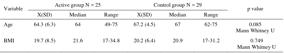
The active group consisted of 25 males (92.6%) and 2 females (7.4%), whereas in the control group were 27 males (96.1%) and 2 females (6.9%). Patients distribution based on gender in both group did not indicate any significant difference (p = 0.941). Distribution of COPD severity in the active group was 11 patients (40.75%) with mild COPD and 16 patients (59.3%) with moderate COPD. In the control group, there were 10 patients (34.5%) with mild COPD and 19 patients (65.5%) with moderate COPD. There was no statistical difference (p = 0.629). Table 3 shows patient’s characteristic based on age and body mass index (BMI). The age mean value in the active group was 64.3 years (SD 6.3) and 67.2 years (SD 4.5) in the control group, and there was no statistical difference (p = 0.085). BMI mean value in the active group was 19.6 (SD 8.5) and in the control group was 20.2 (SD 6.4), and there was no significant difference between two groups (p = 0.749). Table 4 shows the value of blood gas analysis prior to study in active group. The mean value of PO2 was 78.79 (SD 8.8) and PCO2 was 33.28 (SD 5.75).
Table 3. Patients’ characteristics based on age and body mass index (BMI)
Variable Active group N = 25 Control group N = 29 p value
X(SD) Median Range X(SD) Median Range
Age 64.3 (6.3) 64 49-75 67.2 (4.5) 67 62-75 0.085
Mann Whitney U
BMI 19.7 (8.5) 21.6 17-34.8 20.2 (6.4) 20.9 17-31.2 0.749
Table 4. Patients’ characteristics of PO2 and PCO2 values in active group
Active group
Mean SD Range
PO2 initial 78.79 8.80 94.20 – 55.90 PCO2 initial 33.28 5.75 48.10 – 18.60
Initial values of FEV1, 6 meters walking distance,
VO2max and St George’s Respiratory
Questionnaire (SGRQ)
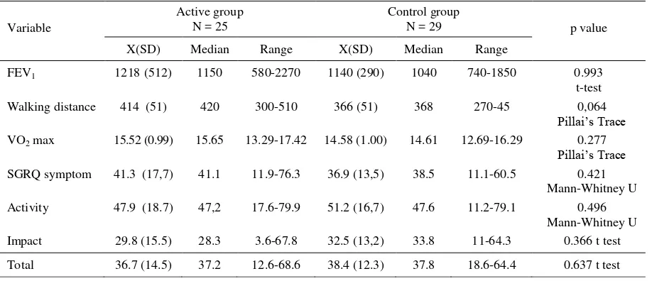
Table 5 shows the initial value of FEV1 in the active group was 1218 ml (SD 512) and in the control group 1140 ml (SD 290). Statistically, there was no significant difference (p = 0.492). The mean value of 6 minutes walking distance in the active group was 414 meters (SD 51), whereas in the control group was 366 meters (SD 51), and statistically it did not indicate any significant difference (p = 0.064). Mean value of VO2 max in the active group was 15.52 (SD 0.99), and in the control group was 14.58 (SD 1.00). The difference was statistically also not significant (p
= 0.277). The values of SGRQ were divided into 4 items namely symptom, activity, impact and total mean value. The mean value of symptom SGRQ of active group was 41.3 (SD 17.7), whereas in control
group was 36.9 (SD 13.5), and statistically it did not indicated significant difference (p = 0.421). Mean value of SGRQ activity in active group was 47.9 (SD 18.7), while in control group was 51.2 (SD 16.7) which statistically did not indicate any significant difference (p = 0.496). The mean value of impact SGRQ in active group was 29.8 (SD 15.5), while in control group was 32.5 (SD 13.2) which statistically did not indicate any significant difference (p = 0.366). Total mean value of SGRQ in active group was 36.7 (SD 14.5) and 38.4 in control group (SD 12.3), there was no significant difference (p = 0.637).
Figure 1. Alteration FEV1 values between two groups
Table 5. Initial values of FEV1, 6 minutes walking distance, VO2 max and SGRQ
Variable
Active group N = 25
Control group
N = 29 p value
X(SD) Median Range X(SD) Median Range
FEV1 1218(512) 1150 580-2270 1140 (290) 1040 740-1850 0.993
t-test
Walking distance 414 (51) 420 300-510 366 (51) 368 270-45 0,064
Pillai’s Trace
VO2 max 15.52 (0.99) 15.65 13.29-17.42 14.58 (1.00) 14.61 12.69-16.29 0.277
Pillai’s Trace
SGRQ symptom 41.3 (17,7) 41.1 11.9-76.3 36.9 (13,5) 38.5 11.1-60.5 0.421
Mann-Whitney U
Activity 47.9 (18.7) 47,2 17.6-79.9 51.2 (16,7) 47.6 11.2-79.1 0.496
Mann-Whitney U
Impact 29.8 (15.5) 28.3 3.6-67.8 32.5 (13,2) 33.8 11-64.3 0.366 t test
Total 36.7 (14.5) 37.2 12.6-68.6 38.4 (12.3) 37.8 18.6-64.4 0.637 t test
1050 1100 1150 1200 1250 1300
Active Control
Initial FEV1
Changing of FEV1, 6 minutes walking distance, VO2 max and St George’s Respiratory
Questionnaire values
On table 6 and Figure 1, the changing value of FEV1 in the active group was 41 ml (SD 163), while in the control group was 13 ml (SD 175) from initial value, which statistically no difference (p = 0.421). Changing value of 6 minutes walking distance in the active group was 50 (SD 15), while in the control group was 7.9 (SD 22) from initial value (see Figure 2). There was significant difference in distance between two groups (p = 0.000). Figure 3 shows changing value of VO2 max in both groups was statistically significant (p = 0.000). Changing SGRQ value of symptom in the
active group was ─21.3 (SD 11.2), while in the
control group was 0.2 (SD 4.2) which statistically indicated significant difference (p = 0.000). Changing SGRQ value of impact in the active group was ─13.9 (SD 9.9), while in the control group was ─3.5 (SD 7.9) which statistically indicated significant difference (p = 0.000). Changing total SGRQ value in the active group was ─15.1 (SD 8.7), while in control group was ─2.2 (SD 3.9) which statistically indicated significant difference (p = 0.000) (see Figure 4).
Figure 2. Alteration of 6 minutes walking distance
Figure 3. Alteration of VO2 max (ml/Kg/minute)
Figure 4. Alteration symptom, activity, impact and SGRQ total values (%)
Table 6. Changing of FEV1, 6 minutes walking distance, VO2 max and SGRQ values
Variable X(SD) Active group Median Range X(SD) Control group Median Range p value
FEV1 41 (163) 30 ─240 - 420 13 (175) 20 ─380 - 350 0.421
Mann-Whitney U
distance 50 (15) 52.5 22.5 - 87.5 7.8 (22) 10 ─30 - 60 0.000
Mann-Whitney U Δ VO2 max 0.95 (0.45) 1.08 1.73 - ─0.59 0.22 (0.50) 0.29 1.59- ─0.59 0.000
T-test
SGRQ symptom ─21.3 (11,2) ─17.4 ─48 - ─8.1 0.2 (4,2) 0 ─11 – 14.4 0.000
Mann-Whitney U
Activity ─14.5 (10) ─12.2 ─32 - 0.0 ─5.3 (6,8) 6.8 ─26.0 - 5.4 0.000
Mann-Whitney U
Impact ─13.9 (10) ─11.4 ─36.4 – 0.9 ─3.5 (7.9) ─3.4 ─30.6 - 8.3 0.000
Mann-Whitney U Total ─15.1 (8.7) ─14.1 ─32.8 - 2.4 ─2.2 (3.9) ─2.3 ─11.6 - 8.1 0.000
Mann-Whitney U
0 100 200 300 400 500
Active Control
Initial distance End distance
0 10 20 30 40 50 60
Active Control
Initial sym ptom End sym ptom
Initial activity End activity
Intial im pact End im pact
Intial total End total
13,5 14 14,5 15 15,5 16 16,5
Active Control
Unexpected condition
There were unexpected conditions during the 6 weeks study in the active and control groups such as exacerbation occurred during exercise and exercise complication. Other unexpected conditions were not found.
Medication administered before and after study
Medications administered before and after study were similar in most of patients. This condition indicated that there was no exacerbation, or patients were at stable condition.
DISCUSSION
Subjects characteristics
There were 60 subjects, consists of 2 groups, namely 30 subjects of mild to moderate COPD who had pulmonary rehabilitation and 30 subjects of mild to moderate COPD without pulmonary rehabilitation. There were 56 subjects who have completed the present study, whereas 3 subjects of the active group and 1 subject of the control group were excluded. There was no significant difference in subject gender and was greater amount of male than female. This fact was in accordance with Yunus study in Persahabatan Hospital, which indicated greater amount of male (86.2%) than female (13.6%).2
Wihastuti et al. found 95% of male and 5% of female in study about correlation of pulmonary physiologic value and quality of life in COPD.9 Subject distribution of age indicated no significant difference between the active group (64.3 years) and the control group (67.2 years). This was in keeping with Yunus study, which found greatest age at 61-80 years old.2 Wihastuti et al. found mean age of COPD patients was 65.4 years old.9 Subject distribution of COPD severity indicated no significant difference between these two groups. Subject distribution of BMI also indicated no significant difference between the active and control group. Harik-Khan et al. found that the lower BMI value, the greater COPD risk.10
PO2 characteristic of the active group was 78.79 mmHg (SD 8.8) and PCO2 was 33.28 (SD 5.75) mmHg. The present study showed no exacerbation condition in the active group. Ortega et al. conducted study on COPD patient and they found strength and endurance training for 12 weeks. The PO2 characteristics
was 71±7 mmHg and PCO2 was 44±7 mm Hg for strength, PO2 65 ± 10 mmHg and PCO2 45±7 mmHg for endurance, PO2 73 ±9 mmHg and PCO2 44±6 mmHg for both. They found 4 subjects being hospitalized because of exacerbation.11 Spruit et al. compared resistance and endurance training with PO2 characteristic was 63±14 mmHg and PCO2 characteristic was 42±9 mmHg for resistance, PO2 70±20 mmHg and PCO2 43±8 mmHg for endurance.12 In this study there were 7 exacerbations of 24 resistance group and 5 exacerbations of 24 endurance group.
Pulmonary physiologic changes
There was no significant difference of pulmonary function before study in the active and control group. In the active and control group after study, there was an increase of FEV1 value, but statistically was not significant. Wijkstra et al. studied about FEV1 changes of patients who had home rehabilitation for 12 weeks. They found no changes of FEV1 value in the active group and significant decrease of FEV1 value about 200 ml (p < 0.05) in the control group. Wijkstra could not explain about FEV1 decrease in the control group even though they recognize that FEV1 value of COPD decreases about 50 ml/year.13 Berry et al studied FEV1 changes in patients who had exercise program for 12 weeks. They found no significant results.14 Giiell et al. studied about benefits of long-term rehabilitation in COPD patients. They found significant result of Forced Vital Capacity (FVC) increase and no significant value of FEV1 changes. Those were caused by breathing technique exercise and physiotheraphy that would produce increase flexibility of chest wall and respiratory muscles strength. Giiell stated that the strength of respiratory muscle only affect the FVC value.15 Wouters explained about 3 types of COPD patients’ muscles, namely myosin heavy chain-1 (MHC-1), MHC-2A and MHC-2B. Muscle type of MHC-2B mostly was found in COPD than other muscles. In their study, there were no significant correlation between MHC-2B and FEV1 value level (p = 0.38) and significant correlation between MHC-2A and FEV1 (p= 0.05).
These explain that COPD patient without
improve ventilation muscles function, so it might reduce pulmonary hyperinflation.17
Alteration of 6 minutes walking distance
Before study the value of six minutes walking distance between the active group and the control group had no significant difference. Alteration of walking distance after study in the active group was 50 meters and in the control group was 7.8 meters. The comparison of walking distance between two groups indicated statistically significant difference (p
= 0.000). Bendstrup et al. studied on patients with exercise, education, occupational therapy and stop-smoking program for 12 weeks. They found increased 6 minutes walking distance which was significantly different between the active and control group. After 6 weeks, they found increase walking distance by 79.8 meters in the active group and 21.6 meters in the control group. This was caused by exercise program which increased anaerobic threshold and maximal oxygen consumption.18
Finenerty et al. studied on patients with 6 weeks exercise program. They found significant increase between the active and control group. In the active group, they found increase walking distance after 12 weeks of 51 meters. In such study, an exercise monitoring 1 hour/week was continued to home exercise program. Finenerty concluded that those exercises would improve dyspnea so it would increase the exercise tolerance and walking distance.19 Wijkstra et al. studied on patients with 12 weeks home exercise program. They found significant difference in 6 minutes distance between the active and control group. Exercise is the best intervention, psychologically and physiologically for dyspnea. Exercise tolerance and adaptation is the best mechanism to reduce dyspnea although generally the lungs and heart functions remain.13
Berry et al. studied on patients with 12 week exercise program. They found increase walking distance, which significantly different on mild COPD group (p < 0.01), moderate COPD group (p < 0.0001) and severe COPD group (p < 0.01).14 Lacasse et al. conducted meta-analysis about COPD rehabilitation. They found mean value of 6 minutes walking distance increase by 55.7 meters. They concluded that minimum clinically important difference (MCID) was 50 meters. Minimum clinically important difference (MCID) is the shortest distance that should be attained in order to have
significant clinical improvement.20 British Thoracic Society (BTS) recommended significant clinical improvement of minimum distance was 54 meters.4 Redelmeier et al. conducted meta-analysis and stated that 6 minutes walking distance clinically significant at 54 meters.21 Weiner et al. studied patients with long-acting bronchodilator and exercise program. They found significant increase of 6 minutes walking distance by 50 meters.22 Ortega et al. compared the benefit of strength, endurance training and both combination. Strength training is an exercise with various techniques to strengthen the respiratory muscles, whereas endurance is persisting exercise on a certain technique. Ortega concluded that the best result was obtained with combination of both techniques. This was caused by dynamic hyper-ventilation occurred in COPD that reduce the tidal volume during exercise, which cause dyspnea and reduce exercise tolerance. Combination of both techniques will improve ventilation, reduce end expiration volume, and dyspnea.11 Marin et al. described that there was strong correlation between reduced dynamic hyper-ventilation and increased 6 minutes walking distance.23
Alteration of VO2 max
Alteration of VO2 max in the active group was 0.95 (SD 0.45) and in the control group was 0.22 (SD 0.50). The comparison of VO2 max value of both groups statistically indicated significant difference (p
= 0.000). Wijkstra et al. studied on COPD patient with home rehabilitation for 12 weeks. They found significant increase of VO2 max in the active group. The increase of VO2 max was also followed by significant reduces of lactate, dyspnea and increase of work performance.13 Spruit et al. found increased VO2 max in the resistance training group. Resistance training will cause increase component of oxygen transportation (increase amount of capillary and oxidative enzyme), increase density volume of mitochondria and increase speed of phosphocreatine recovery, so that finally causes VO2 max increase.12
Alteration of St. George’s Respiratory Questionnaire (SGRQ)
significantly different between two groups (p = 0.000). Finnerty et al. studied on patient with 6 weeks exercise and education program. They found significant difference between the active and control group (p < 0.001). In the active group, there was decreased symptom value by 20.5, activity value by 12.1, impact value by 9.6 and total value by 12.5. In control group, there was decreased symptom value by 4, impact value by 0.7, total value by 0.8 and increased activity value by 0.7. They concluded that there was alteration of SGRQ value, which was significantly different between the active and control group. Those values indicate that the lesser SGRQ value, the better quality of life. Significant minimal alteration was if there was 4% decreased of SGRQ value (minimally clinically significant change).19 British Thoracic Societies recommended utilization of SGRQ because it is more sensitive to evaluate clinical changes.4 Wijkstra et al. studied on patients with 12 weeks home exercise program. They found significant different changes for quality of life in the active and control group (p < 0.001).13 Berry explained that rehabilitation would cause increase maximum oxygen consumption and maximum work capacity, therefore it would increase functional capacity and quality of life.14 Lacasse et al. described that by meta-analysis, pulmonary rehabilitation would improve dyspnea and
increase the patient’s ability. Therefore, functional
capacity and quality of life will increase.20
CONCLUSIONS
1. Pulmonary rehabilitation in COPD patient may improve quality of life, namely by improving symptom, activity, impact and total SGRQ value 2. Pulmonary rehabilitation of COPD patient will
increase the functional capacity, namely by significant increase of 6 minutes walking distance
REFERENCES
1. Soemantri S, Budiarso RL, Suhardi, Sarimawar, Bachroen C. Survei kesehatan rumah tangga (SKRT). Jakarta: Dep Kes RI; 1995. p. 96-125.
2. Yunus F. Gambaran penderita PPOK yang dirawat di bagian Pulmonologi FKUI/SMF Paru RSUP Persahabatan Jakarta. J Respir Indo 2000;20:64-8.
3. Sherrill DL, Lebowitz Md, Burrows B. Epidemiology of chronic obstructive pulmonary disease. Clin. Chest Med 1990; 11:375-87.
4. British Thoracic Society. Pulmonary rehabilitation. Thorax 2001;56:827-34
5. Ries AL. Rehabilitation in chronic obstructive pulmonary disease and other respiratory disorder. In: Fishman AP,
Elias JA, Fishman JA, Grippi MA, Kaiser RM, Senior RM, editors. Pulmonary disease and disorder, 3rd ed. New York: McGrow-Hill; 1998. p. 709-19.
6. ATS. Pulmonary rehabilitation-1999. Am J Respir Crit Care Med 1999;159:1666-78.
7. Jones PW, Quirk FH, Baveystock CM. The St George’s respiratory questionnaire. Respir Med 1991;85:25-31. 8. ATS. Guidelines for the six-minute walk test. Am J Respir
Crit Care Med 2002;166:111-7.
9. Wihastuti R, Wiweka IBS, Yunus F, Manuhutu EJ. Hubungan antara nilai faal paru dengan kualiti hidup penderita penyakit paru obstruksi kronik. J Respir Indo 2001;21:147-51.
10. Harik-Khan RI, Fleg JI, Wisw RA. Body mass index and risk of copd. Chest 2002;121:370-6.
11. Ortega F, Toral J, Cejudo P, Villagomez R, Sanchez H, Castillo J, at al. Comparison of effects of strength and endurance training in patients with chronic obstructive pulmonary disease. Am J Respir Crit Care Med 2002; 166:669-74.
12. Spruit MA, Gosselink R, Troosters T, De Paepe K, Decramer M. Resistance versus endurance in patients with copd and peripheral muscle weakness. Eur Respir J;2002:1072-8.
13. Wijkstra PJ, Van de mark TW, Kraan J, Van altena R, Koeter GH, Postma DS. Effects of home rehabilitation on physical performance in patients with chronic obstruction pulmonary disease (COPD). Eur Respir J;1996:104-10. 14. Berry MJ, Rejeski WJ, Adair NE, Zaccaro D. Exercise
rehabilitation and chronic obstructive pulmonary disease stage. Am J Respir Crit Care Med 1999;160:1248-53. 15. Giiell R, Casan P, Belda J, Sangenis M, Morante F,
Guyatt GH, et al. Long-term effect of outpatient rehabilitation of COPD. Chest 2000;117:976-83.
16. Wouters EFM. Muscle weakness in chronic obstructive pulmonary disease. Eur Respir Rev 2000;1074: 349- 53. 17. Ries AL, Ellis B, Hawkins RW. Upper extremity exercise
training in chronic obstructive pulmonary disease. Chest 1988;93:688-92.
18. Bendstrup KE, Jensen JI, Holm S, Bengtsson B. Outpatient rehabilitation improves activities of daily living, quality of life and exercise tolerance in chronic obstructive pulmonary disease. Eur Respir J 1997;10:2801-6. 19. Finnerty JP, Keeping I, Bullough I, Jones J. The
effectiveness of outpatient pulmonary rehabilitation in chronic lung disease. Chest 2001;110:1705-10.
20. Lacasse Y, Wong E, Guyatt GH, King D, Cook DJ, Goldstein RS. Meta-analysis of respiratory rehabilitation in chronic obstructive pulmonary disease. Lancet 1996; 348:1115-9.
21. Redelmeier DA, Ahmed M, Bayoumi, Roger S, Goldstein. Interpreting small differences in functional status : the six minute walk test in chronic lung disease patients. Am J Respir Crit Care Med 1997;155:1278-82.
22. Weiner P, Azgad Y, Ganam R. Inspiratory muscle training combined with general exercise reconditioning in patients with COPD. Chest 1992;139;4-7.