Eye Closure and Open Detection Using Adaptive
Thresholding Histogram Enhancement (ATHE)
Technique and Connected Components Utilisation
Masrullizam Mat Ibrahim, Azmi Awang Md Isa, and Abd Majid Darsono
Faculty of Electronics and Computer Engineering
Universiti Teknikal Malaysia Melaka
Hang Tuah Jaya, 76100 Durian Tunggal, Melaka, Malaysia.
{ masrullizam, azmiawang, abdmajid}@utem.edu.my
Abstract-Eye closure detection is an important operation
prior to carry out the main algorithm such as iris recognition algorithms, and eye tracking algorithms. This paper introduces a method to detect eye closure using Adaptive Thresholding Histogram Enhancement (ATHE) technique and connected component utilisation. The ATHE technique is a combination of histogram enhancement and estimation threshold technique. Firstly, in this proposed method the eye region is required to be localised. The ATHE technique enhances the eye region image then and yield the threshold value to segment the iris region. Based on the segmentation result, the connected components of binary image are used to classify the state of eye whether open or close. This classification is based on the shape and size of segmented region. The performance of the proposed technique is tested and validated by using UBIRIS, MMU and CASIA iris image database.
Keywords— Eye closure, histogram enhancement, adaptive threshold, segmentation.
I. INTRODUCTION
Eye tracking algoritm, iris recognition biometric system and fatigue detection system are amongst the applications which demand an accurate eye closure detection in preliminary operation. Prior to carry out of primary algorithm, the state of the eye is needed to be known in advance, so that the appropriate action could be taken. For example, in iris recognition algoritm, the algorithms are to be carried out only whenever the eye is absolutely detected open [1]. Similarly for the fatigue detection in which signs of fatigue are measured based on the state of the eye, which is very particular with size of eye open, the main algoritm can only be performed when the open eye is detected [2]. Therefore, an accurate eye's state detection in preliminary operation is crucially required.
This paper introduces a new method which will be shown to be able to classify between eye closure and eye open. In general, eye state detection methods can be classified into three categories; knowledge-based methods which encode human knowledge regarding the eye state [3]; feature based methods which employ features from the eye such as colours, shapes, and edges [4]; and appearance based methods which use the intelligence of machine learning algorithms to evaluate the eye state from specifically
extracted features [3]. There are several machine learning classifiers commonly deployed in these applications such as boosted classifiers, Support Vector Machine (SVM) and Neural Networks.
Knowledge-based methods customarily employ template matching algorithms which use pre-prepared eye model templates. Wu et al.,[5] created the open eye template in which the eye is already located in order to track the eyes and the correlation of the template regions in between two frames. The state of the eye is determined by calculating the correlation with a high correlation score signifying an opened eye and a low score for a closed eye. However, this approach requires two frames to determine the current state of the eye. In papers [6, 7] statistical active appearance shape models are used that require a set of the annotated eye open and closed images. These images are to be applied then in a statistical model training algorithm.
One of the eye specialties, it’s rich with various features such as colour, texture and edges that can be utilised to evaluate the eye state. For example, Liying et al.,[8] use of skin colour to differentiate between eye open and close. While Moris et al.,[9] compute the variance map of the eye to obtain the distinctive features of the eye state. However, these approaches are particularly sensitive to variation of illumination and varieties of skin colour from different races of people. Techniques that are based on appearance and use machine intelligence require a large number of eye images to train the classifiers. For instance, Wang et al.,[10] extract the eye features using the Gabor wavelet technique and classify them using Neural networks, while papers [11, 12] employ the Adaboost classifier to determine a closed eye. Research in [13] determines the eye closure and open based on rectangular eye position pixels; this position is trained and classified using SVM. The performance of this method relies on the features and the number of images for training.
In this paper, a novel technique ATHE is introduced that integrates a histogram enhancement, and estimation threshold in order to segment region of interest. Then, the connected components of segmented region are utilised to determine the state of the eye. This proposed method requires no images to be trained, and it is also developed to adapt to illumination variation. The remainder of this paper
is organised as follows. Section 2 discusses the eye region that to be used in ATHE. Section 3 explains the ATHE algorithm in detail. Section 4 describes the connected components utilization. Chapter 5 discusses the experimental results. The conclusions of the paper are provided in Section 6.
II. EYE REGION
In this proposed method, eye closure is detected by examining the size and shape of the darkest area in the eye region. Therefore, the eye region is an important element that needs to be localised precisely before ATHE technique is implemented. In this paper, to localise the eye region of the face we proposed using a cascade classifier with Haar-like features [14, 15]. The cascade classifier trained pair of eyes features and the detection result as shown in Fig.1(a) which pairs of eyes are localised together.
(a) (b)
Fig.1. (a) Pair of eyes is detected by the cascade classifier (b) The region of the eye is localised by splitting the detected eyes pair region into two.
By applying a pair of eye detection, the characteristics of the eye are clearly visible, compared to single eye detection, in which the eye is possibly detected even the face indirectly facing forward. This may cause characteristics of the eye are presented inappropriately. In the proposed technique, only single eye region to be processed. Therefore, in order to obtain a single eye region, the detected pair of eye region is split into two regions as shown in Fig.1(b). This single region is used in the next ATHE operation.
III. ADAPTIVE THRESHOLDING HISTOGRAM ENHANCEMENT (ATHE)
Adaptive Thresholding Histogram Enhancement (ATHE) is a technique which aims to produce threshold value to segment the iris in the eye region. This technique is a combination of histogram enhancement and estimation threshold technique. In this technique the histogram of a detected eye region is enhanced firstly. Then, by applying estimation of the iris area in the eye region, an appropriate threshold to segment iris region is obtained.
A. Histogram Enhancement
The first process of ATHE when eye region is obtained, the histogram of eye region
g
′
(
x
,
y
)
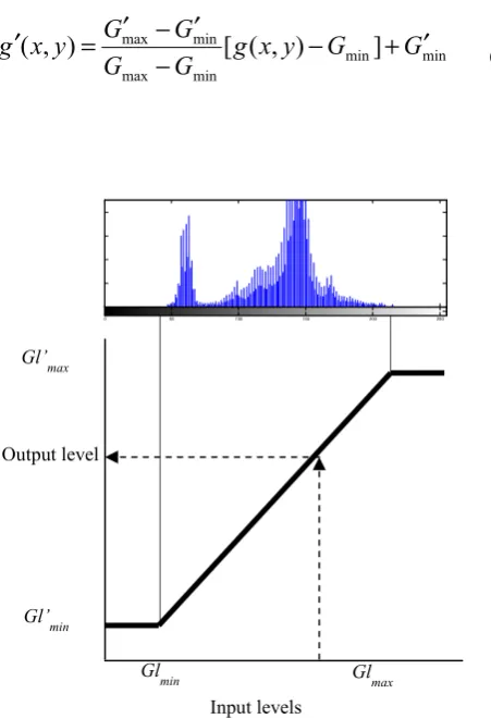
is enhanced by transforming the histogram of the image to spread the level of colour values evenly but without changing the shape of the input histogram. This is computed as follows [16]:min min
min max
min
max
[
(
,
)
]
)
,
(
g
x
y
G
G
G
G
G
G
y
x
g
−
+
′
−
′
−
′
=
′
(1)Fig. 2. Transformation of enhancement histogram
This linear transform illustrated in Fig. 2 stretches the histogram of the input image into the desired output intensity level G’ of the histogram between Gmin′ and Gmax′ , whereGminand Gmaxare the minimum and maximum input image intensities G respectively. From this enhanced histogram (Fig. 3(a)), the cumulative histogramHi which generates total numbers of index pixels in histogram bins between zeros until 255 (Fig. 3(b)) is calculated as follows:
∑
== i
j j
i h
H
1
(2)
Gl
min Glmax
Gl’
min
Gl’
max
Input levels
0 50 100 150 200 250
0 50 100 150 200 250 0
10 20 30 40
0 50 100 150 200 250
0 1000 2000 3000
(a) (b)
Fig. 3. (a) Enhanced image histogram; (b) Cumulative histogram.
B. Adaptive Threshold
In this proposed technique, eye is classified as open or closure based upon the size of the iris. Visually, the eye is classified open when more than half of the total iris circle area is visible. Therefore, ATHE technique introduces an adaptive threshold to segment the iris region, which is a darker object located in the middle of the eye region image. Based on experimental observation over 450 eye region images that obtained from MMU, UBIRIS, CASIA, and face detect database as shown in Fig. 4, iris area of eye opening is identified constitutes approximately within 8% to 16% of the entire extracted eye region.
.
Fig. 4. Example of the extracted eye region
From this estimation iris area, the iris in the histogram of the extracted eye region are situated between 0 and ‘X’ intensity levels of image as stated in (3). The intensity value X can thus be obtained by determining the area of the first (within 8% to 12%) of the total number of pixels, contained in the enhanced histogram image (Fig. 3 (a)). After counting the first ( 8% - 16%) of intensity value, the X value is corresponding intensity value off the x-axis ( as shown in Fig. 5)
255
0< X < (3)
Then, the adaptive threshold Ta value is calculated as
follows:
min max
min
P P
P X Ta
− −
= (4)
where the Pmaxand Pmin are the maximum and minimum
pixel value respectively.
Fig. 5. X value in x-axis
IV. CONNECTED COMPONENT OF BINARY IMAGE
The ATHE technique converts the input image to a binary image with the interest iris region is segmented. The segmented region needs to be examined in order to determine eye state status whether open or close. Therefore, connected components of segmented region is applied in which the size and shape of segmented region are measured. Fig. 6 shows the segmentation result of ATHE technique with the left indicate the segmented iris region in bounded by rectangular which is formed from connected component.
Fig. 6. Segmentation result of ATHE technique.
Instead of iris denote the darkest region in the eye region, eyebrow also can be segmented as region of interest. This eyebrow region interferes the process to determine eye state. Hence, the region in the boundary of the eye region image which constitute 20% of total length and high of image (as shown in Fig. 7) are eliminated first.
Fig. 7. Elimination region for initial process
In order to determine the state of the eye, the region of interest iris is examined in terms of the aspect ratio of the bounding box and the area of the region of interest within it. Firstly, the aspect ratio of the bounding box shape S of height (H) and width (W ) are calculated (5) (as shown in Fig. 8). Based on carried out experimental involved 420 eye closed images taken from ZJU [11] , MMU [17], CASIA [18], and Strathclyde Fatigue Facial (SFF) database, the best ratio of the bounding box that represent eye closed is 0.4 and
X
less. Otherwise the region of interest in the bounding box needs to be examined further.
Fig. 8. Width and height of the region of interest
⎪⎩ ⎪ ⎨
⎧ <
=
otherwise W
H S
1
4 . 0 0
(5)
V. EXPERIMENTAL RESULTS
The main aim of the proposed method is to determine the status of eye whether is close or open. The eye is still categorised as closed even though the eye is half opened as shown in Fig. 9. This figure shows the result of region of interest segmentation in three states of the eye; eye opening, eye half opening, and eye closed. The bounding box that formed around the region interest clearly indicates the shape that represents eye open.
Fig. 9. The result of iris segmentation from three states of the eye; eye opening (on top) , half eye opening (in the middle), and eye fully closed.
In order to evaluate the performance of the proposed method, extensive experiments have been performed. These experiments focused on evaluating the performance of ATHE for segment the iris region, and the overall performance of eye closure and open detection. For these experiments, there were four set data had been used to evaluate the performance; UBIRIS iris database, MMU iris database, and CASIA iris database. Table 1 shows the experimental results of iris segmentation over five percentage (8%- 16%) region of interest. This experiment aims to find the best percentage that can be used in ATHE technique to produce the threshold value. Based this experiment over 70 eye images randomly taken from
above-mentioned databases, 10% is the best percentage to be used in ATHE technique for segmenting the region of the iris.
Table 1. Experimental results of iris segmentation
Iris area Eye region area
8% 10% 12% 14% 16%
Segmentation rate 95% 96% 95% 94% 92%
In order to evaluate the overall performance, the proposed method is compared to other methods as indicated in Table 2. For the appearance based, the method that employed cascade classifier as implemented [12] is used. While the technique that introduced by Liying is used for present the features based method [8]. There are 100 eye images are employed in this experiment, and the proposed method show the best detection rate compared with others two methods.
Table 2. Comparison results of overall performance
Algorithm Proposed
Method
Appearace Based
Features Based
Detection rate 98% 97% 93%
VI. CONCLUSION
A new method to detect eye closure and open was presented. This method is a combination of Adaptive Thresholding Histogram Enhancement (ATHE) technique and connected components. ATHE is a new technique to segment the region of interest based on histogram enhancement and estimation threshold. Based on the conducted experiment, the 10% is the best percentage of the region interest estimation to segment the iris region. From segmention result, the connected component of segmented region are used to determine the status of eye. The bounding box is formed covering the iris region. Then, the aspect ratio of a bounding box is computed in order to determine the state of the eye. For overall performance of the proposed method, eye images are randomly taken from UBIRIS, MMU and CASIA database are employed for testing and evaluation. The comparison has been made with the other two methods, and the proposed method show the better result.
REFERENCES
[2] M. M. Ibrahim, J. J. Soraghan, and L. Petropoulakis, "Non-rigid eye movement tracking and eye state quantification," in Systems, Signals and Image Processing (IWSSIP), 2012 19th International Conference on, 2012, pp. 280-283.
[3] Y. Ming-Hsuan, D. J. Kriegman, and N. Ahuja, "Detecting faces in images: a survey," Pattern oAnalysis and Machine Intelligence, IEEE Transactions on, vol. 24, pp. 34-58, 2002.
[4] E. Hjelmås and B. K. Low, "Face Detection: A Survey," Computer Vision and Image Understanding, vol. 83, pp. 236-274, 2001.
[5] Q. Wu, B. Sun, B. Xie, and J. Zhao, "A PERCLOS-Based Driver Fatigue Recognition Application for Smart Vehicle Space," in Information Processing (ISIP), 2010 Third International Symposium on, 2010, pp. 437-441. [6] F. Sukno, S.-K. Pavani, C. Butakoff, and A. Frangi,
"Automatic Assessment of Eye Blinking Patterns through Statistical Shape Models
Computer Vision Systems." vol. 5815, M. Fritz, B. Schiele, and J. Piater, Eds., ed: Springer Berlin / Heidelberg, 2009, pp. 33-42.
[7] I. Bacivarov, M. Ionita, and P. Corcoran, "Statistical models of appearance for eye tracking and eye-blink detection and measurement," Consumer Electronics, IEEE Transactions on, vol. 54, pp. 1312-1320, 2008. [8] L. Liying and Q. Haoxiang, "The Study of Driver Fatigue
Monitor Algorithm Based on Skin Color Segmentation," in Intelligent Information Technology Application Workshops, 2008. IITAW '08. International Symposium on, 2008, pp. 463-466.
[9] T. Morris, P. Blenkhorn, and F. Zaidi, "Blink detection for real-time eye tracking," J. Netw. Comput. Appl., vol. 25, pp. 129-143, 2002.
[10] R.-b. Wang, K.-y. Guo, S.-m. Shi, and J.-w. Chu, "A monitoring method of driver fatigue behavior based on machine vision," in Intelligent Vehicles Symposium, 2003. Proceedings. IEEE, 2003, pp. 110-113.
[11] P. Gang, S. Lin, W. Zhaohui, and L. Shihong, "Eyeblink-based Anti-Spoofing in Face Recognition from a Generic Webcamera," in Computer Vision, 2007. ICCV 2007. IEEE 11th International Conference on, 2007, pp. 1-8. [12] I. Fasel, B. Fortenberry, and J. Movellan, "A generative
framework for real time object detection and classification," Comput. Vis. Image Underst., vol. 98, pp. 182-210, 2005.
[13] R. Senaratne, D. Hardy, B. Vanderaa, and S. Halgamuge, "Driver Fatigue Detection by Fusing Multiple Cues," presented at the Proceedings of the 4th international symposium on Neural Networks: Part II--Advances in Neural Networks, Nanjing, China, 2007.
[14] P. Viola and M. Jones, "Rapid object detection using a boosted cascade of simple features," in Computer Vision and Pattern Recognition, 2001. CVPR 2001. Proceedings of the 2001 IEEE Computer Society Conference on, 2001, pp. I-511-I-518 vol. 1.
[15] M. M. Ibrahim, J. S. Soroghan, and L. Petropoulakis, "Mouth covered detection for yawn," in Signal and Image Processing Applications (ICSIPA), 2013 IEEE International Conference on, 2013, pp. 89-94.
[16] E. R. Davies, Machine vision: theory, algorithms, practicalities: Morgan Kaufmann, 2004.
[17] M. U. (MMU). (November 2011). iris image database (2005). Available: http://pesona.mmu.edu.my/ccteo
[18] C. A. o. S. (CASIA). Biometrics Idea Test. Available: