Steroid response as prognostic factor and its correlation with
molecular assessment of childhood acute lymphoblastic leukemia
Keywords: acute lymphoblastic leukemia, molecular assessment, prognostic factor, steroid
pISSN: 0853-1773 • eISSN: 2252-8083 • http://dx.doi.org/10.13181/mji.v24i4.1177 • Med J Indones. 2015;24:215–20 • Received 30 Dec 2014 • Accepted 24 Dec 2015
Correspondence author: Murti Andriastuti, [email protected]
Copyright @ 2015 Authors. This is an open access article distributed under the terms of the Creative Commons Attribution-NonCommercial 4.0 International License (http://creativecommons.org/licenses/by-nc/4.0/), which permits unrestricted non-commercial use, distribution, and reproduction in any medium, provided the original author and source are properly cited.
Murti Andriastuti,1
Djajadiman Gatot,1
Riadi Wirawan,2 Rianto Setiabudy,3
Muchtaruddin Mansyur,4
I Dewa G. Ugrasena5
1 Department of Child Health, Faculty of Medicine, Universitas Indonesia, Cipto Mangunkusumo Hospital, Jakarta, Indonesia
2 Department of Clinical Pathology, Faculty of Medicine, Universitas Indonesia, Cipto Mangunkusumo Hospital, Jakarta, Indonesia
3 Department of Pharmacology, Faculty of Medicine, Universitas Indonesia, Jakarta, Indonesia
4 Department of Community Medicine, Universitas Indonesia, Jakarta, Indonesia
5 Department of Child Health, Faculty of Medicine, University of Airlangga, Dr. Sutomo Hospital, Surabaya, Indonesia
C l i n i c a l Re s e a rc h
ABSTRAK
Latar belakang: Angka kesintasan leukemia limfoblastik akut
(LLA) anak di Indonesia masih rendah. Ketepatan stratifikasi
risiko merupakan hal penting dalam meningkatkan kesintasan.
Di negara maju, stratifikasi risiko dibuat berdasarkan
pemeriksaan fusi gen yang terkait dengan resistensi steroid. Respons steroid hari ke-8 berhubungan dengan prognosis. Pemeriksaan ini dapat diaplikasikan di pusat rujukan yang belum dapat melakukan pemeriksaan molekular secara rutin. Penelitian ini bertujuan untuk menilai apakah respons steroid berhubungan dengan pemeriksaan molekular.
Metode: Studi potong-lintang dilakukan di Departemen Ilmu Kesehatan Anak, FKUI-RSCM (Januari 2013–Maret 2014 dengan 73 subjek penelitian). Steroid diberikan selama 7 hari. Sel blas darah tepi diperiksa pada hari ke-8, respons
dikatakan baik bila blas <1000 /µL dan buruk jika ≥1000 /µL.
Pemeriksaan fusi gen dilakukan sebagai standar baku. Data dianalisis menggunakan SPSS versi 20.0.
Hasil: Fusi gen ditemukan pada 45 subjek. Sebanyak 26/32 (81%) subjek berusia 1–10 tahun menunjukkan respons baik, sementara 75% subjek <1 tahun dan 7/9 (78%) subjek ≥10 tahun menunjukkan respons buruk. Sebanyak 5/7 (71%) subjek dengan leukosit >100.000 /µL dan 7/8 (88%) dengan sel-T memiliki respons buruk. Usia, jumlah leukosit, dan sel-T berhubungan dengan respons steroid (p<0,05). Fusi gen E2A-PBX1 adalah yang tersering 19/45 (42%), diikuti TEL-AML1 17/45 (38%), BCR-ABL, 5/45 (17%), dan MLL-AF4 1/45 (3%). Sebanyak 4 dari 5 (80%) subjek dengan BCR-ABL dan 1 subyek dengan MLL-AF4 menunjukkan respons buruk. Sebaliknya, 12/19 (63%) subjek dengan E2A-PBX1 dan 13/17 (77%) dengan TEL-AML1 memiliki respons baik. Tidak terdapat hubungan antara respons steroid dengan pemeriksaan molekular.
Kesimpulan: Respons steroid berhubungan bermakna dengan usia, jumlah leukosit, dan jenis sel-T namun tidak dengan pemeriksaan molekular.
ABSTRACT
Background: Survival rate of children with acute lymphoblastic leukemia (ALL) in Indonesia remains low. Risk
stratification accuracy is important to improve survival. In developed countries, risk stratification is determined based
on gene fusion that is known related to steroid resistency. Steroid response at day-8 correlates with prognosis. The assessment can be applied in centers that cannot perform molecular assessment. This study aims to evaluate whether steroid response correlated to molecular assessment. Methods: A cross-sectional study was performed at Child Health Department, Cipto Mangunkusumo Hospital (January 2013-March 2014), a total of 73 patients were enrolled. Steroid was given for 7 days. Peripheral blast count at day 8 was evaluated, good response if blast count <1000 /µL and
poor if ≥1000 /µL. Fusion gene detection was also performed.
The data was analysed using Statistical Package for Social Sciences (SPSS) version 20.0.
Results: Fusion gene was detected in 45 patients. In 1–10 years age group, 26/32 (81%) subjects had good response,
while 75% in <1 year age group and 7/9 (78%) in ≥10
years age group had poor response. 5/7 (71%) subjetcs had leukocyte count >100,000 /µL and 7/8 (88%) with T-cell showed poor response. Age, leukocyte count, and T-cell were statistically correlated with steroid response (p<0.05). E2A-PBX1 fusion gene was the most common 19/45 (42%), followed by TEL-AML1 17/45 (38%), BCR-ABL 5/45 (17%),
and MLL-AF4 1/45 (3%). Four of five subjects (80%) with BCR-ABL and one subject with MLL-AF4 had poor steroid
response. On the other hand, 12/19 (63%) with E2A-PBX1 and 13/17 (77%) with TEL-AML1 had good response. There was no correlation between steroid response and molecular assessment.
Acute lymphoblastic leukemia (ALL) is the most common type of childhood leukemia which accounts for 75–80% of all cases.1 From 2007 to 2012, there were 1,957 cases from 15 referral hospitals in Indonesia.2 In Cipto Mangunkusumo Hospital (CMH), Jakarta, there were 579 cases during 2007–2013 with approximately 80 new cases each year.3,4
The event free survival (EFS) rates in developed countries is 80–90% but it is very different in developing countries.5 Gatot and Windiastuti3 reported EFS rates of 53% from ALL patients in CMH from 1998–2004, 53% while Mulatsih et al6 reported that in Dr. Sardjito Hospital, Yogyakarta, Indonesia, the EFS rates were 53%. A high relapse rate in ALL is also another major problem. Windiastuti7 found that relapse rates in CMH was 29%, almost three times higher compared to developed countries. These differences can be attributed to numerous factors, such as early detection, referral system, diagnostic tools, treatment choice, remission monitoring, nutritional status, evaluation of side effect from chemotherapy, and supportive facilities which in turn influences the treatment outcome.5
Risk stratification in developing countries is based on the National Cancer Institute (NCI) criteria; age 1–0 years old and leukocyte count <50,000 /µL.8 Pediatric Hematology Oncology Division, Child Health Department, Faculty of Medicine Universitas Indonesia (FMUI-CMH) is using the following criteria; age, leukocyte count, mediatinal mass, and central nervous system (CNS) infiltration. This criteria stratifies ALL patients into high risk or standard risk.7 Meanwhile, developed countries are already using molecular risk stratification for ALL patients.5
Molecular assessment shows that certain fusion gene plays a role in leukemic cell proliferation and differentiation, affect the clinical characteristics and now becomes one of important prognostic factors. Some important fusion genes of leukemia are TEL-AML1, BCR-ABL, MLL-AF4, E2A-PBX1 in B-cell ALL.9 Patient with TEL-AML1 has an excellent prognosis with EFS rates of approximately 90%, regardless age and leukocyte count.10 Infant <1 year old was originally categorized as high risk based on NCI criteria, but studies have found that if there is no MLL-AF4 gene changes, the prognosis is similar
to those aged >1 year.11 Fusion gene BCR-ABL and MLL-AF4 are related to cytostatic drug resistence including steroid.12 E2A-PBX1 needs intensive chemotherapy to reach good EFS. This indicates the importance of genetic factors in determining prognosis. Based on fusion gene, molecular stratification can be divided into three categories; high risk (BCR-ABL and MLL-AF4), intermediate risk (E2A-PBX1) and standard risk (TEL-AML1).
Reduction or clearance of peripheral blast after one week steroid administration is known as a significant prognostic factor to predict treatment outcome.13 Good response is defined as a blast count of <1,000 /µL and has 2–3 times better prognosis compared to poor response, defined as a blast count of ≥1,000 /µL.14 This assessment is easy, inexpensive, and can be used in centres in Indonesia. Moreover, it is significant in predicting prognosis and has yet to be routinely performed in Indonesia. The aim of this study was to evaluate if steroid response at day-eight has a significant correlation with molecular assessment as the gold standard.
METHODS
A comparative cross sectional study was performed at CMH from January 2013 to March 2014. A total of 73 patients were enrolled. Inclusion criteria were newly diagnosed ALL children (age 0-18 year old). Patients were excluded from this study if they
had ALL L3 subtype and Down syndrome. Bone
marrow aspiration was performed for molecular assessment. Patients were given steroid and the response was evaluated at day eight. The protocol for the present study was approved by the Ethics Committee of Faculty of Medicine Universitas Indonesia/Cipto Mangunkusumo Hospital (No. 594/H2.F1/ETIK/2013).
Clinical laboratory parameters
Hematology parameters, peripheral blast count, and immunophenotyping were examined in Clinical Pathology Department CMH, and Dharmais Cancer Hospital.
Molecular assessment
fusion genes examined were TEL-AML, BCR-ABL, E2A-PBX1, and MLL-AF4. Cell line for each
Characteristics Poor response (n=16) Good response (n=29) Total (n=45) p Demographic
Age (mean 3.5 years) <0.001#
<1 year 3 1 4
1–< 10 years 6 26 32
≥10 years 7 2 9
Gender 0.84*
Male 10 19 29
Female 6 10 16
Clinical
Pallor 14 24 38 1.00#
Fever 12 19 31 0.74#
Bone pain 13 17 30 0.12*
Bleeding 7 14 21 0.77*
Limphadenopathy 7 14 21 0.77*
Hepatomegaly 10 21 31 0.52#
Splenomegaly 7 14 21 0.77*
Mediastinal mass 3 1 4 0.12#
Laboratory
Hemoglobin (mean 8.4 g/dL) 0.58#
<7 8 12 20
7–11 5 12 17
>11 3 5 8
Leukocyte (mean 72,036 /µL) <0.001#
<50,000 9 27 36
50,000–100,000 2 0 2
>100,000 5 2
7
Platelet (mean 40,217 /µL) 0.79*
<20,000 6 12 18
20,000–100,000 9 14 23
>100,000 1 3 4
Morphology 0.33#
L1 13 27 40
L2 3 2 5
Immunophenotyping 0.03#
T-cell 7 1 8
B-cell 9 25 34
Mixed 0 3 3
Molecular 0.19#
TEL-AML1 4 13 17
E2A-PBX1 7 12 19
BCR-ABL 4 1 5
MLL-AF4 1 0 1
*Chi-square test #Fisher exact test
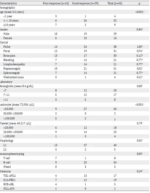
Table 1. Clinical and laboratories characteristics in steroid poor and good response patients
Statistical analysis
The data was analysed using statistical package for social sciences (SPSS) version 20.0. Chi-square or fisher exact test were done as appropriate to compare between good and poor response.
RESULTS
Due to sampling quantity, molecular assessment was only performed in 52 of 73 patients. From them, the fusion gene was only detected in 45 patients. Table one shows the initial clinical and laboratory characteristics of both groups of patients. The mean age was 3.5 years with the number of male patients 29/45 (66%) being almost two times that of female patients. This research found 26/32 (81%) of patients aged one to <10 years old were good responders, while 3/4 (75%) of patient <1 year old and 7/9 (78%) patient aged ≥10 years old were poor responders. Age had significant influence on steroid response (fisher exact test, p=<0.001). Pallor, fever, bone pain, bleeding, limphadenopathy, hepatomegaly, and splenomegaly as common presenting features were more prominent in poor response group. Mediastinal mass was only found in four patients, and 3/4 (75%) of them in poor response group. None of patients presented with testis or CNS infiltration.
The mean value for hemoglobin was 8.4 g/dL, platelet was 40,217 /µL, and leukocyte was 72,036 /µL. Most patients 36/45 (80%) with leukocyte count <50,000 /µL. All patients (100%) with leukocyte count between 50,000–100,000/µL were good responses while 5/7 (71%) patients with leukocyte count >100,000/µL were poor responses, which showed leukocyte count as significant factor to steroid response (fisher exact test, p<0.05).
Based on morphology, 40/45 (89%) subjects
had L1 type and 5/45 (11%) had L2 type.
Immunophenotyping showed 34 subjects (76%) with B-cell and only three subjects (7%) with mixed cell. There were eight subjects (27%) with T-cell, and seven of them (88%) showed poor steroid response while 74% of B-cell and 100% of mixed-cell showed good steroid response. Statistical analysis showed that ALL cell had significant correlation with steroid response (fisher exact test, p<0.05).
Molecular assessment showed that the most common fusion gene in this study was E2A-PBX1 19/45 (42%), followed by TEL-AML1 17/45 (38%), BCR-ABL 5/45 (17%), and MLL-AF4 1/45 (3%). From all response, 13/17 (77%) patients with TEL-AML1 and 12/19 (63%) patients with E2A-PBX1 showed good steroid response while 4/5 (80%) patients with BCR-ABL and all patients with MLL-AF4 fusion gene showed poor steroid response.
In this study, fusion gene TEL-AML1 was stratified into standard risk group while BCR-ABL, MLL-AF4, and E2A-PBX1 were stratified into high risk group. Table 2 showed that 25% patients from standard risk group of molecular had poor steroid response. Otherwise, there were 55% patients from high risk group with good steroid response. Statistical analysis showed that steroid response has no correlation with molecular assessment (p>0.05).
DISCUSSION
The mean age of patients in this study was 3.5 years, similar to what has been reported in literature that the peak incidence of childhood ALL is between the ages of two to five years old.15 The age distribution was similar with previous study in Oman that found 13% childhood ALL patient were aged <1 year, 77% were aged between one to 10 years, and 11% were aged >10 years.16
In this study, 81% of patient aged one to <10 years old had good response, while 75% of patient <1 year old and 78% patient aged ≥10 years old had poor response. Age is known as an important prognostic factor, with many studies showing that patient aged <1 and >9 years old had poor prognosis. This is related to certain
Steroid response
Molecular
p RR
(CI 95%) High risk
(n=28) n (%)
Standard risk (n=17)
n (%)
genetic abnormalitites. MLL fusion gene is more common in patient aged <1 year old and BCR-ABL in patient aged >9 years old. TEL-AML1 is related with good prognosis and commonly found in ALL children aged between one to nine years.17,18
This study found that ALL incidence in male were almost two times higher than female (64% vs 36%), similar with many previous studies in Myanmar,17 Oman,16 Argentina,13 and Pakistan.18 Pallor, fever, bone pain, bleeding, lymphadenopathy, hepatomegaly, and splenomegaly as main clinical symptoms in ALL were more prominent in poor responders. Testicular and CNS infiltration as early clinical sign were very rare. Yasmeen and Ashraf18 found only 2% of ALL patients presented with testicular infiltration and 5% with CNS infiltration. Felice, et al found 1–3% patient with CNS infiltration and no testicular infiltration.13 In this study, there were no patients that presented with testicular or CNS infiltration.
There were four patients (13%) with mediastinal mass, Pui19 found 10%–18% of patient presented with mediastinal mass. Three of four patients (75%) were poor responders. Many studies found that mediastinal mass is a poor prognosis factor that related to thymus hyperplasia and resulter in T-cell. Clinically, T-cell was correlated with high leucocyte count and high incidence of relaps and CNS infiltration.20
This study found that most patient (75%) with leukocyte count <50,000 /µL and all patient (100%) with leukocyte count between 50,000-100,000 / µL were good responders, while 71.4% patient with leukocyte count >100,000 /µL were poor responders. High leukocyte count is related with poor prognosis and need more intensive therapy.21
Morphology examination from bone marrow aspiration showed that L1 type (88.9%) was more
common than L2 type (11.1%). These results
were similar with Mulatsih, et al6 who found 70% L1 and 30% L2 and also Onciu and Pui1 who found 82% L1 and 15% L2. Immunophenotyping assessment showed B-cell as most common ALL type (76%), followed by T-cell 27% and mixed cell 7%. This result was quite similiar with Supriyadi et al22 who found 63% patient with B-cell, 67% with T-cell and only 0.2% with mixed cell. From all eight patients with T-cell,
seven of them (88%) showed poor steroid response. Felice et al13 found 80% patient with T-cell were poor responders. Clinically, T-cell is correlated with age >9 years, leukocyte count >50,000 /µL, mediastinal mass, CNS infiltration, and more common in male, which all lead to poor prognosis.
Fusion gene detection showed that E2A-PBX1 was the highest percentage (42%), followed by TEL-AML1 (38%), BCR-ABL (17%), and MLL-AF4 (3%). This result is different with Mulatsih et al6 who found TEL-AML1 as the most common (23%), followed by BCR-ABL (11%), E2A-PBX1 (9%), no patient with MLL-AF4, and 57% patients without fusion gene. Pui19 also found that TEL-AML1 was the most common fusion gene both in white and black children (19% and 13%), followed by E2A-PBX1 (11%) in black children.
Many studies found that BCR-ABL and MLL-AF4 fusion gene are related to high leukocyte count and steroid resistence.12 In this study, 80% patient with BCR-ABL and 100% patient with MLL-AF4 fusion gene had poor steroid response. Meanwhile, 63% patient with E2A-PBX1 fusion gene showed good steroid response. Although E2A-PBX1 fusion gene was initially reported as poor prognostic factor, the cure rates is increasing with the use of more intensive chemotherapy such as high dose methotrexate.12 It is already known that TEL-AML1 fusion gene has good prognosis with high EFS rate of approximately 90%.12,23 Borkhardt et al24 found that this fusion gene were more common in female, aged one to five years, and without hyperleukocytosis. In this study, 77% patients with TEL-AML1 had good steroid response. Similar findings were found in a study by Uckun et al25 where 84.0% patients with TEL-AML1 showed good response, directly related to its sensitivity to steroid.25
In conclusion, age, leukocyte count, and immunophenotyping were all correlated with steroid response. Although some of fusion gene did show an implication in patient’s steroid response, but there is no significant correlation with molecular assessment statistically.
Conflicts of interest
REFERENCES
1. Onciu M, Pui CH. Diagnosis and classification. In: Pui CH, editor. Childhood leukemias. 2nd ed. New York: Cambridge University Press; 2006. p. 21–47.
2. Windiastuti E, Nency YM, Hagung P, Supriyadi E. Mulatsih S, Sjakti HA, et al. Five years evaluation of the Indonesian acute lymphoblastic leukemia: 2006 protocol of the Pediatric Hematology Oncology Working Group. In: Yeoh A, Blair S, Yen SS, editors. Malignancies
in children. 7th St. Jude-Viva Forum; 2013. p. 186. 3. Gatot D, Windiastuti E. Treatment of childhood acute
lymphoblastic leukemia in Jakarta: results of modified
Indonesian National Protocol 94. Paediatr Indones. 2006;46:179–88.
4. Registered data of cancer patient Hematology Oncology Division Child Health Department Faculty of Medicine Universitas Indonesia/Cipto Mangunkusumo Hospital. Jakarta: 2014. Indonesian.
5. Hunger SP, Sung L, Howard SC. Treatment strategies and regimens of graduated intensity for childhood acute lymphoblastic leukemia in low-income countries: a proposal. Pediatr Blood Cancer. 2009;52:559–65. 6. Mulatsih S, Sumadiono, Sutaryo, Purwanto. The result
of treating children’s acute lymphoblastic leukemia
(LLA) in Dr. Sardjito Hospital with WK-LLA Protocol
1999–2002. Buletin Ilmu Kesehatan Anak FK UNAIR. 2005;17:808–19. Indonesian.
7. Windiastuti E. Childhood acute leukemia: Cipto Mangunkusumo Hospital experiences. Presented in Perhimpunan Hematologi dan Transfusi Darah Indonesia. Medan: 2011.
8. Donadieu J, Auclerc MF, Baruchel A, Leblanc T,
Landman-Parker J, Perel Y, et al. Critical study of prognostic factors in childhood acute lymphoblastic leukaemia: differences in outcome are poorly explained by the
most significant prognostic variables. Fralle group. Frech acute lymphoblactic leukaemia study group. Br J
Haematol. 1998;102(3):729–39.
9. Okuda T, Fisher R, Downing JR. Molecular diagnostics in pediatric acute lymphoblastic leukemia. Mol Diagn. 1996;1(12):139–51.
10. Rubnitz JE, Downing JR, Pui CH, Shurtleff SA, Raimondi SC, Evans WE, et al. TEL gene rearrangement in acute lymphoblastic leukemia: a new genetic marker with prognostic significance. J Clin Oncol. 1997;15(3):1150–7.
11. Raimondi SC, Behm FG, Roberson PK, Williams DL, Pui CH, Crist WM, et al. Cytogenetics of pre-B-cell acute lymphoblastic leukemia with emphasis on prognostic implications of the t(1;19). J Clin Oncol. 1990;8(8):1380–8.
12. Gutierrez A, Armstrong SA, Look AT. Pathobiology of acute lymphoblastic leukemia. In: Hoffman R, Benz EJ, Silberstein LE, Heslop H, Weitz J, Anastasi, editors. Hematology: basic principles and practice. 6th ed. Elsevier; 2013. p. 935–50.
13. Felice MS, Zubizarreta PA, Alfaro EM,
Sackmann-Muriel F. Childhood acute lymphoblastic leukemia:
prognostic value of initial peripheral blast count in good responders to prednisone. J Pediatr Hematol Oncol. 2001;23(7):411–5.
14. Manabe A, Ohara A, Hasegawa D, Koh K, Saito T, Kiyokawa
N, et al. Significance of the complete clearance of peripheral blasts after 7 days of prednisolone treatment in children with acute lymphoblastic leukemia: the Tokyo Children’s Cancer Study Grup Study L99-15. Haematologica. 2008;93(8):1155–60.
15. Spector LG, Ross JA, Robison LL, Bhatia S. Epidemiology and etiology. In: Pui CH, editor. Childhood leukemias. 2nd ed. New York: Cambridge University Press; 2006. p. 48–65.
16. Udayakumar AM, Bashir WA, Pathare AV, Wali YA,
Zacharia M, Khan AA, et al. Cytogenetic profile of
childhood acute lymphoblastic leukemia in Oman. Arch Med Res. 2007;38(3):305–12.
17. Ei E, Ko Y, Thet K, Pe K, Myint T, Sein WK, et al. Clinical
profile of childhood leukemia in Mandalay. St Jude Viva Forum. 2011.
18. Yasmeen N, Ashraf S. Childhood acute lymphoblastic leukaemia; epidemiology and clinicopathological features. J Pak Med Assoc. 2009;59(3):150–3.
19. Pui CH, Howard S. Current management and challenges of malignant disease in the CNS in pediatric leukaemia. The Lancet Oncol. 2008;9:257–68.
20. Smith M, Arthur D, Camitta B, Carroll AJ, Crist W,
Gaynon P et al. Uniform approach to risk classification
and treatment assignment for children with acute lymphoblastic leukemia. J Clin Oncol. 1996;14:18–24. 21. Smith M, Arthur D, Camitta B, Carroll AJ, Crist
W, Gaynon P, et al. Uniform approach to risk classification and treatment assignment for children with acute lymphoblastic leukemia. J Clin Oncol. 1996;14(1):18–24.
22. Supriyadi E, Widjajanto PH, Veerman AJ, Purwanto I, Nency YM, Gunawan S, et al. Immunophenotypic patterns of childhood acute leukemias in Indonesia. Asian Pac J Cancer Prev. 2011;12:3381–7.
23. Meijerink, JP, den Boer ML, Pieters R. New genetic abnormalities and treatment response in acute lymphoblastic leukemia. Semin Hematol. 2009;46(1):16–23.
24. Borkhardt A, Cazzaniga G, Viehmann S, Valsecchi MG, Ludwig WD, Burci L, et al. Incidence and clinical relevance of TEL/AML1 fusion genes in children with acute lymphoblastic leukemia enrolled in German and Italian multicenter therapy trials. Associazione Italiana Ematologia Oncologia Pediatrica and the
Berlin-Frankfurt-Münster Study Group. Blood.
1997;90(2):571–7.
25. Uckun FM, Pallisgaard N, Hokland P, Navara C, Narla