Vol7, No 2, April - June 1998 Transgenic and gene
disruption
55Transgenic
and
gene
disruption
techniques
from a
concept
to a
tool in
studying the basic
pathogenesesis
of various human
diseases
Ahmad Aulia Jusuf
Abstrak
Teknologi transgenik
kini
lam mengknji dan menganalisis fungsi gen dan elemen regulntori yang berperan pada perkembangan dandifuren
ngan teknik ini dengan-teknik disrupsi gen, rekayasa genetik dan teknik laùt seperti teknik biologi molekular, histologi dan lain-lain, banyak digunakan untik mengkaji pangeiesis dasâr dari berbagai penyakit man'.usia. Artikel ini menyarikan teknik yang paling popular dalam modifikasi genom mamalia in vivo. Selain itu, artikel ini jugamenerangkan tentang keuntungan teknik ini dalam mempelajari penyakit manusia, dan juga beberapa contoh patogenesis dasar penyakit manusia yang ditemukan menggunakan teknik ini, serta kemungkinan penggunaan i"*ii* ini aotom mengoiati Tienyakit manusia di masa depan.
Abstract
Transgenic
tech
tudying andan
and regulatoryelements involved
in
n,, ,,1,--,r-disruprion
techniques,
,:,1*;t;::r:::,
i:,:lri;1,;i:,
etc, now many researchers have used it to study the basic patho technique for modification of the mammalian genome in vivo. human diseases and some illustration of basic pathogenesis of humt the future potentiaL of these techniques in the treatment of human diseases.
By
definition, transgenic organismis
a
life
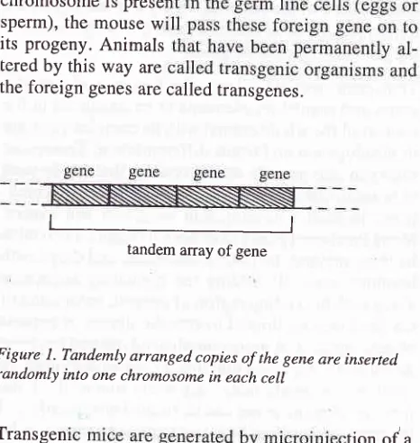
being whose germ line genome has been artificially modified by means of genetic engineering.A
foreign DNA can be randomly integrated into thegenome. In mammals for example, linear DNA frag_ ments introduced into cells are rapidly ligated end to end
by
intracellular enzymesto form long tandem
arrays,which usually
become integratedinto
achromosome at an apparently random site.
A mouse egg injected with 200 copies of linear DNA molecules
will
often develop into a mouse, containingin
manyof its
cells, tandem arrays of copiesof the
injected gene integrated at apparently ranJom site in one site of one of its chromosomes.If
the modifiedchromosome is prese sperm), the mouse w its progeny. Animals tered by this way are
the foreign genes are called transgenes.
ll
tandem anay of gene
The Department of Histology, Faculty of Medicine University of Indonesia, Jakarta, Indonesia
/
ICMR, School of Medicine, Kobe University/Division of
[image:1.595.297.530.450.696.2]Developmental Genetics, School of Medicine, Chiba U niv e rs i ty, Jap an
Figure l. Tandemly arranged copies of the gene are inserted randomly into one chromosome in each cell
Transgen
tion od acloned D
with
subs
Ïli,""ii;
germ
line
can be determined laterby
analysis of genomic DNA in the offsprings. In very early embryothe foreign
DNA
can be incorporatedinto
the chromosome of some embryonic cells and maintainedin them as they proliferate and differentiate into adult
tissues. When the foreign DNA have been integrated
into the genome of the recipipent embryo, the embryo
or the adult animal can be analyzed to determine the
pattem of expression and the phenotypic effect of the
introduced gene.
Transgenic mice had been generated for the first time
in 1981 by Palmitter and Brinster, while Spradling and
Rubin produced transgenic fruit flies.'
This article reviews the most popular technique for modification of the mammalian genome in vivo, i.e. microinjection of fertilized eggs, targeted gene dele-tion through homologous recombination, and other techniques. This article also explains advantages of these techniques in studying human diseases and some illustration of basic pathogenesis of human diseases that had been discovered by using these techniques, and the future potential
of
these techniquesin
thetreatment of human diseases.
THE USE OF TRANSGENIC
MICE
In most cases the transgenic approach aims at specific
modification of the genome, i.e. by introducing whole transcriptional units into the genome, or by
inactivat-ing pre-existinactivat-ing cellular genes.
Transgenic method allows the function
of
specificgenes and regulatory elements to be examined in the
context of the whole animal with its complex program
of development and tissue differentiation. Transgenic mice can also provide model systems that can be used to broaden our understanding of human disease
condi-tions, including development
of
defect and cancer.Many functional genes have been introduced into mice by these method, including oncogenes and the growth hormone gene.
By
adding the regulatory sequences along with the coding region of interest, expression of the gene may be limited to specific tissues or periodsof development. A more complicated method has been developed to replace a functjonal gene with an inactive
allele in transgenic mice.' By using this method, the
function of many genes can be studied more easily and the basic mechanism of many human diseases can be explored and analyzed more extensively, increasing our understanding
of
the pathogenesisof
varioushuman diseases.
The generation of animal models for human diseases
by means of transgenesis has promoted a leap forward
in
our understanding of the pathogenesisof
varioushuman diseases. However, translation
of
thesecon-cepts into experimental and clinical applications is
proceeding slowly, because the development of effi-cient strategies for gene transfer in vivo is not easy.
Now
gene transferis already
being tested as a therapeutic approach in animal models and in a grow-ing number of human disorders. In addition, the time lag between advancesin
basic understanding and progress in applications has been steadily decreased in recent years. The potential of transgenic technologiesfor
the advancementof
medical science has en-couraged molecular biologists to learn about the basic factsof
medicine, while at the same time clinical researchers are increasingly making use of the tools provided by the new technologies.Transgenic technology can be used as a "bridge" be-tween genetic engineering, molec-ular biology, and
other basic medical science's concept in one side and the "real" phenomena in in vivo biological study and clinical research. Finally transgenic technologies can act as communicator
in
the interaction of manydis-ciplines
of
knowledges such as molecular genetics,histology, pathology, and clinical researches.
MANIPULATION OF THE
MAMMALIAN
GENOME IN VIVOCompared
to the transfer
of
genetic information to prokaryotic organismor
cultured eukaryotic cells, genetic modification of a multicellular organism is acomplex and inefficient process.
In
the last decade three approaches have evolvedin
manipulating the mammalian genome:(1)
introductionof
completetranscriptional unit or minigene into the germline of
mice, rat and sheep, to generate a transgenic animal;
(2) introduction
of
foreign genes into specific somatic cells; (3) ablation or subtle site directed modification of the endogenous genetic stock (colloquially termed the "knock out" approach).INTRODUCTION OF TRANSGENES
Rudolf Janish and his colleagues in 1985 showed that
infection of pre-implantation mouse embryos with an
ectopic murine retrovirus can lead to proviral
Vol7, No 2, April - June 1998
material
to
thefilial
generationsin
a Mendelianfashion.
Shortly
thereafter,Erwin
Wagner, Tim Stewart and Beatrice Mintz introduced an alternative technique allowingfor
a higher
flexibility
in
the generation of transgenicmice, i.e., pronuclear microin-jection of zygotes, followed by tranifer to the oviduct.3 This techniques has been slightly modified in the sub-sequent years.4SETTING UP A COLONY FOR THE PRODUC.
TION OF TRANSGENIC
MICE
The animals maintained in a colony for the production
and analysis of transgenic mice can be divided into 5
categories: (1) female mice
for
matingto
producefertilized eggs, (2) fertile stud male mice, (3) sterile male mice
for
the productionof
pseudopregnant female mice, (4) female mice to serve as pseudopreg-nant recipients and foster mothers, (5) transgenic mice lines derived from these founders.The most important thing
in
preparing female for mating to produce fertilized eggs is the strain of mice. Mice usedin
generating transgenic mice must have good genetic background. Superovulated females, i.e., female mice induced by using hormons substituf-ing/similar to FSH and LH. Such female mice are usedalmost exclusively over naturally ovulating females
for the production of fertilized eggs in large numbers.
The stud male mice used in generating transgenic mice
should be in good reproductive performance to fertilize the females' eggs in large number. Male mice reach
ssxual maturity between 6 to 8 weeks of age and can be used as stud for l-2 years depending on the strain,
but it is better to replace the stud at 8-10 months of age,
as their reproductive performance tend
to
decreaseafter that age. Each superovulated female is placed
individually with a stud and is checked for the copula-tion plug the next morning. If a stud fails to plug a
superovulated female several times in a row, or
if his
plugging average is less than 60-80Vo, he should be
replaced. Because the sperm count
will
be depressedfor several days after mating, a stud should not be used
fpr about a week after plugging a female.
Sterile male mice are required for mating to generate pseudopregnant resipient, and usually are produced by vasectomy. For vasectomy, males of at least 2 months of age from any strain with good breeding performance
will
be suitable. Before using a vasectomized male in an experiment,it should
be testedfor
sterility by mating it with females to obtain at least three plugs.If
Transgenic and gene
disruption
57the vasectomy was successful, most of the plugged females should not become pregnant.
Pseudopregnant mice are prepared by mating females
in
natural estruswith
vasectomized males. The qualification of female as a foster mother are (a) at least 6 weeks of age, (b) more than2} grams in body weight,(c) already succesfully reared a litter. Many inves-tigators use coat color differences between the strain of the egg donor and the strain of the pseudopregnant
recipient
to be certain that any mice born
actually derived from the donor eggs. Most mice will give birth on day 19 of pregnancy (counting the day of the plugas day 0), although this may vary between strains.
Potential transgenic mice are usually screened for the presence of the injected gene by Southern or Dot blot hybridization to DNA extracted from the tail. Mice that
develop
from
injected eggs are often termed as"founder mice". As soon as it has been determined that
a given "founder
mice"is
transgenic, it is usually mated to start establishing a transgenic line.If
thefounder is a female and we want to develop a trans-genic line from her, it is necessary to wait until she has
given birth and raised at least one litter before she can be sacrified.
If
the founder is a male, he can be placed with two females which are checked every day andwill
be replaced with a new female as soon as each one is plugged. In this way, the male can generate many litters within a few week. As soon as a male has plugged asufficient number of females (6-8) he may be sacrified,
if necessary, for the analysis of gene expression.
How-ever, if one wants to be sure of establishing atransgenic line, the founder should not be sacrified until positif transgenic progeny has been identified.
Although most transgenic founder
will
transmit theforeign gene to 50Vo of theft offsprings, approximately
20Vo of transgenic founder are mosaic and transmit the gene at a low frequency (i.e. 5-I0Vo instead of 50Vo). In addition, a proportion of females that are plugged
will
fail
to get pregnant,for
exampleif
the male is sterile or semisterile or will not raise a littersuccesful-ly. Homozygote transgenic mice are produced by set-ting up heterozygous intercrosses. Teoretically, one
quarter of the progeny from such a cross should be
homozygous with respect
to
the integrated foreignDNA; the rest, one half should be heterozygous, and one quarter non transgenic.
GENETRATING TRANSGBNIC
MICE
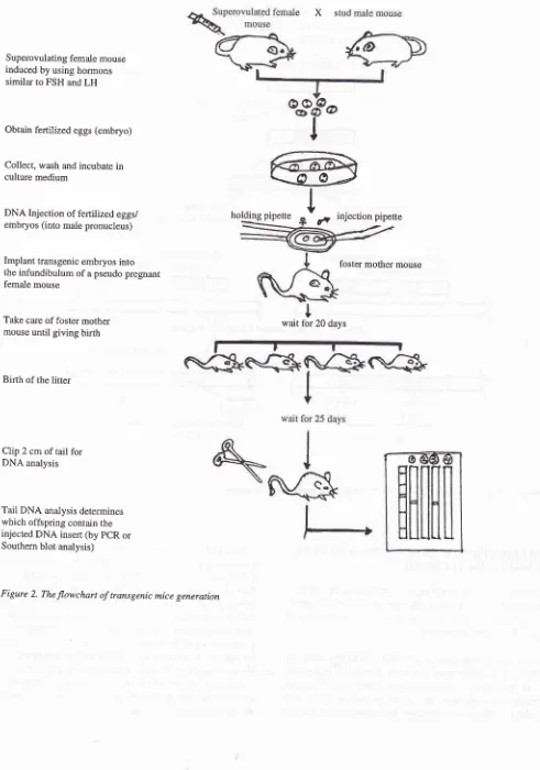
geneiate transgenic mice. Figure 2 shows the flow-chart of numerous steps required for transgenic mice generation.
The
first
stepis
to prepare the clonedDNA
(DNA insert) for injection. The DNA insert should contain apromoter, polyadenylation site, co-mplete protein coding region and at least one intron.) DNA fragment afterrestriction enzyme digestion of plasmidDNA was
purified by agarose gel electrophoresis and electroelu-tion. Although prokaryotic cloning vector sequences have no apparent effect on the integration frequency of microinjected genes,
it
is important to note that they can severely inhibit the expression of eukaryotic genesintroduced into the mouse.o It is not known whether a
specific sequence is responsible for this inhibition or whether
it
is a general property of prokaryotic DNA.So it is advisable to remove all vector sequences from a cloned gene before introducing it into the mouse gene line, if optimal expression of the introduced gene is desired.
The second step
is to obtain
the fertilized
eggs (embryos) from a superovulated female mouse. This superovulated female mice are mated with the stud male mice good in reproductive performance to produce the fertilized eggs in large numbers. Thesefertilized
eggsthen
are collected, washed, and prepared for injection by incubating them in culture medium.The
third
stepis
to inject
the male pronucleus of fertilized eggs (embryos) with DNA insert. For this procedure a special microscope/micromanipulator setup
is
required. Each fertilized eggsis
individually immobilized by a holding micropipette using gentle suction, while DNA insert, are injected into the male pronucleus througha
second injection pipette. The microinjection of DNA directly into the pronuclei of fertilized mouse eggs result in the stable chromosomal integration of foreign DNA in I}-4OVo of the resulting mic;.3'7-9 In most fases, integration appears to occur at the one-cell stage because the foreignDNA
is presentin every cell of the transgenic animal, including all primordial germ cells. Following injection, eggs(embryos) containing DNA insert are transfered into the infundibulum of pseudopregnant female mice by using transfer pipette. Then these foster mothers are
bred for about 20 days until giving birth. After birth, the litters of these female mice and their mothers are
bred together.
At
about 3 weeks after birth, 2 cmof
their taiis are clipped for tail DNA isolation, and finally
the integration of the transgene within the genome are
screened
by
using Southernblot
analysisor
PCR analysis. Then these transgenic mice can be used for further breeding or for analyzing the phenotypic ex-pression.INACTIVATING THE FUNCTIONAL GENES USING TRANSGENIC TECHNIC
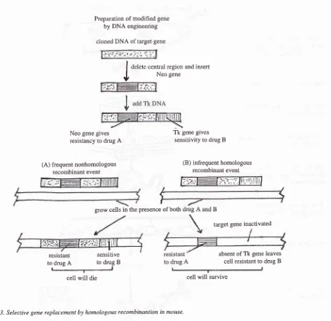
A method had been developed to replace a functional gene with an inactivated allele in transgenic mic-e; this method is called the gene disruption tùh.tiqo".2'g
If
aDNA molecule carrying mutated mouse gene is trans-fered into a mouse cell,
it
usually inserted into the chromosomes at random sites and these nearly always include both ends of the engineered DNA fragment. However, about one in a thousand times, it will replace oneof
the
two copies
of
the normal
gene by homologous recombination. Homologous recom-binant incorporates the center of the engineered DNA fragment without the ends. The rare colonies of cells in which a homologous recombination event has oc-cured and caused a gene replacement are isolated by double drug selection. Then the correct colonies among these are identified by Southern blotting. This show that the correct colonies contain recombinant DNA sequence, i.e. the inserted fragment has replaced all or part of one copy of the normal gene. By exploit-ing this rare "gene targeting" event, any specific genein
a mouse cell can be inactivatedby
direct genereplacement.
In
thefirst
stepof this
procedure,a
cloned DNA containing the geneto be
mutatedis
modified by genetic engineering, so that it contains a bacterial genewithin it, called Neo. The integration of Neo gene into a mouse chromosome renders resistancy
to
a drug (usually antibiotic) that otherwise kills the mouse cells. Thena viral
gene calledTk
(thymidine kinase) is added, attached to one end of the mouse DNA. Then the modified DNA. fragment is introduced into aspe-cial line of mouse embryo derivated stem cell (called embryonic stem cell or ES cell). The integration of Tk into a mouse chromosome makes the cells sensitive to
a different drug. The addition of these two drugs
will
give us the easy way to select the correct colonies of embryonic stem (ES) cells which contain therecom-binant
DNA
sequences (mousemutant
gene), generated by homologous recombination (Figure 3).In the second step, individual cells from the identified colony are inserted into an early mouse embryo, to produce the ES cell-embryo chimaeras. Then these
Vol7, No 2, April - June 1998
Superovulating female mouse
induced by using hormons similar to FSH and LH
Obtain fertilized eggs (embryo)
Collect, wash and incubate in culture medium
DNA Injection of fertilized eggs/
embryos (into male pronucleus)
Implant transgenic embryos into
the infundibulum of a pseudo pregnant female mouse
Take care of foster mother
mouse until giving birth
Birth of the litter
Clip 2 cm of tail for DNA analysis
[image:5.595.36.527.76.776.2]Tail DNA analysis determines which offspring contain the injected DNA insert (by PCR or Southern blot analysis)
Figure 2. The flowchart of transgenic mice generation
Transgenic and gene disruption 59
eXs
@
I
-!--\
.---O
@-z
I
holding pipette
o.
injectionpipettefoster mother mouse
I
Neo gene gives resistancy to drug A
(A) frequent nonhomologous recombinant event
Tk gene gives sensitivity to drug B
(B) infrequent homologous recombinant event
; " ";''li!
liril;ii i
resistant
Preparation of modified gene
by DNA engineering
cloned DNA of target gene
)'iÆl',t' .-.t..:4t,.-1.,
grow cells in the presence of both
sensitive to drug
A
to drug Bcell will die
I
I
delete central region and insertI
Neo sene [image:6.595.80.553.76.535.2]ry
Figure
j.
Selective gene replacement by homologous recombinantion in mouse,APPLICATION OF TRANSGENIC AND GENE
DISRUPTION TECHNIQUE
The ability to prepare transgenic mice lacking aknown
normal gene
is
a major advance, and the technique isnow being widely used
to
dissect the ftrnctions ofspecific mammalian genes.
Since the generation
of
thefirst transgenic
mice inl98l,l
especially after the gene disruption technique was developed to replace a functional gene,''' these techniques had been the most commonly used anduseful techniques
to
analyze the function of specificB
target gene inactivated
cell will survive
genes and regulatory elements. Furthermore, these
techniques were
usedin
exploring the
basicpathogenesis of many human diseases, including
me-tabolic diseases, immune system disorder, cancer, degenerative diseases, defect
in
tissue developmentand differentiation, etc. Transgenic and gene
disrup-tion
techniques arenot only
usedto
study the pathogenesis of human diseases, but also can be usedto search
an alternative methodin
the treatment ofthose diseases. Following description
will explain
the correlation of particular genes with some human dis-eases, which are studied by using transgenic and gene disruption techniques as a tool.resistant absent of Tk gene leaves
VoL7, No 2, April - June 1998
Correlation between Connexin 37, gapjunction
and
infertility
At birth the ovary contains primordial follicles
consist-ing of meiotically arrested oocytes surrounded by a
single layer
of
granulosa cells. Periodically severalprimordial follicles undergo further development, and each cycle, one of
it
proceeds until ovulation. After ovulation, oocytes resume meiosis and the granulosa cells differentiate into steroidogenic cells, forming thecorpus luteum.
Until
nowlittle is
known about the signals that control the follicle development,ovula-tion,
differentiationof
granulosa cellsto
become steroidogenic cells forming the corpus luteum and the relationship between oocyte and granulosa cells.By using transgenic and gene disruption technique,
it
was shown that Connexin 37 played the important role in passing the signal that controlled the follicle growth,luteinization and oocyte maturation. Connexin 37 is a
subunit
of
the proteins that form the gap junctionbetween oocytes and granulosa cells. Loss of Conexin 37 in female mice causes infertility.l0
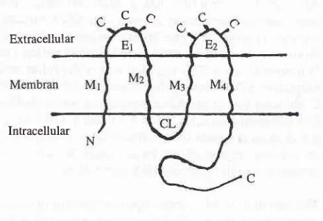
Connexin
is
a
family
of
the integral membrane proteins. Until now at least 12 members of connexinprotein family were discovered. Each of them is en-coded by
its
own gene, located at several differentchromosomes. The expression of these genes on
tis-sues are also different one from another. The structure of connexin consists of 4 transmembrane hydrophobic molecules (M1-M4), 2 extracellular loops
(El-82),1
cytoplasmic loop (CL), amino and carboxyl termini (figure 4). Extracellular, transmembrane, and N ter-minal cytoplasmic domains are well conserved among
family members, while rytoplasmic loop and
C-ter-minal cytoplasmic domain are highly variable in both sequence and size.
Connexin 37 is encoded by connexin3T gene located at chromosome 4. This gene encodes a 1.5 kb mRNA
and is expressed in several organs, such as heart, ovary,
testis,
lung,
skin,ll
and blood vessel endothelialcells. I 2'l 3
All
female mice lacking Connexin 37 (Cx 37) wereinfertile.l0 Although Cx37 -/- mice showed no
exter-nal
abnormalities and appeared healthy,Cx
37 -l-females were foundto
be completely incapable ofovulation, even in gonadotropin stimulation. By using
microinjection of neurobiotin into oocyte and electron
microscopic analysis,
it
was found that gap junctions between oocytes and neighbouring granulosa cells were intact in wild type mice and completely absent inTransgenic and gene
disruption
6lmutant mice. Further,
it
was discovered thatabnor-malities in follicular growth, control of luteinization
and oocyte maturation underlay Cx37 -l- female
infer-tility. In Cx 37 -/- female mice follicular development
consistenly arrested before
full
maturation, and the inappropriate formation of corpora lutea was occured.In
normal animal, luteinization occured following ovulation caused by the lost of oocyte-granulosa cellcommunication. The premature luteinization observed
in Cx 37 -/- females suggests a model in which junc-tional communication plays an important role in
trans-fering the inhibitory signal from oocyte to granulosa
cells and results
in the
preventionof
corpus luteum formation.Finally they concluded that cell-cell signalling rhrough
intercellular channels critically regulated the highly
coordinated set ofcellular interaction required for suc-cessful oogenesis and ovulation.
CC
Extracellular [image:7.595.300.533.315.474.2]Intracellular
Figure 4. The structure of Cbnnexin protein. The protein struc-ture consists of4 transmernbrane hydrophobic molecules (Ml-M4),2 extracellular loops (El-82), cytoplasmic loop (CL), amino and carboryl termini
Lack of transcription factor AP-2 causes defect in
cranial closure and cranial development
Regulation of transcription is fundamental to dynamic
process
underlying
cellular
differentiation
and development. The rate of gene transcription dependson
its
unique constellationof
cis linked regulatory elements and on the combinatorial effect of transcrip-tion factors, both general and cellular type specific that binds to this element.AP-2 is one of the transcription factors that may play
a role in development. This nuclear protein was first identified and purified from Hela cells
as
50 kDa inelement
of
SV-40 and human metallothioneinIIA
gene. The binding sites for AP-2 was also found in avariety of gene promoters, including pro-enkephalin, collagenase, growth hormone, keratin K-14, murine
major histocompatibility, C-myc and acetylcholin
esterase. AP-2 amino acid sequence
is
highly con-served among frog, mice and human, indicating that this protein likely plays a fundamental role indevelop-ment. During the closure
of
neural tubein
mice, transcription factor AP-2 is expressed in ectoderm andneural crest cells migrating from the cranial neural
fold.la Cranial n"u.ui cresi cells provide patterning
information for craniofacial morphogenesis, generate
most
of
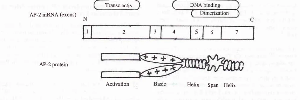
the skull bones, and together with placodal ectoderm, form the cranial ganglia.The transcription factor AP-2 is encoded by a gene located on chromosome 6 near HLA locus. This gene consists of 7 exons and has a 18 kb genomic DNA.
AP-2
protein structure has2
main domains, thetranscriptional activation domain and DNA binding domain (Figure
5).
The transcriptional activationdomain is encoded by exon 2 and located within the
N-terminal region. This region is rich with proline and glutamine. DNA binding domain is located within the C-terminal half of the AP-2 protein and is encoded by
four different exons, i.e., exon 4,5,6 and 7. DNA bind-ing domain is composed of dimerization domain and
an adjacent region
of
net basic charge to achieve a sequence specific protein-DNA interaction.The function of AP-2 transcriptional protein in tissue
development during embryogenesis was tudied by using disruption technique combined with transgenic method. Exon 6, which was important for dimerization and binding to DNA binding site in promoter area of the targeted gene was ommited. Mice lacking AP-Z
Transc.activ AP-2 mRNA (exons)
N
AP-2 protein
Activation
gene dicd perinatally with cranio-abdominoschisis and severe dismorphogenesis
of
the face, skull, sensory organs and cranial ganglia. Failure of cranial closure between 9 and 9.5 days post-coitum coincided withincreased apoptosis in the midbrain, anterior hindbrain and proximal mesenchyme of the first branchial arch,
but did not involve the loss of expression of Twist or
Pax-3, two other regulatory genes known to be re-quired for cranial closure.
Mice lacking NCX/HOX
llL-l
gene showed themegacolon phenomena, a novel pathogenesis
for
megacolonHirsprung's disease is a congenital disorder charac-terized by the congenital absence of the enteric nerve
ganglia
from
the hindgut, resultingin intestinal
obstruction, and causes the megacolon phenotype.
Enteric nerve ganglia were derived.from neural crest cells. Neural crest cells that form a transient population
of
cells, emerged from the neural tube epithelium (neuroepithelium), locatedin
the dorsal partof
the neural tube, at the junction between neuroepithelium and surface ectoderm. These cells then migrate fromits
original sites at a define stageof
development, disperse within the embryo and finally settle in many embryonic regions to yield ganglia and a large varietyof connective tissues in the head. The migration and
colonization of neural crest cells is mainly controlled
by signals
through the c-Ret protooncogene and theendothelin-B receptor gene.
Most
experimentalmodels of Hirsprung's disease, including mutants with loss offunction ofthe tyrosine kinase Ret receptor and its ligand, or the endothelin receptor and its ligand, also demonstrate loss of myenteric neurons.
Helix
Span Helix [image:8.595.53.550.542.708.2]I 2 _t 4 5 6 7
Vol7, No 2, April - June 1998
However, megacolon also results from neuronal intes-tinal dysplasia (NID), a human congenital disorder that
often presents similarly to Hirsprung's disease i.e. a narrow rectum and distended bowel extending through
hind gut into small bowel.
The neuronal intestine dysplasia is characterizedby a
normal number
of
ganglia or hyperplasiaof
entericneuron.
Experiments revealedthat
the
humanhomologue
of
the NC)UHox 11 L-l gene can be a candidat gene causingNID in
human. NC)OHox ll
L-1 is a member of the Hox 1l homeobox gene family.
NCX gene has 87Vo similarity
in
homeodomain se-quences with Hox 11, and the helix 3 sequence that is important for determining the DNA binding spesificityis identical between them.r) Homeobox geneproducts
have
a
conserved domain (homeodomain)of
60 aminoacids, and are known to bind to specific DNAsequences. These genes are classified into several gene
families depending on their homology of homeobox sequences and their locations on chromosomes in
human and mice.
NCXlHox 11 L-1 deficient mice developed myenteric neuronal dysplasia and megacolon after 3-5 weeks of
age. Histochemical analysis revealed that the number
of NADPH diaforese positive neurons in the proximal
colon was higher in the NCX deficient mice than in the
wild type mice. The number
of
substance-p positive neuronal cells and fibers also increased in the proximalcolon
of
NCX deficient mice. NADPH diaforese is identical to nitric oxide synthase and nitric oxidefunc-tions as an inhibitory neurotransmitter to relax smooth
muscle,
while
substance-Pis
an excitatory neuro-transmitter to contract circular smooth muscles.There-fore, histochemical analysis revealed that the enteric
neuron was hyperinnervated in the proximal part of the colon.
The pathogenesis of megacolon with the absence of enteric neurons can be explained
by
the abnormal movement of colon. In case of NID, thehyperinnerva-tion may cause inappropriate action of enteric neurons and may result in functional abnormality of the colon.
The amelioration of the dystrophic phenotype of
mdx mice using a truncated transgene
Duchene and Becker muscular dystrophies (DMD and
BMD) are allelic X-linked recessive disorders which result
in
a distinctive pattern of progressive muscle wasting, and patient usually will die in early twenties,due to respiratory and or cardiac failure. The main symptoms and signs of this disease are muscle
weak-Transgenic and gene
disruption
63ness, muscle pain and mental retardation; the value of
serum creatinin kinase is elevated to 50-100 times of normal.
Histological
analysis shows progressive musclefiber
degeneration and regeneration. EMGanalysis is characteristic with reduction in duration and amplitude of the wave. Immunohistochemical staining
of dystrophin protein shows negative staining in DMD
patient, faint in BMD disease, and mosaic pattern in DMD carrier.
The disease is caused by lack of dystrophin, a large membrane-associated protein expressed in muscle and
brain that
is
localized to the inner faceof
the cell membrane. This cytoskeletal protein (427 kDa in molecular weight)is
encoded by the largest knownhuman gene with a 14-kB RNA transcripts containing
79 exons. Dystrophin has many isoforms generated through differential promoter usage and or alternative
splicing at the 3'end of the gene (until now at least 5 distinct isoforms had been discovered).
Utrophin, another member of cytoskeletal protein (395
kDa in molecular weight) encoded by utrophin gene
which is located at chromosome 6q24 in human and
chromosome 10
in
mouse,with a
13-kb RNAtranscript, has an amino acid sequence that
js
80Vosimilar to the amino acid sequence of dystrophin. Like
dystrophin, utrophin also consists of 4 domains, i.e. N
and C terminal domain, rod domain and cysteine rich
domain.
The
amino
acid
differences
betweendystrophin and utrophin
is
located only on the rod domain. Rod domain of dystrophin has 24 repeats of ,109 amino acid sequence separated by 4 proline rich
hinge regions, while utrophin only contains 22 repeats and 2 proline rich regions.
Utrophin was found in all tissues, particularly in the lung, bood vessel, and nerves. In normal adult muscle,
utrophin is localized at the membrane
of
neuromus-cular junctions. In fetal muscle development, utrophinis
expressed before dystrophin.Like
dystrophin,utrophin is localized at the sarcolemma and neuromus-cular junctions, showing maximum expression at
l7-18 weeks of gestation. However, when dystrophin is expressed, utrophin
is
gradually depleted from the sarcolemma, replaced by dystrophin and leaving thejunc
onl
rophincaus
ny
uggestsion
ge
reasedthe dystrophic muscle phenotype
of
dystrophin dis-ease.related protein, utrophin, which might be able to
com-pensate for the dystrophic phenotype. For this purpose a truncated utrophin transgene has been expressed at
high level in the muscles of the dystrophin deficient mdx mice. By this experiment, it had been shown that
overexpression of utrophin gene could cause ameliora-tion ofihe dystrophic ptr"n6typ" of mdx
-ice.l6
For-thermore, because utrophin is normally expressed in all tissues including muscle, the use of this utrophintransgene rather than a dystrophin transgens in con-ventional gene therapy approaches using viruses or liposomes may avert any potential immunological
respons against the transgene.
CONCLUSION
Transgenic technology now has developed rapidly from concept to a commonly used and useful technique in studying and analyzing the function of many genes and regulatory elements involved in the orchestra of development and differentiation
of
many cells and tissues. By combination with gene disruption and any other techniques,now transgenic
technology is popular in studying the basic pathogenesisof
various human diseases. Furthermore, overexpression of closely related gene might be able to compensate the impairmentof
particular genein
various human genetic based diseases. Finally, combination betweentransgenic technology, gene disruption and other
methods can be used in the research for those purposes.
REFERENCES
1. Palmiter RD, Chen HY, Messing A, Brinster RL. SV40 enchancer and large-T antigen are instrumental in develop-ment of choroid plexus tumors in transgenic mice. Nature 1985;316:457 -60.
2. Capecchi M. Altering the genome by homologous recom-bination. Science 1989:'244:1288-92.
3. Wagner EF, Stewart TA, Mintz B. The human beta globin gene and a functional viral thymidine kinase gene in
developing mice. Proc Natl Acad Sci USA 1981;78:5016-20.
4. Hogan B, Constanstini F, Lacy E. Manipulating the mouse embryos: A laboratory manual. 2nd Ed. new York: Cold Spring Harbor;1994.
5. Brinster R, Allen J, Behringer R, Gelimas R, et al. Introns increase transcriptional efficiency in transgenic mice. Proc Natl Acad Sci USA 1988;85:836-40.
6. Krumlauf R. Hox genes in vertebrate developrnent. Cell 1994;78:l9l-2Ol,
7. Brinster R, Chen H, Trunbauer M, et al. Somatic expression of herpes thymidine kinase in mice, following injection of a
fussion gene into eggs. Cell l98l;27:223-3I.
8. Gordon J, Ruddle F. Gene transfer into mouse embryos: production of transgenic mice by pronuclear injection. Method Enzymol 1983 ; 102:41 1-33.
g.Zimmer A. Manipulating the genome by homologous recombination in embryonic stem cells. Annu Rev Neurosci
1992;15:Il5-37.
10. Simon AM, Goodenogh DA, Li E, Paul DL. Female
infer-tility in mice lacking connexin 37. Nature 1997;385:525-9. 11. HaefligerJA, BruzzoreR, Jenkins NA, GilbertDJ, Copeland
NG, Paul DL. Four novel members of the Connexin family of gap junction protein. J Biol Chem 1992;267:2057-64. 12. Reed KE, Westphale EM, Larson DM, Wang HZ, Veeristra
RD, Beyr EC. Molecular cloning and functional expression
of human connexin 37, an endothelial cell gap junction protein. J Clin Invest 1993:91;997-1004.
13. Willecke K, Heynkes R, Dahl E, Stutenkempr R, Hen-nemann H, Jungbluth S, et al. Mouse connexin 37: Cloning and functional expression of a gap junction gene highly expressed in lung. J Cell Biol l99l;ll4:1049-57.
14. Mitchell PJ, Timnom PM, Hebert JM, Rigby PW, Tjian R. Transcription factor AP-2 is expressed in neural crest cell
lineages during mouse embryogenesis. Genes Dev
I 99 l;5: I 05- 1 9.
15. Dear TN, Colledge WH, Carlton MB, Lovenir I, Larson T, Smith AJ, et al. The hox 1l gene is essential for cell survival during spleen development. Development
1995:.12l;2909-15.
16. Tinsley JM, Potter AC, Phelps SR, Fischer R, Trickett JI, Davies KE. Amelioration of the dystrophic phenotype of