.
Indonesian Association of Food Technologistsfjl:'s,tei
Perhimpunan Ahll Teknologi Pengon IndonesiaOrganizers
efEAFAfT
-
CENTER-: • • 0
in conjunction with
• Department of
セN[[ N@ Food Science
Title Page
Title Diterpenoid geranylgeraniol
inflammation in rat
supplementation
Running title GGOH inhibits NF-KB by suppressing IRAK1 and TRAF6
suppresses LPS-induced
Authors PE Giriwonol, H Shirakawa1, Y Ohsakil, S Hata2, H Kuriyama2, S Satol, T Gotol and M Komail
Affiliation
Background
1. Laboratory of Nutrition, Graduate School of Agricultural Sciences, Tohoku University,
Sendai, JAPAN
2. Tama Biochemical Co., Ltd ., Tokyo, JAPAN
Abstract
Geranylgeraniol (GGOH) is a natural diterpenoid occurring virtually in all dietary sources which possess
the mevalonate pathway, including rice. Rich sources of GGOH include Bixa orellana commonly known for
its annatto food colorant which has been used abundantly in many forms of food for daily consumption.
One functional aspect of GGOH is its reported anti-inflammatory effect. In this study, we examined the
mechanism of GGOH in suppressing inflammation induced by lipopolysaccharide.
Materials and methods
Eight weeks old male Wistar rats were supplemented with or without GGOH at incremental doses (48.3
-4830 mg/kg diet). After ten days of feeding, the rats were ip. injected with 0.5 mg/kg bw LPS or vehicle
then fasted for 18 h before sacrifice. Levels of inflammatory cytokines HilMQセ L@ IL-6 and TNFa) were
measured .
Results
Rats supplemented with GGOH at dosages 483 and 4830 mg/kg diet showed substantial suppression in
plasma concentrations of ilMQセL@ IL-6 and TNFa. The liver of these rats were also protected from damage as
indicated by lower plasma ALT and AST activity. Livers of GGOH treated rats showed down-regulation of
inflammatory and NF-KB signal transducer genes, specifically lrakl, Traf6 and Takl. This suppression,
including inhibition of NF-KB, was observed up to protein level as indicated by western blot assay. It
appears that GGOH involves the degradation of IRAK1 to effectively inhibit activation of NF-KB after LPS
stimulation.
Conclusions
We observed that GGOH treatment in vivo effectively inhibited NF-KB activation after LPS stimulation. We
obtained results that indicate GGOH suppresses NF-KB signal transduction molecules and maintaining
them at low levels to inhibit excessive inflammation.
Keywords: isoprenoid; geranylgeraniol; anti-inflammation; lipopolysaccharide; IRAK1 suppression;
NF-KB inhibition; THP-1 cell; rat liver.
1 Introduction
Natural bioactive compounds in dietary plants such as fruits, vegetables, grains and legumes have
been epidemiologically shown to inhibit and prevent degenerative diseases. It has become increasingly
essential knowledge that the bioactive substances in these products, or quite often referred to as chemo
preventive agents, have an important role in the control and maintenance of inflammation [1, 2]. Results
from epidemiological studies and animal experiments may elucidate some aspect of the mechanisms
involved, and investigations on more comprehensive and detailed mechanism are still ongoing and our
understanding will continue to evolve. Of this vast chemo protective agents gaining recent interest are
isoprenoids or terpenoids, a class of secondary metabolite from the mevalonate pathway consisting of
over 22,000 constituents showing very potent inflammatory gene modulation [1-3].
It has been postulated that the action of isoprenoids in inflammatory suppression lies in the
modification of mevalonate pathway which may also be involved in the biosynthesis of these compounds.
Indeed, several observations have reported increased expression of 3-hydroxy-3-methylglutaryl coenzyme
A (HMG-CoA) reductase and down-regulation of squalene synthase that result in cholesterol production
after inflammatory stimulation with LPS, ilMQセ@ and TNF-a [4-6). Paradoxically, it has also been reported
that dysregulation to the cellular mevalonate pathway, which results in the decrease of both cholesterol
and isoprenoids synthesis, account for inflammation in auto-inflammatory diseases such as the case for
mevalonate kinase deficiency or hyperimmunoglobulin-D syndrome [3,4,7,). Thus it was recommended
that treatment with exogenous isoprenoids may help in the alleviation of these and possibly other
auto-inflammatory diseases. Furthermore, many emerging evidence point to other potentially direct interaction
between isoprenoids with the NF-KB activation cascade [2,3,8], an essential mediator and transcription
factor for inflammation [8-11]. Thus isoprenoids may provide immune-modulatory effect.
Geranylgeraniol (GGOH) has been demonstrated to exhibit anti-inflammatory actions in human
peripheral blood mononuclear cells (PBMCs) [12,13] and a mouse model of alendronate induced
.
inflammation [7]. It has been observed that the geranylgeranyl pyrophosphate (GGPP) precursor, GGOH,and other hydrophobic isoprenoid derivatives, e.g. farnesol (FOH) are able to permeate the cell easily as
the former pyrophosphate groups are prevented from entering living cells [12,14] . In the cell, GGOH and
FOH are subsequently converted into their respective pyrophosphate moieties (GGPP and farnesyl
pyrophosphate) by two successive monophosphorylations [12,15).
There are several proposed mechanisms as to how GGOH may inhibit inflammation. As an
intermediate of the mevalonate pathway, treatment with exogenous GGOH was able to replete the low
levels of de novo synthesized isoprenoids commonly observed in inflammatory conditions and/or
mevalonate dysregulation such as ubi qui nones [7,12,14,16] with little explanation on how it mediates this
effect. Additionally, GGOH may be used for the synthesis of GGPP that is required for post-translational
attachment to other proteins or isoprenylation, proper translocation of various signaling proteins, and is
key to secretion of ilMQセ@ in stimulated BPMCs [13,16] . Furthermore, it has been demonstrated that GGOH
inhibits NF-KB activation, along with other isoprenoid such as ursolic acid, in a manner independent of
redox change; however, the clear mechanism of this action remains elusive [17,18). Thus the objectives of
result in the inhibition of NF-KB activation induced by LPS stimulation. In this study, we observed that
GGOH shows a more direct role in suppressing signal transducers particularly lRAK1 and TRAF6 to
substantially inhibit NF-KB activation.
2 Methods
2.1 Materials
GGOH was provided by Tama Biochemical Co. Ltd, (Tokyo, Japan) and stored at -20°C. LPS [E.coli
0111:B4 (Cat. #L2630)] used throughout this study was purchased from Sigma (St. Louis, MO, USA) and
dissolved in sterilized saline as stock solution (1 mg·mL·l). Phorbol 12-myristate 13-acetate (PMA)
purchased from Sigma was dissolved in ethanol to promote differentiation of THP-1 cells. We used a
vitamin K deficient diet for base diet (TD97053) purchased from Harlan Teklad (Madison, WI, USA)
re-constituted with 0.75 mg·kg·1 phylloquinone.
2.2 Cell culture
Human monocytic THP-1 cells were obtained from RIKEN BioResource Center (Tsukuba, Japan)
and cultured in RPMl 1640 (Sigma) supplemented with 10% fetal bovine serum (Hyclone, Logan, UT, USA),
2 mM L-glutamine, 100 U·mL·1 penicillin, and 100 llg·mL·l streptomycin at 37 °C and 5% C0 2 atmosphere.
The THP-1 cells were differentiated for 48 h with the addition of 10 ng·mL·l PMA before further
treatments. Upon differentiation, the medium was exchanged with fresh medium and treated with GGOH
(10 f.lM) for 24 h incubation. Cells were then treated with LPS (1 llg·mL·l ) and 3 h incubation followed by
mRNA isolation.
2.3 Animal experiment
Eight weeks old male Std:Wistar rats, weighing 130-150g, were purchased from SLC Japan
(Shizuoka, Japan) and maintained in 12 h light dark cycle (08:00 - 20:00 light), 23 ± 1°C, and 50 ± 5% RH .
For acclimatization, standard pellet feed (F2, Funabashi Farm, Chiba, Japan) was given for three days with
.
distilled water. The rats were then divided into four groups; two control groups fed base diet with orwithout LPS challenge (0.5 mg·kg-1 body weight, Con- and Con+); and two groups fed base diet supplemented with 483 mg· kg-l diet GGOH with or without LPS challenge (GG- and GG+). Experimental
feed was given freely for ten days, followed by intraperitoneal injection of LPS and 18 h fasting prior to
sacrifice. Euthanasia was by abdominal aorta exsanguination under diethyl ether anesthesia, from which
plasma was obtained. Livers were promptly excised; snap frozen in liquid nitrogen and stored under -65 °C
until further analysis.
All procedures involving animals were conducted humanely in accordance to the approval of the
Animal Research-Animal Care Committee at the Graduate School of Agricultural Sciences, Tohoku
University.
2.4 Blood and liver biochemical markers assay
Blood taken by exsanguination were centrifuged in Na2EDTA prepared tubes (final concentration
1.5 mg·mL-l blood) at 1,870 x g for 15 min at 4°C; and the resulting plasma was divided into aliquots and
stored at -30°C. Plasma alanine aminotransferase (ALT) and aspartate aminotransferase (AST) activities,
Chemical Industries, Osaka, Japan) according to the manufacturer's instruction. Plasma concentrations of
inflammatory cytokines were analyzed by Quantikine ELISA (R&D Systems, Minneapolis, MN, USA)
according to the manufacturer's protocol.
2.5 RNA preparation and quantitative RT-PCR
Total RNA was isolated from excised liver, previously stored in RNAlater (Ambion, Tokyo, Japan),
by tissue disruption in guanidine isothiocyanate-based reagent (Isogen, Nippon Gene, Tokyo, Japan) with
bead type homogenizer Micro Smash MS-100 (Tomy Seiko, Tokyo, Japan) according to the manufacturer's
instructions. For cell culture, cells were homogenized by repeated pipetting in Isogen. Isolated RNA was
analyzed qualitatively by agarose gel electrophoresis, whereas absorbance ratio of 260 to 280 nm was
used to determine quantitatively. cDNA was synthesized from 5 セァ@ of total RNA, denatured with
oligo-dT /random primers, 10 mM dNTP at 65 °C. The denatured RNA was then incubated in 50 mM Tris-HCI
buffer (pH 8.3) , 0.1 mM OTT, 50 units of Superscript III reverse transcriptase (Invitrogen, Carlsbad, CA,
USA) and 20 units of RNaseOUT RNase inhibitor (Invitrogen); at 25°C for 5 min, then 50°C for 60 min and
finally 70°C for 15 min in TaKaRa PCR Thermal cycler MP (Takara biomedicals, Shiga, Japan). Aliquots of
the synthesized cDNA were used as template for quantitative PCR by ABI 7300 (Applied Biosystems,
Foster City, CA, USA) in accordance with the manufacturer's instructions. Measurement of expressed
mRNA was first normalized to its eukaryotic elongation factor 1a-1 (EFla1) then compared with the
expression of control to yield relative expression [19]. The sequences of primers used for each gene
expression assay are shown in Table l.
2.6 Western blot
Frozen liver samples were homogenized with polytron homogenizer (Polytron, Basel,
Switzerland) in ice cold extraction buffer [20] containing inhibitors for proteinase (Complete Mini
proteinase inhibitor cocktail tablet, Roche Applied Science, Mannheim, Germany) and phosphatase
(PhosSTOP phosphatase inhibitor cocktail tablet, Roche Applied Science). Tissue homogenate was then
.
centrifuged at 15000 x 9 for 20 min at 4°C and the supernatant was collected. For cell samples, after 24 h
GGOH incubation (10 セmIL@ differentiated THP-1 cells were treated with 1 セァᄋュlMャ@ LPS for the determined
duration then lysed at 4 °C for 30 min with the above-mentioned extraction buffer containing inhibitors
for proteinase and phosphatase. The cell lysate was centrifuged at 15,000 x 9 for 20 min and the
supernatant was collected. The protein concentration was determined using a protein assay kit (Bio-Rad
Laboratories, Tokyo, Japan). Twenty micrograms of protein was mixed with SDS gel loading buffer and
resolved on a 10-20% SDS-polyacrylamide gel electrophoresis (Wako Pure Chemical Industries);
subsequently, the proteins were transferred on to a polyvinylidene fluoride membrane (Millipore, Billerica,
MA, USA). The membrane was subjected to blocking for 1 h with TBS-T (10 mM Tris-HCl at pH 7.4, 150
mM NaCI and 0.1 % Tween 20) containing 5% bovine serum albumin (Sigma), and then incubated with
antibodies against MyD88 (Cell Signaling Technology, Danvers, MA; USA), IRAK1 (Cell Signaling
Technology), phosphorylated IRAK1 (Thr209; Abcam, Tokyo, Japan), TAK1 (Cell Signaling Technology),
phosphorylated TAK1 (Thr184/187; Cell Signaling Technology), phosphorylated ikk。Oセ@ (Ser176/180;
Cell Signaling Technology), TRAF6 (Cell Signaling Technology), NF-KB p65 (Cell Signaling Technology) or
phosphorylated NF-KB p65 (Ser536; Cell Signaling Technology), and detected with the Immobilon Western
Detection Reagent (Millipore) using luminescent image analyzer LAS-4000mini (Fujifilm, Tokyo, Japan).
The relative expression level of each protein was normalized according to the amount of a-tubulin
detected by its antibody (Sigma) .
2.7 Statistical analysis
Values are represented as the mean value with standard errors. One-way analysis of variance,
followed by the Fisher least significant difference test was used to evaluate the differences between groups,
unless otherwise stated. SPSS version 11.0 (SPSS Inc., Chicago, IL, USA) was used for all data computation.
Statistical significance was determined at p < 0.05 or lower.
3 Results
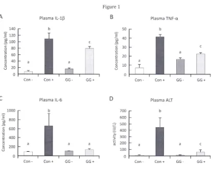
3.1 GGOH suppresses inflammatory cytokines in rat independent of cholesterol alteration
Ten days of experimental feeding supplemented with or without GGOH (483 mg·kg·} diet) did not
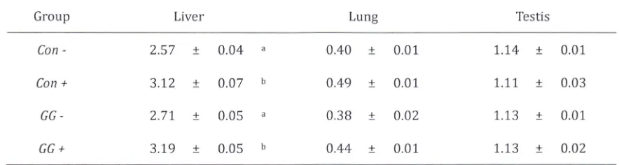
induce change in normalized organ weight among similarly treated rats (with or without LPS stimulation) .
We also observed no significant change in lung and testis weight amongst all groups. It was observed,
however, that livers of LPS challenged rats show increase in weight when compared to its respective
negative controls, indicating acute liver inflammation (Table 2). We obtained significant suppression in the
concentration of plasma inflammatory cytokines ilMQセL@ TNF-a and IL-6 of rats supplemented with 483
mg·kg-} GGOH (Figs. lA-C). Additionally, we observed striking hepatic protection from acute LPS
inflammation in GGOH supplemented rats as indicated by the attenuation of plasma ALT and AST activities
(Figs. 1D&E). The results we observed are in agreement with those observed in other study involving liver
damage induced by diethylnitrosamine with 2-acetylaminofluorene treatment [17].
LPS challenge was observed to increase plasma cholesterol concentrations significantly in both
Con+ and GG+ when compared to non challenged groups (although not significant between challenged
groups) (Fig. IF). This イ・ウセャエ@ confirms observations that LPS treatment may increase plasma cholesterol in
animal models [4,6,17]. hッキ・カ・ャセ@ due to the short duration of feeding with GGOH, no cholesterol change or
improvement was observed among GGOH supplemented group. Similar result was observed in plasma
triglyceride concentration (Fig. IG). Despite no improvement in plasma cholesterol of GG+ rats, substantial
inflammatory suppression indicates GGOH inhibits NF-KB activation by other mechanism .
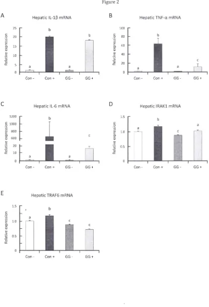
3.2 GGOH modulates inflammatory genes in rat liver
The striking suppression of acute inflammation-induced liver damage observed in GGOH
supplemented rats intrigued us to further investigate changes in inflammatory gene expression modulated
by GGOH. Messenger RNA isolated from livers of rats with or without GGOH supplementation was
quantified by RT-PCR. We observed that liver of GGOH supplemented rats showed down-regulation of
inflammatory genes (Figs. 2A-C), strongly correlated with the decrease in plasma inflammatory cytokines
(Figure 1A-C). We presumed GGOH treatment interacts and affects suppression of molecules closely
regulating NF-KB activation and observed further down-regulation of signal transducer genes upstream of
IKK complex, notably Irakl and Traf6, in the liver of GGOH supplemented groups (Figs. 2D&E). This
suppression was further observed in western blots of GGOH supplemented rat livers thus inhibiting of
supplemented rats without LPS challenge thus indicating possible modulation of these gene expressions
by GGOH. Suppression of TRAF6 and IRAKI proteins was also observed in human HepGZ cells incubated
with 10 11M GGOH for 24 hours (Supplementary Figure 1).
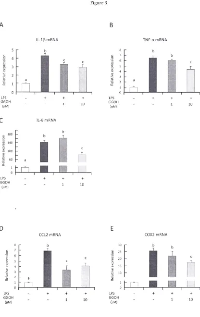
3.3 Pre-incubation with GGOH suppresses inflammatory genes in human THP-l cells
In an attempt to elucidate the molecular mechanism of GGOH in suppressing TLR4-NF-KB
inflammatory response to LPS stimulation, we turn to an established in vitro model using human
differentiated monocytic THP-l cells. One rationale is that differentiated THP-l cells behave like native
monocyte-derived macro phages more than other human myeloid cell lines, such as HL-60, U937, KG-I, or
HEL cell lines; thus providing a valuable model for studying the mechanisms involved in regulation of
macrophage-specific genes as they relate to physiological functions such as inflammatory responses [21].
The other reason is that hepatic tissue contains resident macrophagic cells, or Kupffer cells, and is
subjected to further macrophagic infiltration during the onset of inflammatory challenge.
Differentiated THP-l cells pre-incubated with GGOH for 24 h (111M or 10 11M) then treated with 1
I1g·mL·l LPS showed significant suppression of inflammatory cytokine mRNA expressions, primarily ilMャセL@
TNF-a and IL-6 (Figs. 3A-C) . The suppressions of these genes were observed clearly substantial (p < 0.05)
in the cells pre-incubated with higher GGOH concentration. However, time course pre-incubation of GGOH
did not show strong correlation of its inflammatory suppression effects (data not shown) . Quantification
of other NF-KB target genes involved in inflammatory response, such as CCL2 and COX2, also showed
significant down-regulation (Figs. 3D&E). To further validate our observation that GGOH effectively
abolished NF-KB activation after LPS stimulation, results from western blot of phosphorylated NF-KB p65
(activated form) and totallKBa indicate clear inhibition by 10 11M GGOH 24 h pre-incubation (Fig. 3F) .
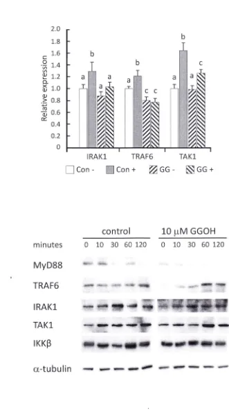
3.4 GGOH suppresses iセkャ@ expression and subsequent phosphorylation of NF -KB signaling
molecules
We observed that GGOH was able to suppress gene and protein expressions of Irakl and Traf6 in
rat liver and HepG2 cells. This observation was also confirmed in differentiated THP-l pre-incubated with
10 11M GGOH for 24 h. GGOH significantly suppressed mRNA expressions of IRAKl, TRAF6, and TAKI in
LPS stimulated cells (Fig. 4A). This was further demonstrated in the western blot assay that protein
expressions of IRAKl and TRAF6 were decreased after GGOH 24 h pre-incubation (0 min); and up to 30
min after LPS stimulation in TRAF6 (Fig. 4B). To clarify whether GGOH suppresses expressions of IRAKI
and TRAF6 prior to LPS stimulation, we incubated THP-l cells with GGOH for transiently and observed
mRNA expression change only for IRAKl. However, we acquired substantial protein suppression in
western blot of IRAKI and TRAF6 thus indicating a possible increase of degradation promoted by GGOH
treatment (Supplementary Figure 2).
We further investigated whether GGOH treatment may affect the phosphorylation of these
Signaling proteins. We treated differentiated THP-l cells with 10 11M GGOH for 24 h followed by LPS
stimulation then harvested at indicated times and immediately lysed for western blot analysis. The results
was apparent that GGOH treatment promotes the suppression of IRAKl and TRAF6 resulting in the
decrease of subsequent phosphorylation of TAKI and ikk。Oセ[@ and inhibition of NF-KB activation.
4 Discussion and conclusions
In this study, we have demonstrated the inhibitory effect of GGOH on NF-KB signaling cascade
stimulated by LPS. The anti-inflammatory effect of GGOH treatment have been previously demonstrated in
human PBMCs [12,13], however, to our knowledge this study is the first to report an insight to a more
detailed molecular effect of GGOH on NF-KB inhibition. Supplementation of exogenous isoprenoid has
been numerously cited to affect cholesterol metabolism by post-translational inhibition of HMG-CoA
reductase activity [3,16,17] and has been postulated as a possible anti-inflammatory effect. Although, it
has been reported that 7 weeks of GGOH administration was shown to decrease both plasma cholesterol
and NF-KB activation in rats [17], our ten days feeding results indicate no improvement in plasma
cholesterol and triglyceride concentrations (Figs. IF&G). LPS challenge, however, was observed to elevate
plasma cholesterol in groups treated with or without GGOH in the acute phase period. This elevation was
also reported in previous studies involving, but not limited to, Syrian hamster and has been attributed to
the observation that inflammatory responses greatly decrease hepatic squalene synthase activity [5,6,22].
However, we observed significant anti-inflammatory action with little improvement to plasma cholesterol
concentration in GGOH treated rats. This observation may indicate a more direct effect by GGOH in
suppressing molecules regulating NF-KB activation. Thus we were able to obtain significant
down-regulation of signal transduction genes, in particular IRAKl and TRAF6, in livers of GGOH supplemented
rats with or without LPS challenge (Fig. 2).
NF-KB is an essential mediator of transcription in the immunological response of mammalian cells
[9,23,25]. As a response towards pathogenic agents, such as LPS attaching to TLR4, adaptor proteins
activate Signaling cascade of ubiquitination and phosphorylation in signal transducers starting from
.
recruitments of MyD88 and IRAKs and ultimately leading to degradation of IKB and the nucleartranslocation of both NF-KB subunits [8,9,23]. The binding of NF-KB to its responsive element in target
genes then initiates transcription of hundreds of genes for a variety of functional cellular programs,
including those for inflammatory response [23,24,26]. We have chosen to preliminary determine gene
expressions of key inflammatory proteins ilMャセL@ TNF-a and IL-6 based on the facts that these are rapid
response target genes of NF-KB activation from TLR4 signaling. It has also been reported that LPS induced
secretion ッヲilMャセ@ may be alleviated by GGOH treatment [16].
We obtained results showing dose dependent suppression activity of GGOH on inflammatory gene
expressions (Figs. 3A-C) in agreement with previous report, also showing effective activities at higher dosage [17]. Furthermore, in order to verify the inhibition of NF-KB activation, it was demonstrated that
other target genes including CCL2, COX2, are highly suppressed by GGOH treatment at higher
concentrations (Figs. 3D&E). This observation was also observed in our experiments in which liver mRNA
expressions of these NF-KB target genes were down-regulated in GGOH supplemented rats (Figs 2A-C, and unpublished data of down-regulation of chemokines and cell adhesion molecules). The transcriptional
cytokines. It is apparent that NF-KB was inhibited from nuclear translocation as shown by markedly
decreased NF-KB p65 in nucleus [17] and our western blots of decreased phosphorylated p65, abundance
oflKBa (Fig. 2F&3F) and luciferase reporter assay (data not shown) .
One mechanism that has been shown to inhibit NF-KB Signaling is inhibition of oxidative stress
[27] which would be unlikely in this case as GGOH does not show anti-oxidative properties [28,29].
Another possible mechanism put forward is the interference in the processing of
ras,
one importantactivator of NF-KB [24] or a direct inhibition of IKB phosphorylation, and subsequent ubiquitination, with
an apparent inhibition of NF-KB p-65 phosphorylation as demonstrated with other (cyclic) isoprenoid in
the form of ursolic acid [17,18]. Several small geranyl-geranylated proteins of Ras family, in particular Rho
subfamily, are demonstrated to participate in the Signaling pathways leading to the activation of NF-KB and
subsequent induction of cytokines and chemokines [30,31]. Treatment with statin may inhibit
isoprenylation of these proteins which results in decrease of membrane translocation and increase of
activated GTPase [31]; which negatively affects geranylgeranylation by GGOH treatment. Although
interference to these sma ll G-proteins cannot be ruled out, it would appear that GGOH treatment involves
a different mechanism to inhibit NF-KB activation.
We demonstrated that treatment with GGOH induce suppression of IRAK1 and TRAF6 protein
expression as early as 60 min incubation (Supp. Fig. 2) and more prominently after 24 h incubation. GGOH
was also observed to suppress the transcription of these genes under LPS stimulation (Fig. 4A). Similar observation of TRAF6 gene suppressio n and NF-KB inhibition was reported in RAW264.7 cells by
sesquiterpene lactone parthenolide [32]. GGOH may promote degradation of IRAK1 and TRAF6 as
observed from substantial protein decrease in as early as 60 min after treatment (Supp. Fig. 2).
Proteosomal degradation of TRAF6 may be attained by polyubiquitination of Lys-48, as opposed to Lys-63
for signal transduction, and this degradation has been reported to be promoted by the sesquiterpene
lactone eupatolide [33] . l;I0wever, degradation of IRAK1 has been reported to be initiated by rapid
auto-phosphorylation of IRAK1 as a result of TLRjIL-1R stimulation [34], which correlates with our finding
(data not shown). IRAK1 would then undergo ubiquitination to which its specific E3 ligase has yet to be
elucidated [34,35]. This degradation has been shown to occur in tolerant THP-1 [36] and dendritic cells
[37]. However, as GGOH is not a ligand to TLR or IL1R, this degradation effect is perhaps due to
dysregulation of membrane bound adaptor proteins as GGOH enters cellular space. Nevertheless, as
detailed mechanism of this effect remains unclear and requires further investigation we may conclude that
GGOH suppresses protein expressions of IRAK1 and TRAF6 (Fig. 4d).
Another possible mechanism to inhibit NF-KB activation is interference and suppression of
ubiquitination or phosphorylation. It was observed that GGOH treatment affects substantial inhibition of
TAK1 and ikk。ェセ@ phosphorylation. This may be partly due to GGOH suppressing gene and protein
expression of TRAF6 after LPS stimulation thus inhibiting further signaI' transduction from TLR4. Another
plausible explanation is the inhibition of IKK, NIK and possibly further upstream phosphorylations by
diterpenoid derivatives of GGPP with similar four units of isoprene as mentioned [25]. This may explain
the fact that GGOH shows effective phosphorylation inhibition after 24 h of preincubation in which
does not show inhibited phosphorylation of lRAKl and TAKI (data not shown). A recent report showed
menaquinone-4 (MK-4), one form of vitamin K2, exhibiting similar NF-KB inhibition effect in differentiated
THP-l cells [38] by suppressing ikk。Oセ@ phosphorylation. Apart from its naphthoquinone ring, MK-4 and
GGOH show strikingly similar chemical structures. Other studies have also reported that y-tocotrienol with
its multiple unsaturated isoprene units, but not y-tocopherol, effectively inhibits NF-KB activation by
inhibiting TAKI and all other downstream Signaling protein in a TNF stimulated inflammation without
affecting its DNA binding ability [8](Ahn et al., 2007) . Similar inhibition of TAKI was also observed in
celastrol (a novel isoprenoid) treated human embryonic kidney A293 cells stimulated with TNF-a to
activate NF-KB [39]. We view that more investigations on the details of how GGOH promotes degradation
and inhibition of phosphorylations in NF-KB signaling cascade is required . Recent studies have shown and
will continue to reveal immunomodulatory effect by various isoprenoids and terpenoids, including GGOH,
in the treatment of various diseases that provides alternative use of natural compounds to target signal
transducers.
5 Acknowledgment
This work was partially supported by a grant-in-aid for scientific research from the Japan Society
for the Promotion of Science to HS (#20380071).
6 Conflict of interest
7 References
1. Bengmark, S. Control of systemic inflammation and chronic diseases-the use of turmeric and
curcuminoids, in: Mine, Y., Miyashita, K , Shahidi, F. (Eds.), Nutrigenomics and Proteomics in Health and Disease: Food Factors and Gene Interactions. Wiley-Blackwell, New York 2009, pp. 161-163 .
2. Pan, M. H., Lai, C. S., Dushenkov, S., Ho, C. T, Modulation of inflammatory genes by natural dietary
bioactive compounds.
I
Agric. Food Chem . 2009,57,4467-4477.3. Surh, Y. J., Chun, K S., Cha, H. H., Han, S. S., et al., Molecular mechanisms underlying chemopreventive activities of anti-inflammatory phytochemicals: down-regulation of COX-2 and iNOS through
suppression ofNF-KB activation. Mutat. Res. 2001, 480-481, 243-268.
4. Houten, S. M., Frenkel, J., Waterhama, H. R, Isoprenoid biosynthesis in hereditary periodic fever
syndromes and inflammation. Cell Mol. Life Sci. 2003,60, 1118-1134.
5. Feingold, K R, Pollock, A. S., Moser, A. H., Shigenaga, J. K , Grunfeld,
c.,
Discordant regulation ofproteins of cholesterol metabolism during the acute phase response.
I
Lipid Res. 1995,36, 1474-1482. 6. Memon, R A., Shechter, I., Moser, A. H., Shigenaga, J. K, et al., Endotoxin, tumor necrosis factor and interleukin-1 decrease hepatic squalene sy nthase activity, protein and mRNA levels in Syrian hamsters.I
Lipid Res. 1997,38, 1620-1629.7. Marcuzzi, A., Pontillo, A., De Leo, L., Tommasini, A., el al., Natural isoprenoids are able to reduce inflammation in a mouse model of mevalonate kinase deficiency. Pediatr. Res. 2008, 64, 177-182. 8. Ahn, KS., Sethi, G., Krishnan, K., Aggarwal, B.B., y-Tocotrienol inhibits nuclear factor-KB Signaling
pathway through inhibition of receptor-interacting protein and TAK1 leading to suppression of
antiapoptotic gene products and potentiation ofapoptosis.I Bio/' Chem. 2007,282,809-820.
9. Wang, Y. D., Chen, W. D., Wang, M., Yu, D., et al., Farnesoid X Receptor antagonizes Nuclear Factor KB in hepatic inflammatory response. Hepatology 2008, 48, 1632-1643.
10. Ricote, M., Huang, J. T, Welch, J. S., Glass, C. K., The peroxisome proliferator activated receptor y
(PPARy) as a regulator of monocyte/macrophage function .I Leukoc. Bioi. 1999,66, 733-739.
11. Joseph, S. B., Castrillo, A., Laffitte, B. A., Mangelsdorf, D. J., Tontonoz, P, Reciprocal regulation of
inflammation and lipid metabolism by liver X receptors. Nat. Med. 2003, 9, 213-219 .
12. Frenkel, J., Rijkers, G. T, Mandey, S. H. L., Buurman, S. W. M., et al., Lack of isoprenoid products raises ex vivo interleukin-10 secretion in hyperimmunoglobulinemia D and periodic fever syndrome. Arthritis Rheum. 2002,46,2794-2803 .
13. Ruiz-Velascoa, N., Dominguez, A., Vega, M. A., Statins upregulate CD36 expression in human monocytes,
an effect strengthened when combined with PPAR-y ligands Putative contribution of Rho GTPases in
statin-induced CD36 expression. Biochem. Pharmacol. 2004,67,303-313.
14. Haas, D., Hoffman, G. F., Mevalonate kinase deficiency and autoinflammatory disorders. N. Eng/.
I
Med.2008,356,2671-2673.
15. Crick, D.
c.,
Andres, D. A., Waechter. C. J., Farnesol is utilized for protein isoprenylation and thebiosynthesis of cholesterol in mammalian cells. Biochem. Biophys. Res. Commun. 1995,211,590-599. 16. Mandey, S. H., Kuijk, L. M., Frenkel, J., Waterham, H. R, A role for geranylgeranylation in interleukin-1
17. Espindola, R. M., Mazzantini, R. P., Ong, T. P., de Conti, A., et al., Geranylgeraniol and p-ionone inhibit
hepatic preneoplastic lesions, cell proliferation, total plasma cholesterol and DNA damage during the
initial phases of hepatocarcinogenesis, but only the former inhibits NF-KB activation. Carcinogenesis,
2005,26, 1091-1099.
18. Shishodia, S., Majumdar, S., Banerjee, S., Aggarwal, B. B. Ursolic acid inhibits nuclear factor-KB
activation induced by carcinogenic agents through suppression of IKBa kinase and p65
phosphorylation: correlation with down-regulation of cyclooxygenase 2, matrix metalloproteinase 9,
and cyclin 01. Cancer Res. 2003, 63, 14375-14383.
19. Ohsaki, Y, Shirakawa, H., Hiwatashi, K, Furukawa, Y, et al., Vitamin K suppresses
lipopolysaccharide-induced inflammation in the rat. Biosci. Biotechnol. Biochem. 2006, 70,926-932.
20. Ozaki, I., Zhang, H., Mizuta, T., Ide, Y, et al., Menatetrenone, a vitamin K2 analogue, inhibits
hepatocellular carcinoma cell growth by suppressing cyclin 01 expression through inhibition of
nuclear factor KB activation. Clin. Cancer. Res. 2007,13,2236-2245.
21 . Auwerx,
J.,
The human leukemia cell line, THP-l: a multifacetted model for the study ofmonocyte-macrophage differentiation . Experientia, 1991, 47,22-31.
22 . Feingold, K R., Hardardottir, I., Memon, R., Krul, E.
J.,
et al., Effect of endotoxin on cholesterolbiosynthesis and distribution in serum lipoproteins in Syrian hamsters. I Lipid Res. 1993, 34,
2147-2158.
23. Ghosh, S., Hayden, M. S., New regulators ofNF-KB in inflammation. Nat. Rev.lmmunol. 2008, 8, 837-848.
24. Ravi, R., Bedi, A., NF-KB in cancer-a friend turned foe . Drug Resist. Updat. 2004, 7,53-67.
25. Salminen, A, Lehtonen, M., Suuronen, T., Kaarniranta, K, Huuskonen,
J..
Terpenoids: natural inhibitorsof NF-KB signaling with anti-inflammatory and anticancer potential. Cell Mol. Life Sci. 2005, 65,
2979-2999.
26. Medzhitov, R., Horng, T., Transcriptional control of the inflammatory response. Nat. Rev. Immun. 2009,
9,692-703.
27 . Van den Berg, R., Haenen, G. R., Van den Berg, H., Bast, A, Transcription factor NF-KB as a potential
biomarker for oxidative stress. Br.I Nutr. 2001,86, S121-S127.
28. Seyama, Y, Hayashi, M., Takegami, H., Usami, E., Comparative effects of vitamin K2 and vitamin E on
experimental arteriosclerosis. Int. I Vitam. Nutr. Res. 1999,69,23-26.
29. Mo. H., Peffley, D. M., Elson, C E., Targeting the action of isoprenoids and related phytochemicals to
tumors, in: Heber, D., Blackburn, G. L., Go, V. L. W,. (eds.) Nutritional Oncology. Academic Press: San
Diego 1999, pp. 379-391.
30. Montaner, S., Perona, R., Saniger, L., Lacal,
J.
C, Multiple signaling pathways lead to the activation ofthe nuclear factor KB by the Rho Family of GTPases. I Bioi. Chem. 1998,273, 12779-12785 .
31. Cordle, A, Koenigsknecht- Talboo,
J.,
Wilkinson, B., Limpert, A , Landreth, G., Mechanisms ofstatin-mediated inhibition of small G-protein function . I BioI. Chem . 2005,280,34202-34209 .
32. Yip, K H. M., Zheng, M. H., Feng, H. T., Steer,
J.
H., et al. Sesquiterpene lactone parthenolide blockslipopolysaccharide-induced osteolysis through the suppression of NF-KB activity. I Bone Miner. Res.
33 . Lee, j., Tae, N., Lee, j. j., Kim, T., Lee, j. H., Eupatolide inhibits lipopolysaccharide-induced Cox-2 and
iN OS expression in RAW264.7 cells by inducing proteosomal degradation of TRAF6. Eur. j. Pharmacal.
2010, ahead of print doi :10.1016/j.ejphar.2010.03.021 .
34. Gottipati, S., Rao, N. R, Fung-Leung, W. P, lRAKl: A critical signaling mediator of innate immunity. Cell
Signal. 200S, 20,269-276.
35. Schauvliege, R , janssens, S., Beyaert, R, Pellino proteins are more than scaffold proteins in TLR/IL-1R
signaling: a role as novel RING E3-ubiquitin-ligases. FEBS Lett. 2006,508,4697-4702.
36. Adib-Conquy, M., Cavaillon, j. M. Gamma interferon and granulocyte/monocyte colony-stimulating
factor prevent endotoxin tolerance in human monocytes by promoting interleukin-1
receptor-associated kinase expression and its association to MyDSS and not by modulating TLR4 expression. j.
Bioi. Chem. 2002 , 277,27927-27934.
37. Albrecht, V, Hofer, T. P j., Foxwell, B., Frankenbergec M., Ziegler-Heitbrock, L., Tolerance induced via
TLR2 and TLR4 in human dendritic cells: role of lRAK-l. BMC Immunol. 200S, 9,69.
3S. Ohsaki, Y., Shirakawa, H., Miura, A., Giriwono, P E., et al., Vitamin K suppresses the
lipopolysaccharide-induced expression of inflammatory cytokines in cu ltured macrophage-like cells via the inhibition of
the activation of nuclear factor KB through the repression of I KKa/0 phosphorylation. j. Nutr. Biochem.
2010, ahead of print doi: 10.1016/j.jnutbio.2009.09.011.
39. Sethi, G., Ahn, K. S., Pandey, M. K., Aggarwal, B. B., Celastrol, a novel triterpene, potentiates
TNF-induced apoptosis and suppresses invasion of tumor cells by inhibiting NF-KB-regulated gene products
and TAK1-mediated NF-KB activation . Blood 2007,109,2727-2735.
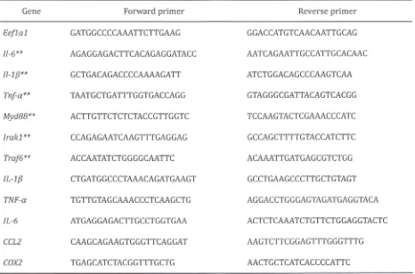
Table
1.Sequences of primers used to detect and quantitated gene expressions from cDNA synthesized
from isolated rat liver and differentiated THP-l cell RNA.
Gene
Eeflal
1I-6**
1I-1f3**
Tnf-a**
Myd88**
lrakl
**
Traf6**
IL-l
f3
TNF-a
IL-6
CCL2 COX2
Forward primer
GATGGCCCCAAATTCTTGAAG
AGAGGAGACTTCACAGAGGATACC
GCTGACAGACCCCAAAAGATT
TAATGCTGATTTGGTGACCAGG
ACTTGTTCTCTCTACCGTTGGTC
CCAGAGAATCAAGTTTGAGGAG
ACCAATATCTGGGGCAATTC
CTGATGGCCCTAAACAGATGAAGT
TGTTGTAGCAAACCCTCAAGCTG
ATGAGGAGACTTGCCTGGTGAA
CAAGCAGAAGTGGGTTCAGGAT
TGAGCATCTACGGTTTGCTG
Reverse primer
GGACCATGTCAACAATTGCAG
AATCAGAATTGCCATTGCACAAC
ATCTGGACAGCCCAAGTCAA
GTAGGGCGATTACAGTCACGG
TCCAAGTACTCGAAACCCATC
GCCAGCTTTTGTACCATCTTC
ACAAATTGATGAGCGTCTGG
GCCTGAAGCCCTTGCTGTAGT
AGGACCTGGGAGTAGATGAGGTACA
ACTCTCAAATCTGTTCTGGAGGTACTC
AAGTCTTCGGAGTTTGGGTTTG
AACTGCTCATCACCCCATTC
**
Sequence specific to detect and quantify rodent gene
(Rattus norvegicus) [image:14.595.69.535.103.411.2]Table 2. Weight of selected organs normalized to 100g body weight after ten days of experimental fe eding,
ip. injection and 18 h fasting.
Group Liver Lung Testis
Con - 2.57 ± 0.04 0.40 ± 0.01 1.14 ± 0.01
Con + 3.12 ± 0.07 0.49 ± 0.01 1.11 ± 0.03
GG- 2.71 ± 0.05 0.38 ± 0.02 1.13 ± 0.01
GG + 3.19 ± 0.05 b 0.44 ± 0.01 1.13 ± 0.02
[image:15.597.79.521.105.223.2]Figure legends.
Figure 1. GGOH suppresses LPS induced inflammation and liver damage independent of cholesterol
alteration in vivo. (A-C) Plasma inflammatory cytokines after LPS challenge were substantially decreased after ten days of 483 mg·kg-1 diet GGOH supplementation (GG +). (0, E) Inflammation caused liver damage
was similarly suppressed by GGOH supplementation as marked reduction in plasma concentrations of ALT
and AST. (F) Plasma concentration of cholesterol was markedly increased in LPS challenged rats, however
no alleviation was observed in GGOH supplemented group. (G) Plasma triglyceride concentration shows
no alteration. All values are mean ± SEM; n=4-8. The values with different letters (a, b, and c) are
significantly different at p<O.OS .
Figure 2. GGOH effectively dampens inflammatory genes and NF-KB in the liver. (A-C) Inflammatory gene expressions were significantly suppressed in livers of rats supplemented with GGOH 483 mg·kg-1 diet, correlating with decreased plasma cytokine concentrations. (0, E) Gene expressions of Irakl and Traf6
were suppressed by GGOH supplementation in both LPS challenged and unchallenged rats. (F) Western
blot of GGOH supplemented rat livers show suppressed Irakl and Traf6 protein levels. All values are mean
± SEM; n=S-8. The values with different letters (a, b, and c) are significantly different at p<O.OS.
Photographs are representative of 5 rats.
Figure 3. GGOH preincubation prevents over-activation ofNF-KB in LPS stimulated THP-l cells. THP-l cells
were differentiated with PMA then pre-incubated with GGOH or not for 24 h before LPS stimulation. After
3 h stimulation, cells were harvested for RNA isolation described in Methods. (A-C) Pre-incubation with high GGOH concentration effectively inhibited over-expression of inflammatory cytokine genes. (0, E)
Over-expressions of other NF-KB target genes involved in further inflammatory enhancement were also
suppressed by GGOH preincubation. All values are mean ± SEM; n=3. The values with different letters (a, b,
and c) are significantly different at p<O.OS. (F) Representative photograph of suppressed NF-KB p6S
phosphorylation and IKB degradation in GGOH pre-incubated cells. Cells were harvested after indicated
time of LPS stimulation. Western blot results are from 3 independent experiments. *The values are
significantly different compared from those of control at p<O .OS.
Figure 4. GGOH preincubation modulates NF-KB signaling molecules. Differentiated THP-l cells were
incubated with or without lOIlM GGOH for 24 h, then stimulated with LPS for indicated times. (A) After 3 h
of LPS stimulation, THP-l cells were harvested followed by RNA isolation and cDNA synthesis. Signal
transduction gene expressions were determined by quantitative RT-PCR. Values are mean ± SEM; n=3 . The
values with different letters (a, b, and c) are significantly different at p<O.OS. Western blot of total signal
transduction proteins (B) and their phosphorylated state (C) were markedly dampened in cells
pre-incubated with GGOH for 24 h. Photographs are representative of three independent experiments. *The
values are significantly different compared from those of control at p<O.OS.
Figure 1
A
Plasma il Mャセ@B
Plasma TNF-a140 b 50 b
E 120 E 40
---
セ@ 100---
OJ).3: c
80 c 30
0 0
-.:0 . ."
[1' 60
セ@ 20
セ@
c c
OJ 40 OJ
u u
C c 10
0 20 0
u u
0 0
Con - Con + GG - GG + Con - Con + GG- GG +
C
Plasma I L-6 D Plasma ALT1000 b 700 b
E
800 600
---
OJ).3: ::::; 500
---c 600 セ@
400 0
. ."
2':
[1'
400 300
C .;;
OJ B 200
a c
u ro a
c 200
0 100
u
0 0
E
Plasma ASTF
Plasma TC2500 b 70
b b
::; 60
2000 "0
--
a::; OJ) 50
--
E:2 1500 c 40
> 0
.'2i 1000
';:; 30
セ@ セ@
u c 20
ro a (lJ
500 u
c 10 0
u
0 0
Con - Con + GG- GG+ Con - Con + GG- GG +
G
Plasma TG180
::; 160
"0 140
--
OJ)E 120
c 100 0 ';:; 80
セ@c 60
(lJ
40 u
c
0 20
u 0
Con - Con + GG- GG +
Fi gu re 2
A
Hepatic IL- l(3 mRNAB
Hepatic TNF-a mRNA25 b 100
c
Q 20 c Q 80 b
V> V>
V> V>
セ@
0. 15 セ@0. 60
x X
Q) Q)
Q)
> 10
';0
Q)
40
>
';0
ro ro
Qj
cr: 5 Qj cr: 20
a
0 0
Ca n - Can + GG- GG + Ca n - Ca n + GG - GG +
C
Hepatic IL-6 mRNA0
Hepati c IRA Kl mRNA1200
セ@
b 1.5 b§ 1000
1
ca
'Vi 'Vi a
V>
800 V>
Q) Q) 1.0
C. x c C.
600 x
Q) Q)
Q) Q)
> 20
f
I
>
';0
0
';0 0.5ro ro
Qj
10 Qj
cr: cr:
a a
0 = 0
Can - Can + GG- GG + Can - Ca n + GG - GG +
E
Hepatic TRAF6 mRNA1.5
b
c
a
'Vi
V> a
Q) 1,0
C. x c
Q) Q)
>
';0 0.5
.!!!
Q)
cr:
0
Ca n - Con + GG - GG +
[image:19.599.77.499.74.694.2]F
1.2
c 1.0
0
Nセ@
i':' 0.8
セ@
'" 0.6
'" >
Nセ@ 0.4 a;
c: 0.2
1.8
c 1.6
0 1.4
i
1.2 1.0'"
'" 0.8 >
Nセ@ 0 .6
a; c: 0.4 0.2
Con
-Irakl
Traf6
P-p65
IKBa
a-tubulin
Liver Irak! WB
Con - Con + GG
-Liver P-p65 WB
Con - Con + GG-GG +
GG +
Con +
GG-
GG+
Liver Traf6 WB
1.2
c 1.0
0
'Bi
i':' 0.8
セ@ 0 .6
'" >
Nセ@ 0.4 a;
c: 0.2
Con · Con + GG - GG +
Liver IKBa WB
1.2
c 1.0
0
Nセ@
i':' 0.8
"-1ij 0.6
'" >
Nセ@ 0.4
a;
c: 0.2
A
5 c
0 4
Nセ@
Q)
a.
3x Q) Q) 2 > B セ@ ro Qj 1 0:: 0 LPS GGOH HセimI@
C
180 c 0 os:: 140 Q)a.
xQ) 100
Q) > B セ@ 60 セ@ Q) 0:: 1 0 LPS GGOH (;tM)
D
c o "Vi '" Q)a.
x Q) Q) > ";:; ro Qj 0:: LPS GGOH (;tM)セ@
8 7 6 4 1 o a0
iャMャセ@ mRNA b + + 1 Il-6 mRNA b b•
+"
+ 1CCl2 mRNA
[image:21.599.97.511.67.721.2]b + + 1 Figure 3 + 10 c
n
+ 10 + 10B
8 c 7"2
'" 6 '" セ@
c.
x
Q) 4 Q) > B セ@ セ@ Q) 0:: 0 LPS GGOH (;tM)
E
30g
25 "Viセ@ 20
c.
(;j 15
Q) £; 10
ro Qj 0:: LPS GGOH (;tM) TNF-a mRNA b + + +
1 10
COX2 mRNA
b
I
b cn
+ + +1 10
F
min
phos.-p65
IKBa
a-tubulin
c 0 '';::::; ro ... +-> c Q) u c 0 u Q) > +-> ro Q) 0:::: c 0 +-> ro ... +-> c Q) u c 0 u Q) > +-> ro Q) 0::::control
o
10 30 60 120
phos .-p65 western
4.0
3.0
2.0
1.0
0.0
0
10
30
time (min)
GGOH
o
10
30 60 120
60
120
• control
o
GGOHIKBa western
1.8
1.4
1.0
0.6
0.2
0.0
0
10
30
60
time (min)
120
A
B
2.0
1.8
1.6
c
0
1.4 Vl Vl
(l) 1.2
[image:23.597.113.444.97.688.2]
'--c.. x 1.0 (l) (l) Nセ@ 0.8 ...
ro
(l) 0.6
a::: 0.4
0.2
0
minutes
MyD88
TRAF6
IRAK1
TAK1
Figure 4
b
a
IRAKl TRAF6
D Can - セ c。ョ@
+
セ ggMTAKl
セ ggK@
control
10
セm@GGOH
o
10 30 60 120o
10 30 60 120a-tubulin
... _ ' ... . ,
セ@ "f!!!itiJIIII>- -1.4 Total MyD88
c 1.2 c
.Q .Q
Vl 1.0 Vl
Vl Vl
(IJ (IJ
.... ....
0.. 0.8 0..
x x
(IJ (IJ (IJ 0.6 (IJ
N セ@
....
>0.4 N セ@
rn セ@
OJ (IJ
.... ....
0.2
0.0
0 10 30 60 120
time (min)
Total IRAK1 2.5
c 2.0 c
.Q .Q
Vl Vl
Vl Vl
(IJ 1.5 (IJ
.... ....
0.. 0..
x x
(IJ (IJ (IJ 1.0 (IJ
> N セ@
N セ@
....
セ@ rn
セ@ 0.5 セ@
0.0
0 10 30 60 120
time (min)
TotallKKp 2.5
c 2.0
0
Vl Vl (IJ 1.5
....
0..
x
(IJ (IJ 1.0
N セ@
....
rn
(IJ 0.5
....
0.0
0 10 30 60 120
time (min) 2.0 1.5 1.0 0.5 0.0 3.5 3.0 2.5 2.0 2.5 1.0 0.5 0.0 Total TRAF6
o
10 30t ime (min)
Total TAK1
0 10
• control
D
GGOH30 time (min)
60 120
*
*
c
6.0
5.0 c 0
'Vi
Vl
4.0 Q)
'-a.
x 3.0
Q) Q) >
',j:; 2.0 ro Q)
0:: 1.0
0.0
0
control
10 /-lM GGOH
minutes
o
10 30 60 120 0 10 30 60 120P-TAKl
P- IKK
。Oセ@
a-tubulin ... _ ....
p-TAKl
*
10 30 60 120
time (min)
• Control
o
GGOH4.0 c 0
'Vi
Vl
3.0 Q)
'-a. x Q) Q) 2.0 >
',j:;
セ@
Q) 1.0
0::
0.0
ーMikk。Oセ@
0 10 30
time (min)
r
Supplementary Figure 1.
10 JlM GGOH
control
12 h
24 h
TRAF6
IRAK1
a-tubulin
TRAF6 western IRAK1 western
1.4 1.4
c 1.2 c 1.2
0 0
Vl
1.0 Vl 1.0
Vl Vl
Q) Q)
...
...
a. 0.8 a. 0.8
x
*
*
xQ) Q)
Q) 0.6 Q) 0.6
.2: ... .2:
0.4 ...
*
rc rc 0.4
Q) (ij
cr::
0.2 cr:: 0.2
0.0 0.0
Control 12 h 24 h Control 12 h 24 h
Supplementary Figure 1. Incubation with GGOH suppresses TRAF6 and IRAK1 expression in HepG2 cells.
HepG2 cells were incubated with GGOH for 0,12 or 24 h in DMEM with 10% FCS at 1.0 x 10⁶ cells·mL·l. After incubation period, cells were rinsed and lysed and immunoblotted with corresponding antibodies.
Supplementary Figure 2,
Incubation time (h)
Control a,s 1 3 24
IRAKlllil!tl.
セ@
セN[[N@
qLm'
TRAF6
- ' I,I!WIIL'
GDMエエセGセゥDセセ[BセE@ セ@.... . . .
a-tubulin • • • セ@• • セ サ@• • • • • •
THP-1I RAKl western
1.4 a
c 1.2 0 'Vi
1.0
Vl Q)
0. x 0,8 ab
Q)
Q) 0,6 b
>
.'"
ro 0.4 Qjc:r:: 0.2
a
Control 0.5 1 3 24
Incubation time (h)
THP-l TRAF6 western
2.5 c
0 2,0
'Vi Vl Q) 0. 1.5
x
Q) Q)
1.0
>
.'"
セ@
Q) a,s
c:r::
a
Control 0.5 1 3 24
Incubation time (h)
Supplementary Figure 2, Short incubation with GGOH promotes degradation and suppression of lRAK1
and TRAF6 expression in THP-1 cells, THP-1 cells were incubated with GGOH for 0 (control) or indicated
times in RPM! 1640 with 10% FCS at 1.0 x 10⁶ cells'mLol, After incubation period, cells were rinsed and
lysed and immunoblotted with corresponding antibodies, Values are mean ± SEM; n=3; p<O,OS ,