HAYATI Journal of Biosciences, September 2007, p 119-122 Vol. 14, No. 3 ISSN: 1978-3019
SHORT COMMUNICATION
Isolation and Characterization of Silaffin that Catalyze Biosilica
Formation from Marine Diatom C
haetoceros gracilis
AGNES IMELDA MANURUNG1, ALBERTA RIKA PRATIWI2, DAHRUL SYAH1, MAGGY THENAWIDJAJA SUHARTONO1∗∗∗∗∗
1Department of Food Science and Technology, Bogor Agricultural University, Darmaga Campus, Bogor 16680, Indonesia 2Department of Food Technology, Soegijapranata Chatholic University, Jalan Pawiyatan Luhur IV/1,
Bendan Duwur, Semarang 50234, Indonesia
Received September 8, 2006/Accepted August 1, 2007
The method of making silica in industries requires extreme conditions. The finding of proteins involved in the forma-tion of biosilica from diatoms, has opened up an alternative way of producforma-tion. Chaetoceros gracilis is one of the diatoms, which is potential in producing silaffin protein. This study aimed to isolate and to characterize the protein. We also analyzed the protein activity toward tetraethoxyorthosilicate (TEOS) substrate in in vitro reaction. Diatom biomass was harvested and further kept in 2% SDS/100 mM EDTA solution. Protein isolation was conducted by dissolving the silica and separating the protein by soaking in 2 M HF/8 M NH4F. Protein concentration was analyzed using Bradford method and the molecular weight was estimated through SDS-PAGE. Protein activity was observed by reacting it with TEOS substrate to form silica polymer and measured by colorimetric molibdate assay. Protein concentration was 1.20 mg/ml and appeared filamentous. The apparent molecular weights consisted of 12, 23, 42, 44 kDa. These protein was able to polymerize the silica at room temperature within 10 min. As much as 85.65 µµµµµmol TEOS was polymerized per 1.4 x 10⁶ silaffin protein per min.
SEM analysis showed the formation of spherical, aggregate biosilica.
Key words: Chaetoceros gracilis, silaffin protein, biosilica, polymerization
___________________________________________________________________________
The application of silica-based material is widely distributed. Silica and silicates are extensively used in cosmetic, paint, food industry, catalyst, semiconductor, and biosensor for various analysis (Perry 2003). Chemical synthesis of silica in industries requires extreme temperatures, pressure, pH, and dangerous chemical compound. In contrast, biological synthesis of silica in nature proceeds (such as occurring in Diatom) at ambient temperatures, pressure, and neutral pH. Diatoms are one of organism that produces nanostructure silica as the component of the cell wall. This organism is unicellular photosynthetic eukaryotes within the class Bacillariophyceae whose peculiarity amongst other microalgae is the silicaceous cell wall.
Diatom silica nanostructure is precisely controlled by the cell, which involves protein as the biocatalyst. The nature of this organic molecule was clarified through the characterization of diatom biosilica-associated peptides (silaffin) which accelerate silica formation from a silicic acid solution in vitro
(Kröger et al. 1999). Finding of silaffin protein that catalyzes the silica condensation is important for the design of technological process for silica production under mild and friendly environment and economically favorable.
Indonesia is a maritim country with high diversity in its marine diatoms. Diatom Chaetoceros gracilis is species that
can be a potential source of silaffin like-proteins, which probably shows unique characteristic in their capability of catalyzing biosilification. This research was aimed to isolate and to characterize the silaffin protein as well as to analyze its activity of toward TEOS substrate.
Culture Condition. The axenic culture of C. gracilis diatom was provided by Mariculture laboratory of Research Centre for Oceanography- Indonesian Institute of Science (LIPI). The culture was grown in modified f/2 medium. The f/2 medium was containing mayor nutrient (0.99 mM NaNO3, 0.07 mM NaH2PO4·2H2O, 5.28 µM Na2SiO3·9H2O), minor nutrient (5.36 µM FeCl3·6H2O and 26.86 µM Na2EDTA), vitamins (0.59 µM vitamin B1, 0.001 µM vitamin B12, 0.004 µM biotin) also trace metal (0.781 µM CuSO4·5H2O, 2.12 µM ZnSO4·7H2O, 0.521 µM NaMoO4·2H2O, 0.005 µM (NH4)6Mo7O24·4H2O, 18.19 µM MnCl2·4H2O, 0.61 µM CoCl2·6H2O). The medium was adjusted to pH 8. The culture was continuous bubbled with sterile air and maintained at 25 oC as well as under constant light at 4000 lux.
Biomass Collection and Silicaceous Cell Wall (Frustule) Extraction. Cell biomasses were harvested by centrifugation at 10,000 x g, 4 oC for 15 min. These biomasses were stored frozen at -20 oC until use.
Extraction of frustule was performed as described by Kröger
et al. (2002). The cell biomasses were boiled twice in 2% SDS/ 100 mM EDTA to remove intracellular component and
_________________ ∗
∗ ∗ ∗
membrane. Frustule were pelleted by centrifugation at 2,800 xg, extracted with acetone twice, washed extensively with distilled water, and dried at room temperature until further analysis.
Isolation and Characteristic of Silaffin Protein. Isolation of C. gracilis silaffin protein was performed as described by Shimizu et al. (1998). Silaffin protein is tightly associated with diatom biosilica and can only be solubilized by complete removal of the silica. The frustule was dissolved in 50 ml 2 M HF (hydrogen fluoride)/8 M NH4F (ammonium fluoride) (pH 5). After incubation for 30 min at 0 oC, HF/NH
4F was evaporated. This extract was dialyzed (2 kDa-cut off) once against distilled water at 4 oC for 4h and repeated 9 times. The dialysate was centrifuged (10,000 xg, 20 min) at 4 oC. The precipitate protein was dissolved with distilled water and stored at 4 oC until further analysis. The morphology of protein was observed by microscopic analysis. Concentrations of protein were measured by Bradford methods (dye-binding method) with protein Bovine Serum Albuminas standard (Dunn 1989). The protein was counted by using Haemacytometer.
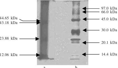
Estimation of protein molecular weight were conducted using SDS-PAGE as described by Laemmli(1970). Protein (10-15 µl) was dissolved in sample buffer solution (0.3 ml of 1 M Tris-HCl pH 6.8, 2.5 ml of 50% glycerol, 1.0 ml of 10% SDS, 0.25 ml of 2-â mercaptoetanol, 0.5 ml of 1% bromephenol blue and 0.45 ml aquadest). These suspension was boiled at 95 oC for 5 min and was run on 10% polyacrilamide gel for 100 Volt for 1.5 h. Low Molecular Weight protein as marker contained phosphorylase b (97.0 kDa), albumin (66.0 kDa), ovalbumin (45.0 kDa), carbonic anhidrase (30.0 kDa), trypsin inhibitor (20.1 kDa), á-lactalbumin (14.4 kDa) (Amersham Biosience, USA). Silver staining was used following SDS-PAGE.
Silaffin Protein Activity in the In Vitro Reaction. Protein solution was reacted in 50 ml of 50 mM CH3COONa/ CH3COOH pH 5.5 and incubated at 18 oC for 30 min. Assay of protein activity was performed using monosilicic acid as a substrate. A stock of substrate solution was prepared by hydrolyzing 10 ml of 1 M TEOS in 100 ml of 1 mM HCl for 10 min at 25 oC. Immediately after incubation, aliquots of the silicic acid solution were withdrawn and added to the reaction mixtures to give a final concentration of 100 mM silicic acid. Subsequently, the mixtures of reaction were incubated with different incubation time (10, 30 min, 1, 12, and 24 h) at 18 oC by shaker incubator (100 rpm).
The reaction mixture were centrifuged and immediately processed either for quantification of silica or silica structure analysis. Silica quantification was conducted by Colorimetric molibdate assay(Strickland & Parsons 1972). The precipitate silica was washed 3 times with ethanol to remove free TEOS (that was not polymerized), and recentrifuged. Subsequently, the precipitated silica was incubated in 1 ml of 1 M NaOH at 85-95 oC for 48 h. The samples of 0.2 ml was reacted with 0.4 ml of 0.8% ammonium molybdate and 12 ml of 36% HCl solution for 10 min, mixed with 0.6 ml reduction reagent (2 ml of metol sulfite containing 1.2% Na2SO3 and 2% p-metylaminophenol sulfate, 1.2 ml of 50% H2SO4, 1.2 ml of 10% saturated oxalate acid, 1.6 ml water distilled), for 3 h. The absorbance was read
at ë 810 nm. Different silicon concentrations (TEOS in DMSO solution) between 20-240 µmol/ml were used as standard.
The silica polymer structure was observed by Scanning Electron Microscope(SEM). Sample was centrifuged at 16,000 xg for 2 min and resuspended in 5 µl aquadest, mounted onto a 200-mesh copper grid (diameter 3.05 mm; Agar Scientific, Stansted, Essex, U.K.), washed and air dried. Photograph with JEOL JSM-5310LV SEM was prepared before photographing with JEOL JFC-1200 Fine Coater film.
Biomass and Frustule. Biomass cell of 19.3 g produced frustules as much as 0.06 g (Figure 1). The silica frustule is composed of the silaffin protein. Protein was tied very strongly with the silica. Therefore, we needed to dissolve the silica to obtain the silaffin molecule.
Characteristic of Silaffin Protein. After extraction using HF/NH4F solvent the macromolecule protein was released. The microscopic analysis of protein showed filamentous appearance filament (Figure 2). As much as 1.4 x l06 protein filaments per ml of the solution was found. We found the concentration of silaffin protein at 1.2 mg/ml by using Bradford methods.
SDS-PAGE analysis of these proteins revealed four distinct protein bands. The molecular weight of each protein was 44.65, 42.18, 23.88, and 12.06 kDa (Figure 3).
Activity of Silaffin Protein. Results of the silaffin protein reaction with the TEOS substrate showed that this substrate was polymerized up to 856.58 µmol/ml with incubation time of ten min. Therefore, the TEOS was polymerized at 8.56 µmol/ ml per min and at 1.5 x 10¹¹ molecules per protein filament (Table 1). Analysis using SEM (3500 x) showed clearly the formation of the silica polymer with round (spherical) shape. In addition, the spherical silica structures also showed aggregate formation (Figure 4).
The silica component in the diatom frustules (silica
frustules) could read up to approximately 90% of the dry cell (Round et al. 1990). However, this concentration depends on the diatom species. Each diatom species possesses specific characteristics of each wall that was marked by the content and the type of silica structure. The process of frustules formation is not yet known clearly but is suspected to involve silica diffusion. The process of this diffusion, known as sintering, is affected by several factors for example pH and temperatures, which can be varied if the diatom is cultivated in different conditions (Parkinson & Gordon 1999).
The protein isolated from diatom C. gracilis in this study was found at 1.2 mg/ml. This protein is strongly attached onto the silica cell wall of the diatom. Therefore, the success in isolating the protein is determined by how far the silica could be dissolved. Frigeri et al. (2006) stated that EDTA plays a role in eliminating Ca2+ from diatom cell wall and SDS disrupt the membrane to expose the silica containing silaffin protein. Addition of HF/NH4F, further dissolves the silica and reveals the silaffin.
Biosilica protein (silaffin protein) in this study appeared filamentous with length in range at 0.036-0.096 mm. As much as 1.4 x 10⁶ protein filament was obtained per ml solution. However, silicatein protein isolated from sponge Tetya aurantia that catalyzed silica polymerization showed needle
or stick shape (Shimizu et al. 1998; Krasko et al. 2000). Polymerization of silica that was formed attached to the protein. Both silaffin and silicatein protein were used as template for silica polymerization (Cha et al. 1999; Kröger et al. 2002).
Estimation of the protein molecular weight based on analysis of SDS PAGE with 10% polyacrilamide showed four bands of 44, 42, 23, and 12 kDa. Poulsen et al. (2003) reported that on the biosilica protein from diatom Cylindrotheca fuciformis found 3 peptides with molecular weight of 6.5, 10, and 40 kDa known as natSil-lA, natSil-lB, and natSil-2 respectively. Based on the weight similarity, the protein with molecular weight of 42 kDa from diatom C. gracilis was probably natSil-2 and the 12 kDa protein was probably similar
Figure 1. Silica frustule of Diatom C. gracilis.
12.06 kDa 43.18 kDa 44.65 kDa
23.88 kDa
97.0 kDa 66.0 kDa
45.0 kDa
30.0 kDa
20.1 kDa
14.4 kDa
Figure 3. Apparent of molecular weight of protein fraction from diatom C. gracilis analysis by SDS-PAGE (a), LMW marker (b). a b
Table 1. Bioactivity of silaffin protein from C. gracilis with TEOS substrate
Silica attached to protein
µmole/ml Molecule per protein per minute filament Incubation time
(minute)
10 30 60 720 1440
856.58 992.10 1094.75 1200.00 1003.95
85.66 33.07 18.25 1.67 0.70
1.5 x 10¹¹
5.8 x 10¹⁰
3.2 x 10¹⁰
2.9 x 10⁹
1.2 x 10⁹
µmole/ml
Number of protein filament adds in each reaction are 1.4 x 10⁶ filament/ml
to natSil-1B. In this study natSil-lA was not detected, possibly this peptide was present at very low concentration. This result is in agreement with the study of biosilica protein from diatom
Thalasiosira pseudonana, which revealed four peptides.
Three peptides have molecular weight of 35 kDa and two peptides have molecular weight of 19 kDa and both of them were isomers (Poulsen & Kröger 2004). The cell wall silica shows specific structure that is controlled by a specific protein biosilica in each diatom cell. According to Sumper and Kröger (2004), the silaffin diverse chemical structure found in various diatoms implied no homology in their protein sequence the proteins found in different kind diatom evidently have no similarity or homology in their amino acid. In nature, each kind of diatom is formed by unique silica structure, which is genetically programmed. Reaction of the protein catalyst from different diatom will be produce precipitation of silica with different structures.
The result of in vitro reaction between biosilica protein from C. gracilis and TEOS substrate in this study proved that the biosilica protein (silaffin) had capability to catalyze the silica polymerization in a very short time (10 mins) at room temperature and pH 5.5. This is clearly advantageous compare to chemical production of silica in industry with temperature of more of hundreds to thousands oC which also required long period of time (Harsono 2002). The formation of silica precipitation in short time is thus promising for production of environment-friendly silica biofabrications. Table 1 showed the highest activity of the protein, which polymerized TEOS at 856.58 ì mol/ml per 1.2 mg protein filament within ten minutes or 85.658 ì mol/ml per min. This result was higher than the polymerization capacity of silicatein (biosilica protein) found in sponge. Research in biosilica protein from sponge ST1
Figure 4. The spherical morphology of silica precipitated forming aggregate after in vitro reaction between silaffin and TEOS substrate.
5 µm
Figure 2. SEM micrograph of silaffin protein with filamentous shape from diatom C. gracilis.
10 µm
(Binuangen) and ST3 (Nias Island) reported polymerization of TEOS as much as 22 and 42 µmol/ml per min (Nurjanah 2005). This showed that the sponge silicatein polymerized the silica slower than the diatom silaffin.
Observation of the silica polymerization by SEM showed that silaffin diatom C. gracilis could polymerize the silica. The polymerized silica on the silaffin was found as aggregate structure with spherical shape. This study result was in agreement with silica precipitation from diatom T.pseudonana
produced spherical silica. However, T. pseudonana produced various spherical forms like silica with the diameter of 230 nm, silica porous sheet with the diameter of 20-200 nm, silica plates of densely packed and silica sphere polydisperse with the diameter of 0.9-42 µm (Poulsen & Kröger 2004).
Cell morphology of T. pseudonana and C. gracilis were centric, however they produce different type of silica form in the in vitro reaction. C.fuciformis with pinnate morphology was reported to produce spherical silica precipitate (Kröger
et al. 1999). Figure 4 shows the formation of silica aggregate by the protein catalyst (silaffin protein) isolated from diatom
C. gracilis. The protein with silica polymers appeared different from the protein before the reaction (Figures 4 & 2).
According to Sumper and Kröger (2004), formation of silica from silicate acid by silaffin protein can be divided into three stages i.e. polymerization of silicate acid through the condensation of siloxane, forming dimer, trimer, and further into cyclic oligomers. Oligosilicate acids tend to polymerize silica by forming the maximal association of (Si-O-Si). In this stage, polysilicate acid was formed as the core of further formation of biosilica. In the second stage, the core of the polysilicate acid grows forming spherical particle with further polymerization. The monomer and olygomer of silicate acid were fused into these particles. During the last stage, nanosphere silica form network of three dimensions from the particle’s cross chains of the associated siloxan.
Silica that was produced by protein silaffin shows diameter of 100-1000 nm and appear as round in shape (Kröger et al.
2002). According to Poulsen et al. (2003), the size of silica particle is determined by the silaffin concentrations. The increasing silaffin concentration will increase the size of the silica particle as well. However, the amount of silica that is precipitated will be decreased and this biosilica layer will be thinner. In this case, the polymerized silica will be more easily broken during SEM analysis. According to Kröger et al. (1999), silaffin could carry out polymerization and precipitation of silica spontaneously with diameter of the particle being ~50 nm when peptides Sil 1A, IB, and 2 are available. However, if only silaffins 1A exist, the size of the silica particle formed will be approximately 700 nm. Moreover, it is known that type of silicatein fraction such as Sil 1A, Sil 1B, and Sil 2 affected silica diameter. To understand further about the activity of
the silaffin fractions, it is necessary to conduct the polymerization reaction in the presence of various composite fractions of isolated silaffin.
ACKNOWLEDGEMENT
This research is part of the project “Biofabrication of nanosilica structure from Indonesian marine sponge and diatom”, supported from Basic Research Project-DIKTI-2005/ 2006 to Dahrul Syah (Department of Food Science and Technology, Bogor Agricultural University).
REFERENCES
Cha JN et al. 1999. Silicatein filaments and subunits from a marine sponge direct the polymerization of silica and silicones in vitro.
Proc Nat Acad Sci USA 96:361-365.
Dunn MJ. 1989. Determination of total protein concentration. In: Harris EL, Angal S (eds). Protein purification methods. Oxford IRL.
Frigeri LG, Radabaugh TR, Haynes PA, Hildebrand M. 2006. Identification of proteins from diatom cell wall fraction of the Thalassiosira pseudonana: insight into silica structure formation.
Mol Cell Proteomic 5:182-193.
Harsono H. 2002. Pembuatan silika amorf dari limbah sekam padi. J
Ilmu Dasar 3:98-103.
Krasko A et al. 2000. Expression of silicatein and collagen genes in the marine sponge Suberites domuncula is controlled by silicate and myotrophin. Eur J Biochem 267:4878-4887.
Kröger N, Deutzmann R, Sumper M. 1999. Polycationic peptides from biosilica that direct silica nanosphere formation. Science 286:1129-1132.
Kröger N, Lorenz S, Brunner E, Sumper M. 2002. Self-assembly of highly phosphorylated silaffins and their function in biosilica morphogenesis. Science 298:584-586.
Laemmli UK. 1970. Cleavage of structural proteins during the assembly of the head of the bacteriophage T4. Nature 227:680-685.
Nurjanah S. 2005. Eksplorasi protein dari sponge asal perairan pulau Nias dan Binuangen yang mengkatalisis polimerisasi silika [Thesis]. Bogor: Bogor Agricultural Univ.
Parkinson J, Gordon R. 1999. Beyond micromachining: the potential of diatom. Trends Biotechnol 17:190-195.
Perry CC. 2003. Silification: The processes by which organism capture and mineralize silica. J Mineral Geochem 54:291-327.
Poulsen N, Kröger N. 2004. Silica morphogenesis by alternative processing of silaffin in the diatom Thalassiosira pseudonana. J
Biol Chem 279:42993-42999.
Poulsen N, Sumper M, Kröger N. 2003. Biosilica formation in diatoms: characterization of native silaffin-2 and its role in silica morphogenesis. Proc Nat Acad Sci 100:12075-12080.
Round FE, Crawford RM, Mann DG. 1990. The Diatom. USA: Cambridge Univ.
Shimizu K, Cha J, Stucky GD, Morse DE. 1998. Silicatein alpha: cathepsin L-like protein in sponge biosilica. Proc Nat Acad Sci
95:6234-6238.
Strickland JDH, Parsons T. 1972. A Practical Handbook of Seawater Analysis. 2nd ed. Ottawa: Fisheries Research Board of Canada. Sumper M, Kröger N. 2004. Silica Formation in Diatoms: the function