Clinical Anatomy
Applied anatomy for students and junior doctors
A revision and applied anatomy
for clinical students
H A R O L D
◊
E L L I S
CBE, MA, DM, MCh, FRCS, FRCP, FRCOG, FACS (Hon) Clinical Anatomist, Guy’s, King’s and
St Thomas’ School of Biomedical Sciences; Emeritus Professor of Surgery, Charing Cross and Westminster Medical School, London;
Formerly Examiner in Anatomy, Primary FRCS (Eng)
Blackwell Publishing, Inc., 350 Main Street, Malden, Massachusetts 02148-5020, USA Blackwell Publishing Ltd, 9600 Garsington Road, Oxford OX4 2DQ, UK
Blackwell Publishing Asia Pty Ltd, 550 Swanston Street, Carlton, Victoria 3053, Australia
The right of the Author to be identified as the Author of this Work has been asserted in accordance with the Copyright, Designs and Patents Act 1988.
All rights reserved. No part of this publication may be reproduced, stored in a retrieval system, or transmitted, in any form or by any means, electronic, mechanical, photocopying, recording or otherwise, except as permitted by the UK Copyright, Designs and Patents Act 1988, without the prior permission of the publisher.
First published 1960 Seventh edition 1983 Second edition 1962 Revised reprint 1986 Reprinted 1963 Eighth edition 1992 Third edition 1966 Ninth edition 1992 Fourth edition 1969 Reprinted 2000 Fifth edition 1971 Tenth edition 2002 Sixth edition 1977 Reprinted 2003, 2004 Reprinted 1978, 1980 Greek edition 1969 Eleventh edition 2006
1 2006
Library of Congress Cataloging-in-Publication Data Data available
ISBN-13: 978-1-4051-3804-8 ISBN-10: 1-4051-3804-1
A catalogue record for this title is available from the British Library
Set in 9/12 Palatino by SNP Best-set Typesetter Ltd., Hong Kong Printed and bound in India by Replika Press Pvt Ltd
Commissioning Editor: Martin Sugden Editorial Assistant: Ellie Bonnett Development Editor: Mirjana Misina Production Controller: Kate Charman
For further information on Blackwell Publishing, visit our website: http://www.blackwellpublishing.com
The publisher’s policy is to use permanent paper from mills that operate a sustainable forestry policy, and which has been manufactured from pulp processed using acid-free and elementary chlorine-free practices. Furthermore, the publisher ensures that the text paper and cover board used have met acceptable
environmental accreditation standards.
Preface, xiii
Acknowledgements, xiv
Part 1:
◊
The Thorax
Surface anatomy and surface markings, 3
◊◊Surface markings of the more important thoracic contents, 3
The thoracic cage, 7
◊◊The thoracic vertebrae, 7 ◊◊The ribs, 7◊◊The costal cartilages, 10 ◊◊The sternum, 11
◊◊The intercostal spaces, 11 ◊◊The diaphragm, 14 ◊◊The pleurae, 18
The lower respiratory tract, 19
◊◊The trachea, 19◊◊The bronchi, 23 ◊◊The lungs, 23
The mediastinum, 28
◊◊The pericardium, 28 ◊◊The heart, 29◊◊The superior mediastinum, 42 ◊◊The oesophagus, 42
◊◊The thoracic duct, 45
◊◊The thoracic sympathetic trunk, 47
On the examination of a chest radiograph, 49
◊◊Radiographic appearance of the heart, 50Part 2:
◊
The Abdomen and Pelvis
Surface anatomy and surface markings, 55
◊◊Vertebral levels, 55◊◊Surface markings, 55
The fasciae and muscles of the abdominal wall, 58
◊◊Fasciae of the abdominal wall, 58◊◊The anatomy of abdominal incisions, 61 ◊◊The inguinal canal, 63
Peritoneal cavity, 65
◊◊Intraperitoneal fossae, 68 ◊◊The subphrenic spaces, 69The gastrointestinal tract, 70
◊◊The stomach, 70◊◊The duodenum, 75 ◊◊Small intestine, 77 ◊◊Large intestine, 78 ◊◊The appendix, 79 ◊◊The rectum, 81
◊◊Arterial supply of the intestine, 86 ◊◊The portal system of veins, 87 ◊◊Lymph drainage of the intestine, 88 ◊◊The structure of the alimentary canal, 88
◊◊The development of the intestine and its congenital abnormalities, 90
The gastrointestinal adnexae: liver, gall-bladder and its
ducts, pancreas and spleen, 93
◊◊The liver, 93
◊◊The biliary system, 98 ◊◊The gall-bladder, 99 ◊◊The pancreas, 101 ◊◊The spleen, 104
The urinary tract, 105
◊◊The kidneys, 105 ◊◊The ureter, 109◊◊The embryology and congenital abnormalities of the kidney and ureter, 110
◊◊The bladder, 112 ◊◊The urethra, 115
The male genital organs, 116
◊◊The prostate, 116◊◊The scrotum, 119
◊◊Testis and epididymis, 119
◊◊Vas deferens (ductus deferens), 123 ◊◊The seminal vesicles, 124
The bony and ligamentous pelvis, 124
◊◊The os innominatum, 124◊◊The sacrum, 125 ◊◊The coccyx, 126
◊◊Joints and ligamentous connections of the pelvis, 127 ◊◊Differences between the male and female pelvis, 128 ◊◊Obstetrical pelvic measurements, 128
◊◊Variations of the pelvic shape, 130
The muscles of the pelvic floor and perineum, 132
◊◊The anterior (urogenital) perineum, 133◊◊The posterior (anal) perineum, 134
The female genital organs, 136
◊◊The vulva, 136◊◊The vagina, 137 ◊◊The uterus, 139
◊◊The Fallopian tubes, 144 ◊◊The ovary, 145
◊◊The endopelvic fascia and the pelvic ligaments, 146 ◊◊Vaginal examination, 147
◊◊Embryology of the Fallopian tubes, uterus and vagina, 148
The posterior abdominal wall, 149
◊◊The suprarenal glands, 151◊◊Abdominal aorta, 151 ◊◊Inferior vena cava, 153
◊◊Lumbar sympathetic chain, 153
Part 3:
◊
The Upper Limb
The female breast, 159
◊◊Structure, 159◊◊Blood supply, 159 ◊◊Lymphatic drainage, 159 ◊◊Development, 161
Surface anatomy and surface markings of the
upper limb, 162
◊◊Bones and joints, 163 ◊◊Muscles and tendons, 164 ◊◊Vessels, 166
◊◊Nerves, 167
The bones and joints of the upper limb, 168
◊◊The scapula, 168◊◊The clavicle, 168 ◊◊The humerus, 169 ◊◊The radius and ulna, 171 ◊◊The bones of the hand, 174 ◊◊The shoulder, 176
◊◊The wrist joint, 183 ◊◊The joints of the hand, 184
The arteries of the upper limb, 186
◊◊The axillary artery, 186◊◊The brachial artery, 187 ◊◊The radial artery, 187 ◊◊The ulnar artery, 188
The brachial plexus, 189
◊◊The segmental cutaneous supply of the upper limb, 191
The course and distribution of the principal nerves of the
upper limb, 191
◊◊The axillary nerve, 191 ◊◊The radial nerve, 192 ◊◊Branches, 194
◊◊The musculocutaneous nerve, 194 ◊◊The ulnar nerve, 194
◊◊The median nerve, 195
The anatomy of upper limb deformities, 197
The spaces of the hand, 200
◊◊The superficial pulp space of the fingers, 200
◊◊The ulnar and radial bursae and the synovial tendon sheaths of the fingers, 201
Part 4:
◊
The Lower Limb
The anatomy and surface markings of the lower limb, 207
◊◊Bones and joints, 207◊◊Bursae of the lower limb, 207 ◊◊Mensuration in the lower limb, 208 ◊◊Muscles and tendons, 211
◊◊Vessels, 211 ◊◊Nerves, 214
The bones and joints of the lower limb, 216
◊◊The os innominatum, 216◊◊The femur, 216 ◊◊The patella, 220 ◊◊The tibia, 223 ◊◊The fibula, 224
◊◊A note on growing ends and nutrient foramina in the long bones, 225 ◊◊The bones of the foot, 225
◊◊The knee joint, 229 ◊◊The tibiofibular joints, 233 ◊◊The ankle, 233
◊◊The joints of the foot, 234 ◊◊The arches of the foot, 235 ◊◊The anatomy of walking, 237
Three important zones of the lower limb— the femoral
triangle, adductor canal and popliteal fossa, 237
◊◊The femoral triangle, 237 ◊◊The fascia lata, 238
◊◊The femoral sheath and femoral canal, 238 ◊◊Femoral hernia, 239
◊◊The lymph nodes of the groin and the lymphatic drainage of the lower limb, 241
◊◊The adductor canal (of Hunter) or subsartorial canal, 242 ◊◊The popliteal fossa, 242
The arteries of the lower limb, 244
◊◊Femoral artery, 244◊◊Popliteal artery, 246 ◊◊Posterior tibial artery, 246 ◊◊Anterior tibial artery, 246
The veins of the lower limb, 247
◊◊Clinical features, 249The course and distribution of the principal nerves of the
lower limb, 249
◊◊The lumbar plexus, 250 ◊◊The sacral plexus, 251 ◊◊The sciatic nerve, 253 ◊◊The tibial nerve, 255
◊◊The common peroneal (fibular) nerve, 255
◊◊Segmental cutaneous supply of the lower limb, 256
Part 5:
◊
The Head and Neck
The surface anatomy of the neck, 261
◊◊The fascial compartments of the neck, 262The thyroid gland, 264
◊◊The parathyroid glands, 267The palate, 270
The tongue and floor of the mouth, 272
◊◊The tongue, 272◊◊The floor of the mouth, 276
The pharynx, 277
◊◊The nasopharynx, 277 ◊◊The oropharynx, 278 ◊◊The palatine tonsils, 279 ◊◊The laryngopharynx, 280◊◊The mechanism of deglutition, 282
The larynx, 284
◊◊Blood supply, 287 ◊◊Lymph drainage, 287 ◊◊Nerve supply, 288The salivary glands, 289
◊◊The parotid gland, 289 ◊◊The submandibular gland, 292 ◊◊The sublingual gland, 293The major arteries of the head and neck, 294
◊◊The common carotid arteries, 294◊◊The external carotid artery, 294 ◊◊The internal carotid artery, 296 ◊◊The subclavian arteries, 298
The veins of the head and neck, 301
◊◊The cerebral venous system, 301◊◊The venous sinuses of the dura, 301 ◊◊The internal jugular vein, 303 ◊◊The subclavian vein, 305
The lymph nodes of the neck, 306
The cervical sympathetic trunk, 308
The branchial system and its derivatives, 310
◊◊Branchial cyst and fistula, 310The surface anatomy and surface markings of the head, 311
The scalp, 312
The accessory nasal sinuses, 318
◊◊The frontal sinuses, 318◊◊The maxillary sinus (antrum of Highmore), 319 ◊◊The ethmoid sinuses, 320
◊◊The sphenoid sinuses, 321
The mandible, 321
◊◊The temporomandibular joint, 322 ◊◊The teeth, 323
The vertebral column, 324
◊◊The cervical vertebrae, 325 ◊◊The thoracic vertebrae, 327 ◊◊The lumbar vertebrae, 327 ◊◊The sacrum, 327◊◊The coccyx, 327
◊◊The intervertebral joints, 328
Part 6:
◊
The Central Nervous System
The spinal cord, 333
◊◊Age differences, 333 ◊◊Structure, 333◊◊Descending tracts, 334 ◊◊Ascending tracts, 336
◊◊The membranes of the cord (the meninges), 337
The brain, 339
◊◊The medulla, 339 ◊◊The pons, 342 ◊◊The cerebellum, 342 ◊◊The midbrain, 344 ◊◊The diencephalon, 346 ◊◊The hypothalamus, 346◊◊The pituitary gland (hypophysis cerebri), 347 ◊◊The thalamus, 349
◊◊The cerebral hemispheres, 349 ◊◊The cerebral cortex, 349 ◊◊The insula, 352
◊◊The connections of the cerebral cortex, 352 ◊◊The basal ganglia, 353
◊◊The long ascending and descending pathways, 354 ◊◊The membranes of the brain (the meninges), 360
◊◊The ventricular system and the cerebrospinal fluid circulation, 361
The cranial nerves, 364
◊◊The olfactory nerve (I), 364◊◊The oculomotor nerve (III), 366 ◊◊The trochlear nerve (IV), 368 ◊◊The trigeminal nerves (V), 369 ◊◊The abducent nerve (VI), 374 ◊◊The facial nerve (VII), 375
◊◊The auditory (vestibulocochlear) nerve (VIII), 377 ◊◊The glossopharyngeal nerve (IX), 379
◊◊The vagus nerve (X), 379 ◊◊The accessory nerve (XI), 381 ◊◊The hypoglossal nerve (XII), 381
The special senses, 383
◊◊The nose, 383◊◊The ear, 384
◊◊The eye and associated structures, 388
The autonomic nervous system, 393
◊◊Visceral afferents, 396◊◊The sympathetic system, 396 ◊◊The sympathetic trunk, 396 ◊◊The parasympathetic system, 399
Glossary of eponyms, 403
Experience of teaching clinical students at five medical schools and of examining them in sixteen cities and in eight countries has convinced me that there is still an unfortunate hiatus between the anatomy which the student learns in the pre-clinical years and that which is later encountered in the wards and operating theatres.
This book attempts to counter this situation. It does so by highlighting those features of anatomy which are of clinical importance using a vertical blue bar, in radiology, pathology, medicine and midwifery as well as in surgery. It presents the facts which students might reasonably be expected to carry with them during their years on the wards, through their final examinations and into their postgraduate years; it is designed for the clini-cal student.
Anatomy is a vast subject and, therefore, in order to achieve this goal, I have deliberately carried out a rigorous selection of material so as to cover only those of its thousands of facts which I consider form the necessary anatomical scaffolding for the clinician. Wherever possible practical appli-cations are indicated throughout the text — they cannot, within the limita-tions of a book of this size, be exhaustive, but I hope that they will act as signposts to the student and indicate how many clinical phenomena can be understood and remembered on simple anatomical grounds.
In this eleventh edition a complete revision of the text has been carried out. New figures have been added and other illustrations modified. Repre-sentative computerized axial tomography and magnetic resonance imaging films have been included, since these techniques have given increased impetus to the clinical importance of topographical anatomy.
The continued success of this volume, now in its forty-seventh year of publication, owes much to the helpful comments which the author has received from readers all over the world. Every suggestion is given the most careful consideration in an attempt to keep the material abreast of the needs of today’s medical students.
Harold Ellis
2006
xiv
I wish to thank the many students who have sent suggestions to me, many of which have been incorporated into this new edition.
To Mrs Katherine Ellis go my grateful thanks for invaluable secretarial assistance. New and revised illustrations were skilfully produced by Jane Fallows and new MR scans were provided by Dr Sheila Rankin of the Department of Radiology at Guy’s Hospital and Professor Adrian Dixon of Cambridge.
I am grateful to the following authors for permission to reproduce illus-trations:
The late Lord Brock for Figs 20 and 21 (from Lung Abscess); and
Professor R. G. Harrison for Figs 12, 32 and 69 (from A Textbook of Human Embryology).
Dr Colin Stolkin gave valuable help in revising the anatomy of the C.N.S.
Finally, I wish to express my debt to Martin Sugden and the staff of Blackwell Publishing for their continued and unfailing help.
surface markings
The experienced clinician spends much of his working life relating the surface anatomy of his patients to their deep structures (Fig. 1; see also Figs. 11 and 22).
The following bony prominences can usually be palpated in the living subject (corresponding vertebral levels are given in brackets):
•◊◊superior angle of the scapula (T2);
•◊◊upper border of the manubrium sterni, the suprasternal notch (T2/3); •◊◊spine of the scapula (T3);
•◊◊sternal angle (of Louis) — the transverse ridge at the manubrio-sternal junction (T4/5);
•◊◊inferior angle of scapula (T8); •◊◊xiphisternal joint (T9);
•◊◊lowest part of costal margin—10th rib (the subcostal line passes through L3).
Note from Fig. 1 that the manubrium corresponds to the 3rd and 4th thoracic vertebrae and overlies the aortic arch, and that the sternum corre-sponds to the 5th to 8th vertebrae and neatly overlies the heart.
Since the 1st and 12th ribs are difficult to feel, the ribs should be enu-merated from the 2nd costal cartilage, which articulates with the sternum at the angle of Louis.
The spinous processes of all the thoracic vertebrae can be palpated in the midline posteriorly, but it should be remembered that the first spinous process that can be felt is that of C7 (the vertebra prominens).
The position of the nipplevaries considerably in the female, but in the male it usually lies in the 4th intercostal space about 4in (10cm) from the midline. The apex beat, which marks the lowest and outermost point at which the cardiac impulse can be palpated, is normally in the 5th inter-costal space 3.5in (9cm) from the midline (just below and medial to the nipple).
The tracheais palpable in the suprasternal notch midway between the heads of the two clavicles.
Surface markings of the more important
thoracic contents
(Figs 2–4)The trachea
The trachea commences in the neck at the level of the lower border of the cricoid cartilage (C6) and runs vertically downwards to end at the level of the sternal angle of Louis (T4/5), just to the right of the mid-line, by divid-ing to form the right and left main bronchi. In the erect position and in full inspiration the level of bifurcation is at T6.
Fig. 1◊Lateral view of the thorax—its surface markings and vertebral levels. (Note that the angle of Louis (T4/5) demarcates the superior mediastinum, the upper margin of the heart and the beginning and end of the aortic arch.)
The pleura
The cervical pleuracan be marked out on the surface by a curved line drawn from the sternoclavicular joint to the junction of the medial and middle thirds of the clavicle; the apex of the pleura is about 1 in (2.5 cm) above the clavicle. This fact is easily explained by the oblique slope of the first rib. It is important because the pleura can be wounded (with consequent
Fig. 3◊The surface markings of the lungs and pleura—posterior view.
pneumothorax) by a stab wound — and this includes the surgeon’s knife and the anaesthetist’s needle—above the clavicle.
The lines of pleural reflexion pass from behind the sternoclavicular joint on each side to meet in the midline at the 2nd costal cartilage (the angle of Louis). The right pleural edge then passes vertically downwards to the 6th costal cartilage and then crosses:
•◊◊the 8th rib in the midclavicular line; •◊◊the 10th rib in the midaxillary line;
•◊◊the 12th rib at the lateral border of the erector spinae.
On the left side the pleural edge arches laterally at the 4th costal carti-lage and descends lateral to the border of the sternum, due, of course, to its lateral displacement by the heart; apart from this, its relationships are those of the right side.
The pleura actually descends just below the 12th rib margin at its medial extremity — or even below the edge of the 11th rib if the 12th is unusually short; obviously in this situation the pleura may be opened acci-dentally in making a loin incision to expose the kidney, perform an adrena-lectomy or to drain a subphrenic abscess.
The lungs
The surface projection of the lung is somewhat less extensive than that of the parietal pleura as outlined above, and in addition it varies quite consid-erably with the phase of respiration. The apexof the lung closely follows the line of the cervical pleura and the surface marking of the anterior border of the right lungcorresponds to that of the right mediastinal pleura. On the left side, however, the anterior borderhas a distinct notch (the cardiac notch) which passes behind the 5th and 6th costal cartilages. The lower borderof the lung has an excursion of as much as 2–3in (5–8cm) in the extremes of respi-ration, but in the neutral position (midway between inspiration and expira-tion) it lies along a line which crosses the 6th rib in the midclavicular line, the 8th rib in the midaxillary line, and reaches the 10th rib adjacent to the vertebral column posteriorly.
The oblique fissure, which divides the lung into upper and lower lobes, is indicated on the surface by a line drawn obliquely downwards and out-wards from 1in (2.5cm) lateral to the spine of the 5th thoracic vertebra to the 6th costal cartilage about 1.5in (4cm) from the midline. This can be rep-resented approximately by abducting the shoulder to its full extent; the line of the oblique fissure then corresponds to the position of the medial border of the scapula.
The surface markings of the transverse fissure(separating the middle and upper lobes of the right lung) is a line drawn horizontally along the 4th costal cartilage and meeting the oblique fissure where the latter crosses the 5th rib.
The heart
1◊◊the 2nd leftcostal cartilage 0.5in (12mm) from the edge of the sternum;
2◊◊the 3rd rightcostal cartilage 0.5in (12mm) from the sternal edge;
3◊◊the 6th rightcostal cartilage 0.5in (12mm) from the sternum;
4◊◊the 5th leftintercostal space 3.5in (9cm) from the midline (correspond-ing to the apex beat).
The left borderof the heart (indicated by the curved line joining points 1 and 4) is formed almost entirely by the left ventricle (the auricular appendage of the left atrium peeping around this border superiorly), the lower border(the horizontal line joining points 3 and 4) corresponds to the right ventricle and the apical part of the left ventricle; the right border (marked by the line joining points 2 and 3) is formed by the right atrium (see Fig. 24a).
A good guide to the size and position of your own heart is given by placing your clenched right fist palmar surface down immediately inferior to the manubriosternal junction. Note that the heart is about the size of the subject’s fist, lies behind the body of the sternum (therefore anterior to tho-racic vertebrae 5–8), and bulges over to the left side.
The surface markings of the vessels of the thoracic wall are of im-portance if these structures are to be avoided in performing aspiration of the chest. The internal thoracic (internal mammary) vesselsrun vertically downwards behind the costal cartilages half an inch from the lateral border of the sternum. The intercostal vessels lie immediately below their corresponding ribs (the vein above the artery) so that it is safe to pass a needle immediately abovea rib, dangerous to pass it immediately below (see Fig. 8).
The thoracic cage
The thoracic cage is formed by the vertebral column behind, the ribs and intercostal spaces on either side and the sternum and costal cartilages in front. Above, it communicates through the ‘thoracic inlet’ with the root of the neck; below, it is separated from the abdominal cavity by the diaphragm (Fig. 1).
The thoracic vertebrae
See ‘vertebral column’, page 327.
The ribs
The greater part of the thoracic cage is formed by the twelve pairs of ribs. Of these, the first seven are connected anteriorly by way of their costal cartilages to the sternum, the cartilages of the 8th, 9th and 10th articulate each with the cartilage of the rib above (‘false ribs’) and the last two ribs are free anteriorly (‘floating ribs’).
articulation with the numerically corresponding vertebra and the vertebra above, a stout neck, which gives attachment to the costotransverse liga-ments, a tuberclewith a rough non-articular portion and a smooth facet, for articulation with the transverse process of the corresponding vertebra, and a long shaft flattened from side to side and divided into two parts by the ‘angle’ of the rib. The angle demarcates the lateral limit of attachment of the erector spinae muscle.
The following are the significant features of the ‘atypical’ ribs.
1st Rib (Fig. 6). This is flattened from above downwards. It is not only the flattest but also the shortest and most curvaceous of all the ribs. It has a prominent tubercleon the inner border of its upper surface for the
Fig. 5◊A typical rib.
insertion of scalenus anterior. In front of this tubercle, the subclavian vein crosses the rib; behind the tubercle is the subclavian groovewhere the subcla-vian artery and lowest trunk of the brachial plexus lie in relation to the bone. It is here that the anaesthetist can infiltrate the plexus with local anaesthetic.
Crossing the neck of the first rib from the medial to the lateral side are the sympathetic trunk, the superior intercostal artery (from the costocervi-cal trunk) and the large branch of the first thoracic nerve to the brachial plexus.
The 2nd ribis much less curved than the 1st and about twice as long. The 10th ribhas only one articular facet on the head.
The 11thand 12th ribsare short, have no tubercles and only a single facet on the head. The 11th rib has a slight angle and a shallow subcostal groove; the 12th has neither of these features.
Clinical features
Rib fractures
The chest wall of the child is highly elastic and therefore fractures of the rib in children are rare. In adults, the ribs may be fractured by direct violence or indirectly by crushing injuries; in the latter the rib tends to give way at its weakest part in the region of its angle. Not unnaturally, the upper two ribs, which are protected by the clavicle, and the lower two ribs, which are unat-tached and therefore swing free, are the least commonly injured.
In a severe crush injury to the chest several ribs may fracture in front and behind so that a whole segment of the thoracic cage becomes torn free (‘stove-in chest’). With each inspiration this loose flap sucks in, with each expiration it blows out, thus undergoing paradoxical respiratory move-ment. The associated swinging movements of the mediastinum produce severe shock and this injury calls for urgent treatment by insertion of a chest drain with underwater seal, followed by endotracheal intubation, or tracheostomy, combined with positive pressure respiration.
Coarctation of the aorta(see Fig. 34b and page 41)
Fig. 7◊Bilateral cervical ribs. On the right side the brachial plexus is shown arching over the rib and stretching its lowest trunk.
Cervical rib
A cervical rib (Fig. 7) occurs in 0.5% of subjects and is bilateral in half of these. It is attached to the transverse process of the 7th cervical vertebra and articulates with the 1st (thoracic) rib or, if short, has a free distal extremity which usually attaches by a fibrous strand to the (normal) first rib. Pressure of such a rib on the lowest trunk of the brachial plexus arching over it may produce paraesthesiae along the ulnar border of the forearm and wasting of the small muscles of the hand (T1). Less commonly vascular changes, even gangrene, may be caused by pressure of the rib on the overlying subclavian artery. This results in post-stenotic dilatation of the vessel distal to the rib in which a thrombus forms from which emboli are thrown off.
The costal cartilages
Clinical features
1◊◊The cartilage adds considerable resilience to the thoracic cage and pro-tects the sternum and ribs from more frequent fracture.
2◊◊In old age (and sometimes also in young adults) the costal cartilages undergo progressive ossification; they then become radio-opaque and may give rise to some confusion when examining a chest radiograph of an elderly patient.
The sternum
This dagger-shaped bone, which forms the anterior part of the thoracic cage, consists of three parts. The manubriumis roughly triangular in outline and provides articulation for the clavicles and for the first and upper part of the 2nd costal cartilages on either side. It is situated opposite the 3rd and 4th thoracic vertebrae. Opposite the disc between T4 and T5 it articulates at an oblique angle at the manubriosternal joint (the angle of Louis), with the body of the sternum(placed opposite T5 to T8). This is composed of four parts or ‘sternebrae’ which fuse between puberty and 25 years of age. Its lateral border is notched to receive part of the 2nd and the 3rd to the 7th costal car-tilage. The xiphoid processis the smallest part of the sternum and usually remains cartilaginous well into adult life. The cartilaginous manu-briosternal joint and that between the xiphoid and the body of the sternum may also become ossified after the age of 30.
Clinical features
1◊◊The attachment of the elastic costal cartilages largely protects the sternum from injury, but indirect violence accompanying fracture disloca-tion of the thoracic spine may be associated with a sternal fracture. Direct violence to the sternum may lead to displacement of the relatively mobile body of the sternum backwards from the relatively fixed manubrium.
2◊◊In a sternal puncture a wide-bore needle is pushed through the thin layer of cortical bone covering the sternum into the highly vascular spongy bone beneath, and a specimen of bone marrow aspirated with a syringe.
3◊◊In operations on the thymus gland, and occasionally for a retrosternal goitre, it is necessary to split the manubrium in the midline in order to gain access to the superior mediastinum. A complete vertical split of the whole sternum is one of the standard approaches to the heart and great vessels used in modern cardiac surgery.
The intercostal spaces
1◊◊the external intercostal, the fibres of which pass downwards and forwards from the rib above to the rib below and reach from the vertebrae behind to the costochondral junction in front, where muscle is replaced by the anterior intercostal membrane;
2◊◊the internal intercostal, which runs downwards and backwards from the sternum to the angles of the ribs where it becomes the posterior intercostal membrane;
3◊◊the innermost intercostal, which is only incompletely separated from the internal intercostal muscle by the neurovascular bundle.
The fibres of this sheet cross more than one intercostal space and it may be incomplete. Anteriorly it has a more distinct portion which is fan-like in shape, termed the transversus thoracis (or sternocostalis), which spreads upwards from the posterior aspect of the lower sternum to insert onto the inner surfaces of the second to the sixth costal cartilages.
Just as in the abdomen, the nerves and vessels of the thoracic wall lie between the middle and innermost layers of muscles. This neurovascular bundle consists, from above downwards, of vein, artery and nerve, the vein lying in a groove on the undersurface of the corresponding rib (remember—
v,a,n).
The vessels comprise the posterior and anterior intercostals.
The posterior intercostal arteriesof the lower nine spaces are branches of the thoracic aorta, while the first two are derived from the superior inter-costal branch of the costocervical trunk, the only branch of the second part of the subclavian artery. Each runs forward in the subcostal groove to anas-tomose with the anterior intercostal artery. Each has a number of branches to adjacent muscles, to the skin and to the spinal cord. The corresponding veins are mostly tributaries of the azygos and hemiazygos veins. The first posterior intercostal vein drains into the brachiocephalic or vertebral vein.
On the left, the 2nd and 3rd veins often join to form a superior intercostal vein, which crosses the aortic arch to drain into the left brachiocephalic vein.
The anterior intercostal arteriesare branches of the internal thoracic artery (1st–6th space) or of its musculophrenic branch (7th–9th spaces). The lowest two spaces have only posterior arteries. Perforating branches pierce the upper five or six intercostal spaces; those of the 2nd–4th spaces are large in the female and supply the breast.
The intercostal nerves are the anterior primary rami of the thoracic nerves, each of which gives off a collateral muscular branch and lateral and anterior cutaneous branches for the innervation of the thoracic and abdom-inal walls (Fig. 9).
Clinical features
1◊◊Local irritation of the intercostal nerves by such conditions as Pott’s disease of the thoracic vertebrae (tuberculosis) may give rise to pain which is referred to the front of the chest or abdomen in the region of the periph-eral termination of the nerves.
2◊◊Local anaesthesia of an intercostal space is easily produced by infiltra-tion around the intercostal nerve trunk and its collateral branch — a proce-dure known as intercostal nerve block.
3◊◊In a conventional posterolateral thoracotomy (e.g. for a pulmonary lobectomy) an incision is made along the line of the 5th or 6th rib; the periosteum over a segment of the rib is elevated, thus protecting the neu-rovascular bundle, and the rib is excised. Access to the lung or medi-astinum is then gained though the intercostal space, which can be opened out considerably owing to the elasticity of the thoracic cage.
4◊◊Pus from the region of the vertebral column tends to track around the thorax along the course of the neurovascular bundle and to ‘point’ to the three sites of exit of the cutaneous branches of the intercostal nerves, which are lateral to erector spinae (sacrospinalis), in the midaxillary line and just lateral to the sternum (Fig. 9).
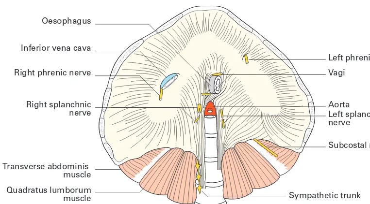
The diaphragm
The diaphragm is the dome-shaped septum dividing the thoracic from the abdominal cavity. It comprises two portions: a peripheral muscular part which arises from the margins of the thoracic outlet and a centrally placed aponeurosis (Fig. 10).
The muscular fibres are arranged in three parts.
1◊◊Avertebral partfrom the crura and from the arcuate ligaments. The right crusarises from the front of the bodies of the upper three lumbar vertebrae and intervertebral discs; the left crusis only attached to the first two verte-brae. The arcuate ligamentsare a series of fibrous arches, the medialbeing a thickening of the fascia covering psoas major and the lateralof fascia overly-ing quadratus lumborum. The fibrous medial borders of the two crura form a median arcuate ligamentover the front of the aorta.
2◊◊Acostal partis attached to the inner aspect of the lower six ribs and costal cartilages.
3◊◊Asternal portionconsists of two small slips from the deep surface of the xiphisternum.
The central tendon, into which the muscular fibres are inserted, is trefoil in shape and is partially fused with the undersurface of the pericardium.
The diaphragm receives its entire motor supply from the phrenic nerve (C3, 4, 5) whose long course from the neck follows the embryological migration of the muscle of the diaphragm from the cervical region (see below). Injury or operative division of this nerve results in paralysis and elevation of the corresponding half of the diaphragm.
Radiographically, paralysis of the diaphragm is recognized by its eleva-tion and paradoxical movement; instead of descending on inspiraeleva-tion it is forced upwards by pressure from the abdominal viscera.
The sensory nerve fibres from the central part of the diaphragm also run in the phrenic nerve, hence irritation of the diaphragmatic pleura (in pleurisy) or of the peritoneum on the undersurface of the diaphragm by subphrenic collections of pus or blood produces referred pain in the corre-sponding cutaneous area, the shoulder-tip.
Openings in the diaphragm
The three main openings in the diaphragm (Figs 10, 11) are:
1◊◊the aortic(at the level of T12) which transmits the abdominal aorta, the thoracic duct and often the azygos vein;
2◊◊the oesophageal(T10) which is situated between the muscular fibres of the right crus of the diaphragm and transmits, in addition to the oesopha-gus, branches of the left gastric artery and vein and the two vagi;
3◊◊theopening for the inferior vena cava(T8) which is placed in the central tendon and also transmits the right phrenic nerve.
In addition to these structures, the greater and lesser splanchnic nerves (see page 49) pierce the crura and the sympathetic chain passes behind the diaphragm deep to the medial arcuate ligament.
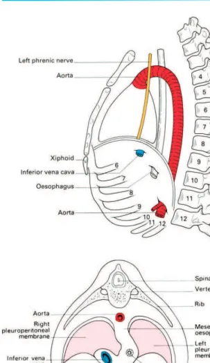
The development of the diaphragm
and the anatomy of diaphragmatic herniae
The diaphragm is formed (Fig. 12) by fusion in the embryo of:
1◊◊the septum transversum (forming the central tendon);
2◊◊the dorsal oesophageal mesentery;
3◊◊a peripheral rim derived from the body wall;
4◊◊the pleuroperitoneal membranes, which close the fetal communication between the pleural and peritoneal cavities.
The septum transversum is the mesoderm which, in early develop-ment, lies in front of the head end of the embryo. With the folding off of the head, this mesodermal mass is carried ventrally and caudally, to lie in its
Oesophagus
Left phrenic nerve Vagi
Aorta
Left splanchnic nerve
Subcostal nerve
Sympathetic trunk Inferior vena cava
Right phrenic nerve
Right splanchnic nerve
[image:30.559.70.451.68.279.2]Transverse abdominis muscle Quadratus lumborum muscle Psoas major muscle
Fig. 11◊Schematic lateral view of the diaphragm to show the levels at which it is pierced by major structures.
definitive position at the anterior part of the diaphragm. During this migra-tion, the cervical myotomes and nerves contribute muscle and nerve supply respectively, thus accounting for the long course of the phrenic nerve (C3, 4 and 5) from the neck to the diaphragm.
With such a complex embryological story, one may be surprised to know that congenital abnormalities of the diaphragm are unusual.
However, a number of defects may occur, giving rise to a variety of con-genital herniae through the diaphragm. These may be:
1◊◊through the foramen of Morgagni; anteriorly between the xiphoid and costal origins;
2◊◊through the foramen of Bochdalek — the pleuroperitoneal canal — lying posteriorly;
3◊◊through a deficiency of the whole central tendon (occasionally such a hernia may be traumatic in origin);
4◊◊through a congenitally large oesophageal hiatus.
Far more common are the acquired hiatus herniae (subdivided into sliding and rolling herniae). These are found in patients usually of middle age where weakening and widening of the oesophageal hiatus has occurred (Fig. 13).
In the sliding herniathe upper stomach and lower oesophagus slide upwards into the chest through the lax hiatus when the patient lies down or bends over; the competence of the cardia is often disturbed and peptic juice can therefore regurgitate into the gullet in lying down or bending over. This may be followed by oesophagitis with consequent heartburn, bleeding and, eventually, stricture formation.
In the rolling hernia(which is far less common) the cardia remains in its normal position and the cardio-oesophageal junction is intact, but the fundus of the stomach rolls up through the hiatus in front of the oesopha-gus, hence the alternative term of para-oesophageal hernia. In such a case
there may be epigastric discomfort, flatulence and even dysphagia, but no regurgitation because the cardiac mechanism is undisturbed.
The movements of respiration
During inspiration the movements of the chest wall and diaphragm result in an increase in all diameters of the thorax. This, in turn, brings about an increase in the negative intrapleural pressure and an expansion of the lung tissue. Conversely, in expiration the relaxation of the respiratory muscles and the elastic recoil of the lung reduce the thoracic capacity and force air out of the lungs.
In quiet inspirationthe first rib remains relatively fixed, but contraction of the external and internal intercostals elevates and, at the same time, everts the succeeding ribs. In the case of the 2nd–7th ribs this principally increases the anteroposterior diameter of the thorax (by the forward thrust of the sternum), like a pump handle. The corresponding movement of the lower ribs raises the costal margin and leads mainly to an increase in the transverse diameter of the thorax, like a bucket handle. The depth of the thorax is increased by the contraction of the diaphragm which draws down its central tendon. Normal quiet expiration, brought about by elastic recoil of the elevated ribs, is aided by the tone of the abdominal musculature which, acting through the contained viscera, forces the diaphragm upwards.
In deep and in forced inspiration additional muscles attached to the chest wall are called into play (e.g. scalenus anterior, sternocleidomastoid, serratus anterior and pectoralis major) to increase further the capacity of the thorax. Similarly, in deep expiration, forced contraction of the abdomi-nal muscles aids the normal expulsive factors described above.
The pleurae
The two pleural cavities are totally separate from each other (Fig. 2). Each pleuraconsists of two layers: a visceral layerintimately related to the surface of the lung, and a parietal layerlining the inner aspect of the chest wall, the upper surface of the diaphragm and the sides of the pericardium and medi-astinum. The two layers are continuous in front and behind the root of the lung, but below this the pleura hangs down in a loose fold, the pulmonary ligament, which forms a ‘dead-space’ for distension of the pulmonary veins. The surface markings of the pleura and lungs have already been described in the section on surface anatomy.
Notice that the lungs do not occupy all the available space in the pleural cavity even in forced inspiration.
Clinical features
2◊◊Fluid can be drained from the pleural cavity by inserting a wide-bore needle through an intercostal space (usually the 7th posteriorly). The needle is passed along the superior border of the lower rib, thus avoiding the intercostal nerves and vessels (Fig. 8). Below the 7th intercostal space there is danger of penetrating the diaphragm.
3◊◊For emergency chest drainage— for example traumatic haemothorax or haemopneumothorax— the site of election is the 5thintercostal space in the
mid-axillary line. An incision is made through skin and fat and blunt dis-section carried out over the upper border of the 6th rib. The pleura is opened, a finger inserted to clear any adhesions and ensure the safety of the adjacent diaphragm before inserting a tube into the pleural space and con-necting it to an under-water drain.
4◊◊Since the parietal pleura is segmentally innervated by the intercostal nerves, inflammation of the pleura results in pain referred to the cutaneous distribution of these nerves (i.e. to the thoracic wall or, in the case of the lower nerves, to the anterior abdominal wall, which may mimic an acute abdominal emergency).
The lower respiratory tract
The trachea
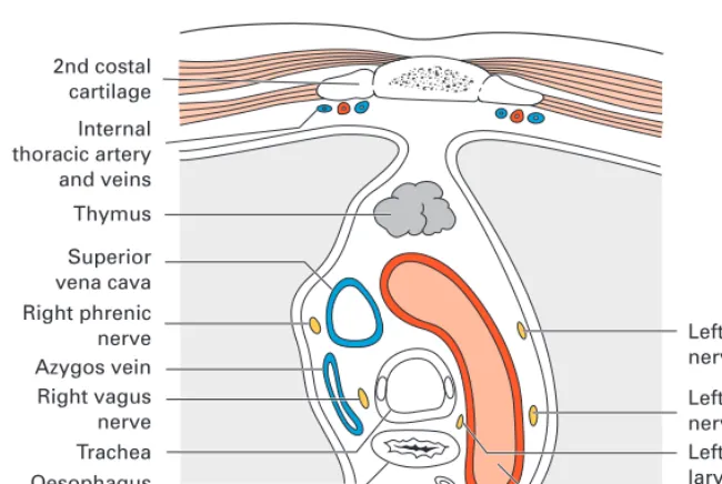
(Figs 14, 15)The trachea is about 4.5in (11.5cm) in length and nearly 1 in (2.5cm) in diameter. It commences at the lower border of the cricoid cartilage (C6) and terminates by bifurcating at the level of the sternal angle of Louis (T4/5) to form the right and left main bronchi. (In the living subject, the level of bifur-cation varies slightly with the phase of respiration; in deep inspiration is descends to T6 and in expiration it rises to T4.)
Relations
Lying partly in the neck and partly in the thorax, its relations are:
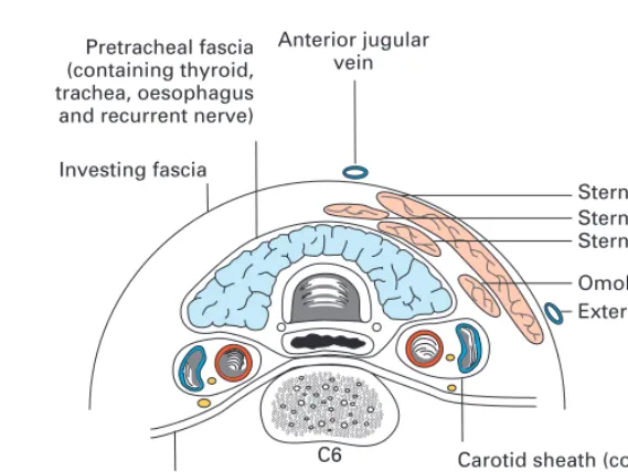
Cervical
•◊◊anteriorly — the isthmus of thyroid gland, inferior thyroid veins, ster-nohyoid and sternothyroid muscles;
•◊◊laterally—the lobes of thyroid gland and the common carotid artery; •◊◊posteriorly—the oesophagus with the recurrent laryngeal nerve lying in the groove between oesophagus and trachea (Fig. 16).
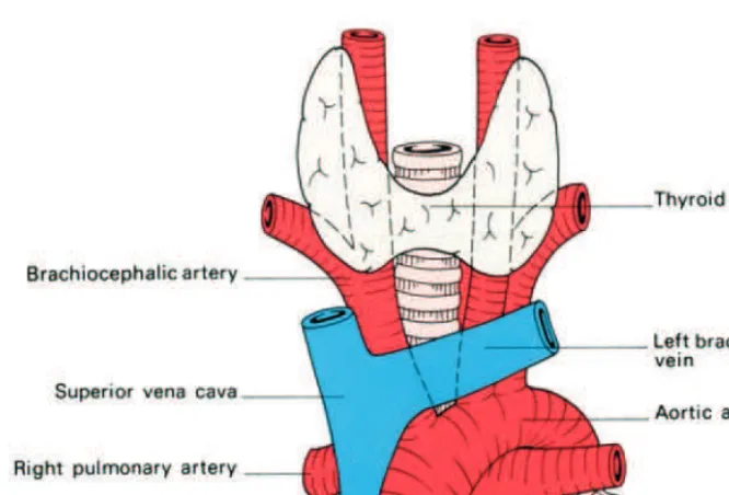
Thoracic
In the superior mediastinum its relations are:
Fig. 14◊The trachea and its anterior relationships.
[image:35.559.39.362.333.605.2]and left carotid artery, both arising from the arch of the aorta, the left bra-chiocephalic (innominate) vein, and the thymus;
•◊◊posteriorly—oesophagus and left recurrent laryngeal nerve;
•◊◊to the left — arch of the aorta, left common carotid and left subclavian arteries, left recurrent laryngeal nerve and pleura;
•◊◊to the right—vagus, azygos vein and pleura (Fig. 17).
Structure
The patency of the trachea is maintained by a series of 15–20 U-shaped car-tilages. Posteriorly, where the cartilage is deficient, the trachea is flattened and its wall completed by fibrous tissue and a sheet of smooth muscle (the trachealis). Within, it is lined by a ciliated columnar epithelium with many goblet cells.
Clinical features
Radiology
Since it contains air, the trachea is more radio-translucent than the neigh-bouring structures and is seen in posteroanterior and lateral radiographs as a dark area passing downwards, backwards and slightly to the right. In the elderly, calcification of the tracheal rings may be a source of radiological confusion.
Displacement
The trachea may be compressed or displaced by pathological enlargement
Sternocleidomastoid
Carotid sheath (containing common carotid artery, internal jugular vein, and vagus nerve) with sympathetic chain behind
Sternohyoid Sternothyroid Omohyoid
External jugular vein Pretracheal fascia
(containing thyroid, trachea, oesophagus and recurrent nerve)
Anterior jugular vein
C6 Investing fascia
[image:36.559.155.440.65.279.2]Pre-vertebral fascia
of the neighbouring structures, particularly the thyroid gland and the arch of the aorta.
‘Tracheal-tug’
The intimate relationship between the arch of the aorta and the trachea and left bronchus is responsible for the physical sign known as ‘tracheal-tug’, characteristic of aneurysms of the aortic arch.
Tracheostomy
Tracheostomy may be required for laryngeal obstruction (diphtheria, tumours, inhaled foreign bodies), for the evacuation of excessive secretions (severe postoperative chest infection in a patient who is too weak to cough adequately), and for long-continued artificial respiration (poliomyelitis, severe chest injuries). It is important to note that respiration is further assisted by considerable reduction of the dead-space air.
[image:37.559.38.363.69.287.2]The neck is extended and the head held exactly in the midline by an assistant. A vertical incision is made downwards from the cricoid cartilage, passing between the anterior jugular veins. Alternatively, a more cosmetic transverse skin crease incision, placed halfway between the cricoid and suprasternal notch, is employed. A hook is thrust under the lower border of the cricoid to steady the trachea and pull it forward. The pretracheal fascia is split longitudinally, the isthmus of the thyroid either pushed upwards or divided between clamps and the cartilage of the trachea clearly exposed. A circular opening is then made into the trachea to admit the tracheostomy tube. 2nd costal cartilage Internal thoracic artery and veins Thymus Superior vena cava Right phrenic nerve Azygos vein Right vagus nerve Trachea Oesophagus Left phrenic nerve Left vagus nerve Left recurrent laryngeal nerve Aortic arch Thoracic duct T4
In children the neck is relatively short and the left brachiocephalic vein may come up above the suprasternal notch so that dissection is rather more difficult and dangerous. This difficulty is made greater because the child’s trachea is softer and more mobile than the adult’s and therefore not so readily identified and isolated. Its softness means that care must be taken, in incising the child’s trachea, not to let the scalpel plunge through and damage the underlying oesophagus.
In contrast, the trachea may be ossified in the elderly and small bone shears required to open into it.
The golden rule of tracheostomy—based entirely on anatomical consid-erations— is ‘stick exactly to the midline’. If this is not done, major vessels are in jeopardy and it is possible, although the student may not credit it, to miss the trachea entirely.
The bronchi
(Fig. 15)The right main bronchusis wider, shorter and more vertical than the left. It is about 1 in (2.5cm) long and passes directly to the root of the lung at T5. Before joining the lung it gives off its upper lobe branch, and then passes below the pulmonary artery to enter the hilum of the lung. It has two important relations: the azygos vein, which arches over it from behind to reach the superior vena cava, and the pulmonary artery which lies first below and then anterior to it.
The left main bronchusis nearly 2 in (5cm) long and passes downwards and outwards below the arch of the aorta, in front of the oesophagus and descending aorta. Unlike the right, it gives off no branches until it enters the hilum of the lung, which it reaches opposite T6. The pulmonary artery spirals over the bronchus, lying first anteriorly and then above it.
Clinical features
1◊◊The greater width and more vertical course of the right bronchus accounts for the greater tendency for foreign bodies and aspirated material to pass into the right bronchus (and thence especially into the middle and lower lobes of the right lung) rather than into the left.
2◊◊The inner aspect of the whole of the trachea, the main and lobar bronchi and the commencement of the first segmental divisions can be seen at bronchoscopy.
3◊◊Widening and distortion of the angle between the bronchi (the carina) as seen at bronchoscopy is a serious prognostic sign, since it usually indicates carcinomatous involvement of the tracheobronchial lymph nodes around the bifurcation of the trachea.
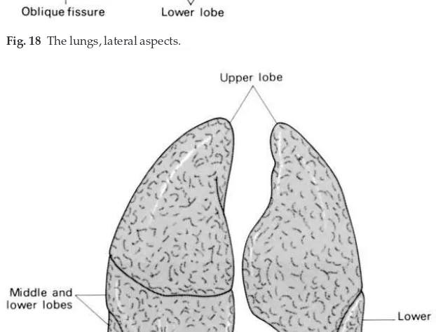
The lungs
(Figs 18, 19)Fig. 18◊The lungs, lateral aspects.
wall and a mediastinal surface which is concave to accommodate the pericardium.
[image:39.559.36.347.301.538.2]The right lung is slightly larger than the left and is divided into three lobes—upper, middle and lower, by the oblique and horizontal fissures. The left lung has only an oblique fissure and hence only two lobes.
Blood supply
Mixed venous blood is returned to the lungs by the pulmonary arteries; the air passages are themselves supplied by the bronchial arteries, which are small branches of the descending aorta. Thebronchial arteries, although small, are of great clinical importance. They maintain the blood supply to the lung parenchyma after pulmonary embolism, so that, if the patient recovers, lung function returns to normal.
Thesuperior and inferior pulmonary veinsreturn oxygenated blood to the left atrium, while the bronchial veinsdrain into the azygos system.
Lymphatic drainage
The lymphatics of the lung drain centripetally from the pleura towards the hilum. From the bronchopulmonary lymph nodesin the hilum, efferent lymph channels pass to the tracheobronchial nodesat the bifurcation of the trachea, thence to the paratracheal nodesand the mediastinal lymph trunks to drain usually directly into the brachiocephalic veins or, rarely, indirectly via the thoracic or right lymphatic duct.
Nerve supply
The pulmonary plexuses derive fibres from both the vagi and the sympa-thetic trunk. They supply efferents to the bronchial musculature (sympa-thetic bronchodilator fibres) and receive afferents from the mucous membrane of the bronchioles and from the alveoli.
The bronchopulmonary segments of the lungs
(Figs 20, 21)
A knowledge of the finer arrangement of the bronchial tree is an essential
Table 1◊The named divisions of the main bronchi.
Apical Upper lobe bronchus
{
PosteriorAnterior Right main bronchus
{
Middle lobe bronchus{
LateralMedial
Medial (cardiac) Lower lobe bronchus
{
Apical AnteriorBasal →
{
Lateral Posterior Upper lobe bronchus Apicoposterior↓
{
Anterior Lingular bronchus Superior Left main bronchus{
{
InferiorApical Anterior Lower lobe bronchus
{
prerequisite to intelligent appreciation of lung radiology, to interpretation of bronchoscopy and to the surgical resection of lung segments. Each lobe of the lung is subdivided into a number of bronchopulmonary segments, each of which is supplied by a segmental bronchus, artery and vein. These segments are wedge-shaped with their apices at the hilum and bases at the lung surface; if excised accurately along their boundaries (which are marked by intersegmental veins), there is little bleeding or alveolar air leakage from the raw lung surface.
The names and arrangements of the bronchi are given in Table 1; each bronchopulmonary segment takes its title from that of its supplying seg-mental bronchus (listed in the right-hand column of the table).
Right Left
Upper lobe Upper lobe ◊1◊◊Apical bronchus ◊1◊◊
Apicoposterior bronchus
◊2◊◊Posterior bronchus ◊2◊◊
}
◊3◊◊Anterior bronchus ◊3◊◊Anterior bronchus
Middle lobe Lingula
◊4◊◊Lateral bronchus ◊4◊◊Superior bronchus
◊5◊◊Medial bronchus ◊5◊◊Inferior bronchus
Lower lobe Lower lobe
◊6◊◊Apical bronchus ◊6◊◊Apical bronchus
◊7◊◊Medial basal
◊◊◊◊(cardiac) bronchus
◊8◊◊Anterior basal ◊8◊◊Anterior basal bronchus
◊◊◊◊bronchus
◊9◊◊Lateral basal ◊9◊◊Lateral basal bronchus
◊◊◊◊bronchus
10◊◊Posterior basal 10◊◊Posterior basal bronchus
[image:41.559.37.340.63.502.2]◊◊◊◊bronchus
The left upper lobe has a lingular segment, supplied by the lingular bronchusfrom the main upper lobe bronchus. This lobe is equivalent to the right middle lobewhose bronchus arises as a branch from the main bronchus. Apart from this, differences between the two sides are very slight; on the left, the upper lobe bronchus gives off a combined apicoposterior segmen-tal bronchus and an anterior branch, whereas all three branches are sepa-rate on the right side.
[image:42.559.109.451.66.496.2]On the right also there is a small medial (or cardiac) lower lobe
bronchus which is absent on the left, the lower lobes being otherwise mirror images of each other.
The mediastinum
The mediastinum is defined as ‘the space which is sandwiched between the two pleural sacs’. For descriptive purposes the mediastinum is divided by a line drawn horizontally from the sternal angle to the lower border of T4 (angle of Louis) into superior and inferior mediastinum. The inferior medi-astinum is further subdivided into the anterior in front of the pericardium, a middle mediastinum containing the pericardium itself with the heart and great vessels, and posterior mediastinum between the pericardium and the lower eight thoracic vertebrae (Fig. 22).
The pericardium
The heart and the roots of the great vessels are contained within the conical fibrous pericardium, the apex of which is fused with the adventitia of the
great vessels and the base with the central tendon of the diaphragm. Anteri-orly it is related to the body of the sternum, to which it is attached by the sternopericardial ligament. The 3rd–6th costal cartilages and the anterior borders of the lungs; posteriorly, to the oesophagus, descending aorta, and vertebra T5–T8, and on either side to the roots of the lungs, the mediastinal pleura and the phrenic nerves.
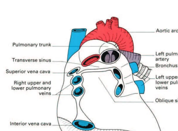
The inner aspect of the fibrous pericardium is lined by the parietal layer of serous pericardium. This, in turn, is reflected around the roots of the great vessels to become continuous with the visceral layeror epicardium. The lines of pericardial reflexion are marked on the posterior surface of the heart (Fig. 23) by the oblique sinus, bounded by the inferior vena cava and the four pul-monary veins, which form a recess between the left atrium and the peri-cardium, and the transverse sinusbetween the superior vena cava and left atrium behind and the pulmonary trunk and aorta in front.
The heart
(Fig. 24)Its great importance means no excuse need be offered for dealing with the heart in considerable detail.
[image:44.559.142.451.384.612.2]The heart is irregularly conical in shape, and it is placed obliquely in the middle mediastinum. Viewed from the front, portions of all the heart chambers can be seen. The right border is formed entirely by the right atrium, the left border partly by the auricular appendage of the left atrium but mainly by the left ventricle, and the inferior border chiefly by the right
ventricle but also by the lower part of the right atrium and the apex of the left ventricle.
[image:45.559.36.387.70.546.2]The bulk of the anterior surface is formed by the right ventricle which is separated from the right atrium by the vertical atrioventricular groove, and from the left ventricle by the anterior interventricular groove.
The inferior or diaphragmatic surfaceconsists of the right and left ventri-cles separated by the posterior interventricular groove and the portion of the right atrium which receives the inferior vena cava.
The base or posterior surfaceis quadrilateral in shape and is formed mainly by the left atrium with the openings of the pulmonary veins and, to a lesser extent, by the right atrium.
Chambers of the heart
Right atrium
(Fig. 25)The right atrium receives the superior vena cava in its upper and posterior part, the inferior vena cava and coronary sinus in its lower part, and the anterior cardiac vein (draining much of the front of the heart) anteriorly. Running more or less vertically downwards between the venae cavae is a distinct muscular ridge, the crista terminalis(indicated on the outer surface of the atrium by a shallow groove — the sulcus terminalis). This ridge sepa-rates the smooth-walled posterior part of the atrium, derived from the sinus venosus, from the rough-walled anterior portion which is prolonged into the auricular appendage and which is derived from the true fetal atrium.
The openings of the inferior vena cava and the coronary sinus are guarded by rudimentary valves; that of the inferior vena cava being contin-uous with the annulus ovalisaround the shallow depression on the atrial septum, the fossa ovalis, which marks the site of the fetal foramen ovale.
Right ventricle
(Fig. 25)The right ventricle is joined to the right atrium by the way of the vertically disposed tricuspid valve, and with the pulmonary trunk through the pul-monary valve. A muscular ridge, the infundibuloventricular crest, between the atrioventricular and pulmonary orifices, separates the ‘inflow’ and ‘outflow’ tracts of the ventricle. The inner aspect of the inflow tract path is marked in the presence of a number of irregular muscular elevations (tra-beculae carneae) from some of which the papillary musclesproject into the lumen of the ventricle and find attachment to the free borders of the cusps of the tricuspid valve by way of the chordae tendineae. The moderator bandis a muscular bundle crossing the ventricular cavity from the interventricular septum to the anterior wall and is of some importance since it conveys the right branch of the atrioventricular bundle to the ventricular muscle.
The outflow tract of the ventricle or infundibulumis smooth-walled and is directed upwards and to the right towards the pulmonary trunk. The pulmonary orifice is guarded by the pulmonary valves, comprising three semilunar cusps.
Left atrium
The left atrium is rather smaller than the right but has somewhat thicker walls. On the upper part of its posterior wall it presents the openings of the four pulmonary veins and on its septal surface there is a shallow depression corresponding to the fossa ovalis of the right atrium. As on the right side, the main part of the cavity is smooth-walled but the surface of the auricle is marked by a number of ridges due to the underlying pectinate muscles.
Left ventricle
(Fig. 26)The left ventricle communicates with the left atrium by way of the mitral valve(so-called because it vaguely resembles a bishop’s mitre), which pos-sesses a large anterior and a smaller posterior cusp attached to papillary muscles by chordae tendineae. With the exception of the fibrous vestibule immediately below the aortic orifice, the wall of the left ventricle is marked by thick trabeculae carneae.
The aortic orifice is guarded by the three semilunar cusps of the aortic valve, immediately above which are the dilated aortic sinuses. The mouths of the right and left coronary arteries are seen in the anterior and left posterior sinus respectively.
The conducting system of the heart
throughout the atrial musculature to reach the atrioventricular nodelying in the atrial septum immediately above the opening of the coronary sinus. The impulse is then conducted to the ventricles by way of the specialized tissue of the atrioventricular bundle(of His). This bundle divides at the junction of the membranous and muscular parts of the interventricular septum into its right and left branches which run immediately beneath the endocardium to activate all parts of the ventricular musculature.
The blood supply to the heart
(Fig. 27)The heart’s blood supply is derived from the right and left coronary arteries whose main branches lie in the interventricular and atrioventricular grooves.
The right coronary arteryarises from the anterior aortic sinus and passes forwards between the pulmonary trunk and the right atrium to descend in the right part of the atrioventricular groove. At the inferior border of the heart it continues along the atrioventricular groove to anastomose with the left coronary at the posterior interventricular groove. It gives off a marginal branchalong the lower border of the heart and theposterior interventricular branch which runs forward in the inferior interventricular groove and to anastomose near the apex of the heart with the corresponding branch of the left coronary artery.
The left coronary artery, which is larger than the right, rises from the left posterior aortic sinus. Passing first behind and then to the left of the pul-monary trunk, it reaches the left part of atrioventricular groove in which it runs laterally round the left border of the heart as the circumflex arteryto reach the posterior interatrial groove. Its most important branch, given off about 2 cm from its origin, is the anterior interventricular arterywhich sup-plies the anterior aspect of both ventricles and passes around the apex of the heart to anastomose with the posterior interventricular branch of the right coronary. Note that the sinuatrial node is usually supplied by the right coronary artery, although the left coronary artery takes over this duty in about one-third of subjects.
Although anastomoses occur between the terminations of the right and left coronary arteries, these are usually inefficient. Thrombosis in one or other of these vessels leads to death of the area of heart muscle supplied (a myocardial infarction).
The venous drainage of the heart
(Fig. 28)The bulk of the venous drainage of the heart is achieved by veins which accompany the coronary arteries and which open into the right atrium. The rest of the blood drains by means of small veins (venae cordis minimae) directly into the cardiac cavity.
The coronary sinus lies in the posterior atrioventricular groove and opens into the right atrium just to the left of the mouth of the inferior vena cava.
It receives:
1◊◊the great cardiac veinin the anterior interventricular groove;
2◊◊the middle cardiac veinthe inferior interventricular groove;
3◊◊the small cardiac vein— accompanying the marginal artery along the lower border of the heart;
4◊◊the oblique vein— descends obliquely on the posterior aspect of the left atrium.
The anterior cardiac veins(up to three or four in number) cross the ante-rior atrioventricular groove, drain much of the anteante-rior surface of the heart and open directly into the right atrium.
Nerve supply
The nerve supply of the heart is derived from the vagus (cardio-inhibitor) and the cervical and upper 5 thoracic sympathetic ganglia (cardio-accelerator) by way of superficial and deep cardiac plexuses.
The development of the heart
The primitive heart is a single tube which soon shows grooves demarcating the sinus venosus, atrium, ventricleand bulbus cordisfrom behind forwards. As this tube enlarges it kinks so that its caudal end, receiving venous blood, comes to lie behind its cephalic end with its emerging arteries (Fig. 29).
The sinus venosus later absorbs into the atrium and the bulbus becomes incorporated into the ventricle so that, in the fully developed heart, the atria and great veins come to lie posterior to the ventricles and the roots of the great arteries.
The boundary tissue between the primitive single atrial cavity and single ventricle grows out as a dorsaland a ventral endocardial cushion which meet in the midline, thus dividing the common atrio-ventricular orifice into a right (tricuspid) and left (mitral) orifice.
The division of the primitive atrium into two is a complicated process but an important one in the understanding of congenital septal defects (Fig. 30). A partition, the septum primum, grows downwards from the poste-rior and supeposte-rior walls of the primitive common atrium to fuse with the
endocardial cushions. Before fusion is complete, however, a hole appears in the upper part of this septum which is termed the foramen secundum in the septum primum.
A second membrane, the septum secundum, then develops to the right of the primum but this is never complete; it has a free lower edge which does,
[image:51.559.36.347.99.526.2]Fig. 29◊The coiling of the primitive heart tube into its definitive form.
however, extend low enough for this new septum to overlap the foramen secundum in the septum primum and hence to close it.
The two overlapping defects in the septa form the valve-like foramen ovalewhich shunts blood from the right to left heart in the fetus (see ‘fetal circulation’ below). After birth, this foramen usually becomes completely fused leaving only the fossa ovalis on the septal wall of the right atrium as its memorial. In about 10% of adult subjects, however, a probe can still be insinuated through an anatomically patent, although functionally sealed foramen.
Division of the ventricle is commenced by the upgrowth of a fleshy septum from the apex of the heart towards the endocardial cushions. This stops short of dividing the ventricle completely and thus it has an upper free border, forming a temporary interventricular foramen. At the same time, the single truncus arteriosus is divided into aorta and pulmonary trunk by a spiral septum (hence the spiral relations of these two vessels), which grows downwards to the ventricle and fuses accurately with the upper free border of the ventricular septum. This contributes the small pars membranacea septi, which completes the separation of the ventricle in such a way that blood on the left of the septum flows into the aorta and on the right into the pulmonary trunk.
The primitive sinus venosusabsorbs into the right atrium so that the venae cavae draining into the sinus come to open separately into this atrium. The smooth-walled part of the adult atrium represents the contri-bution of the sinus venosus, the pectinate part represents the portion derived from the primitive atrium.
Rather similarly, the adult left atrium has a double origin. The original single pulmonary venous trunk entering the left atrium becomes absorbed into it, and donates the smooth-walled part of this chamber with the pul-monary veins entering as four separate openings; the trabeculated part of the definitive left atrium is the remains of the original atrial wall.
The development of the aortic arches
and their derivatives
(Fig. 31)Emerging from the bulbus cordis is a common arterial trunk termed the truncus arteriosus, from which arise six pairs of aortic arches, equivalent to the arteries supplying the gill clefts of the fish. These arteries curve dorsally around the pharynx on either side and join to form two longitudinally placed dorsal aortae which fuse distally into the descending aorta.
The 1st and 2nd arches disappear; the 3rd arches become the carotids. The 4th arch on the right becomes the brachiocephalic and right subclavian artery; on the left, it differentiates into the definitive aortic arch, gives off the left subclavian artery and links up distally with the descending aorta.
The 5th arch artery is rudimentary and disappears.
side this arch retains its connection with the dorsal aorta to form the ductus arteriosus(the ligamentum arteriosum of adult anatomy).
This asymmetrical development of the aortic arches accounts for the different course taken by the recurrent laryngeal nerve on each side. In the early fetus the vagus nerve lies lateral to the primitive pharynx, separated from it by the aortic arches. What are to become the recurrent laryngeal nerves pass medially, caudal to the aortic arches, to supply the developing larynx. With elongation of the neck and caudal migration of the heart, the recurrent nerves are caught up and dragged down by the descending aortic arches. On the right side the 5th and distal part of the 6th arch absorb, leaving the nerve to hook round the 4th arch (i.e. the right subclavian artery). On the left side, the nerve remains looped around the persisting distal part the 6th arch (the ligamentum arteriosum) which is overlapped and dwarfed by the arch of the aorta.
The fetal circulation
(Fig. 32)The circulation of the blood in the embryo is a remarkable example of economy in nature and results in the shunting of well-oxygenated blood from the placenta to the brain and the heart, leaving relatively desaturated blood for less essential structures.
[image:53.559.65.332.64.343.2]Blood is returned from the placenta by the umbilical vein to the inferior vena cava and thence the right atrium, most of it by-passing the liver in the
ductus venosus(see page 95). Relatively little mixing of oxygenated and deoxygenated blood occurs in the right atrium since the valve overlying the orifice of the inferior vena cava serves to direct the flow of oxygenated blood from that vessel through the foramen ovaleinto the left atrium, while the deoxygenated stream from the superior vena cava is directed through the tricuspi