The influence of plaque orientation (pericardial or myocardial) on
coronary arterial remodeling
Michael R. Ward *, Allen Jeremias, Kiyoshi Hibi, Niall A. Herity, Sidney T. Lo,
Steven D. Filardo, David P. Lee, Peter J. Fitzgerald, Alan C. Yeung
Di6ision of Cardio6ascular Medicine,Stanford Uni6ersity Medical Center,300Pasteur Dri6e,Stanford,CA94305-5218,USA
Received 17 September 1999; received in revised form 13 December 1999; accepted 14 February 2000
Abstract
Background: Many systemic, regional and lesion factors have been identified which may influence arterial remodeling, but little is known about the importance of extravascular resistance to vessel enlargement. As myocardial systolic splinting may significantly affect vessel expansion the effect of plaque orientation on arterial remodeling in eccentric coronary atherosclerotic lesions was examined. Methods: Using intravascular ultrasound imaging to obtain cross-sectional vessel area (VA), plaque area (PA) and lumen area (LA), remodeling in eccentric left anterior descending coronary artery lesions was compared which predominantly involved the pericardial or free arc (P, n=25) and the myocardial side (M,n=40) of the vessel wall. Normalized vessel area (NVA, VAlesion/VAreference) was compared as a continuous and categorical variable (positive\1.05, intermediate 0.95 – 1.05,
negativeB0.95) as well as remodeling index (RI, VAlesion−VAreference/PAlesion−PAreference).Results: The two groups were well
matched for clinical and lesion characteristics known to affect remodeling. Reference segments areas were similar in the two groups; while lesion LA was also similar, in the pericardial group there was significantly greater lesion PA (P 12.7890.72, M 10.2690.50 mm2, PB0.05) and VA (P 15.7190.90, M 12.8290.57 mm2, PB0.05) demonstrating enhanced compensatory
remodeling. Outward remodeling was significantly greater in P than in M by both NVA (P 1.0390.03, M 0.8690.03,PB0.01) and RI (P 0.0290.07, M −1.1090.32, PB0.01). Positive, intermediate and negative remodeling occurred in nine, nine and seven lesions in P and in four, ten and 26 lesions inM(PB0.01).Conclusions: Remodeling compensates more for plaque growth in eccentric coronary lesions which are surrounded by the pericardium than those surrounded by the myocardium. Extravascular resistance appears to influence arterial remodeling. © 2001 Elsevier Science Ireland Ltd. All rights reserved.
Keywords:Remodeling; Orientation; Support; Eccentric; Atherosclerosis
www.elsevier.com/locate/atherosclerosis
1. Introduction
Arterial remodeling, or change in cross-sectional ves-sel size, is an important morphological change occur-ring with the growth of atherosclerotic lesions. Compensatory remodeling, where the vessel expands with plaque enlargement, delays the onset of luminal stenosis with progressive atherosclerosis while constric-tive remodeling, or reduction in vessel size, exacerbates stenosis. While large-scale post-mortem studies have found that remodeling response is more predictive of luminal stenosis than plaque size [1], little is known
about the factors that influence the magnitude and direction of atherosclerotic remodeling.
In one of the first experimental descriptions of arte-rial remodeling, compensatory enlargement was associ-ated with localized medial thinning underneath a developing plaque and ‘bulging’ of the plaque into the vessel wall, so that a circular lumen was maintained despite eccentric plaque distribution [2]. Similar mor-phological changes were subsequently observed in a human coronary intravascular ultrasound (IVUS) study [3], prompting these and other authors [4,5] to postulate that localized outward remodeling results from passive mechanical deformation of the vessel in response to atheroinflammatory weakening of the vessel wall.
The hypothesis of this study was that outward re-modeling of arteries at sites of such weakness might be * Corresponding author. Tel.: +1-650-7230180; fax: +
1-650-7256766.
E-mail address:[email protected] (M.R. Ward).
attenuated by external splinting, and specifically in coronary arteries, that the myocardium may provide sufficient splinting to that side of the vessel to affect remodeling whereas the pericardial side would be unre-stricted. It was therefore investigated whether coronary plaque orientation in eccentric lesions (i.e. predomi-nantly pericardial or myocardial) influences the pattern of vascular remodeling.
2. Methods
2.1. Patient population
The Stanford University Medical Center intravascu-lar ultrasound (IVUS) core laboratory database was interrogated to find all preintervention IVUS studies of eccentric coronary lesions (eccentricity index\3, using the index originally described by Mintz [6]). Lesions which were heavily calcified (\90° arc of calcium) and those which involved a bifurcation or where a proximal reference segment was unavailable (ostial lesions) were excluded from the study. The study was limited to those lesions in the left anterior descending coronary artery for two reasons: extravascular landmarks (pericardium, anterior interventricular vein, and diagonal and septal branches) are sufficiently consistent for orientation, and the artery has myocardium and pericardium abutting approximately half of its circumference throughout its course. Clinical information recorded on these patients included age, sex, the presence or absence of atheroscle-rotic risk factors (smoking history, hypertension, hyper-lipidemia, diabetes, family history), and clinical presentation (with or without an acute coronary syndrome).
2.2. IVUS imaging and measurements
Images were obtained after intracoronary administra-tion of 200 mg nitroglycerin using a 30MHz IVUS imaging catheter (2.9 – 3.5 F Microview, CVIS/Boston Scientific) with slow manual pullback. Images were recorded on super VHS tape for off-line analysis of vessel areas using a commercially available image anal-ysis system (Tape Measure, Indec, Capitola, CA). Ves-sel area (VA, area within the external elastic lamina) and lumen area (LA) were measured from end-diastolic frames. Plaque area (PA, plaque+media) was then calculated as the difference between VA and LA. Le-sion site was defined as the site with the smallest LA while proximal reference site was defined as the site within the same segment in which PA was smallest.
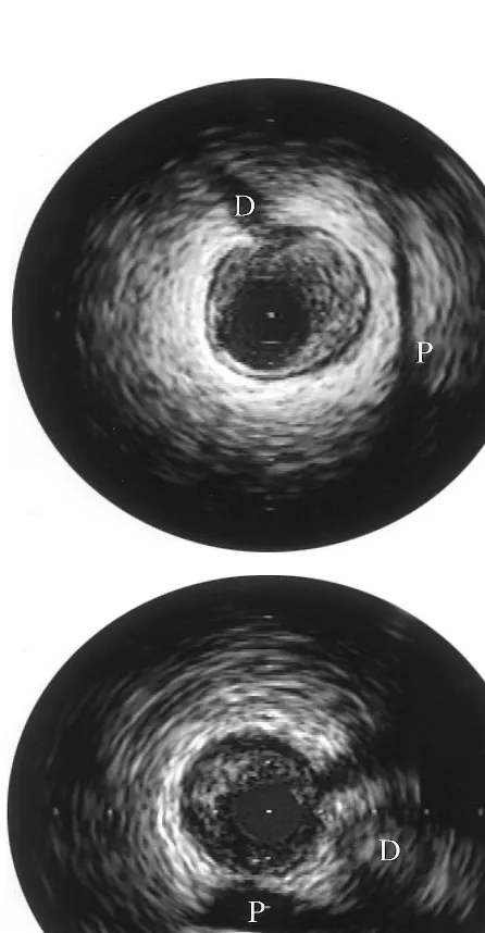
Plaque orientation was assessed using extravascular landmarks as previously described[7] [8] by three inde-pendent observers blinded to the IVUS measurements (examples are shown in Fig. 1). Lesions were classified by the orientation of the majority of the plaque. When plaque was equally distributed between the pericardial and myocardial sides of the vessel (lateral orientation) the case was excluded from the analysis. In cases of disagreement, combined assessment of the tapes was made until agreement could be reached, or the case was excluded from analysis.
Two commonly used measures of remodeling were used:
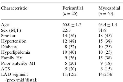
Table 1
Patient and lesion characteristicsa
Pericardial
Characteristic Myocardial
(n=40) (n=25)
Age 65.091.7 63.491.4
Sex (M/F) 22/3 31/9
Family Hx 15 (38)
9 (18)
aAll comparisons between the two groups P\0.30.
the data were normally distributed. Categorical vari-ables were compared byx2testing or Fisher’s exact test where appropriate. Multivariate analysis (multiple lin-ear regression) was used to determine the effect of plaque orientation on NVA and RI when differences in clinical characteristics and position within the vessel were accounted for. Independent variables were entered in the order of the strength of their association with the dependent variable on univariate analysis. P valueB 0.05 was considered statistically significant.
3. Results
3.1. Patient and lesion characteristics
From 584 de novo atherosclerotic lesions in the database, 234 were in the LAD, of which 122 had eccentricity index\3. Seventy-seven of these lesions were suitable for inclusion in the study (not bifurca-tional, ostial or heavily calcified). Of these, eleven were excluded due to lateral orientation and one was ex-cluded due to persistent interobserver disagreement on orientation. Of the remaining 65 cases 40 were predom-inantly myocardial lesions while 25 were predompredom-inantly pericardial. The clinical, angiographic and IVUS mor-phological features of these two groups are shown in Table 1.
3.2. Plaque orientation and remodeling
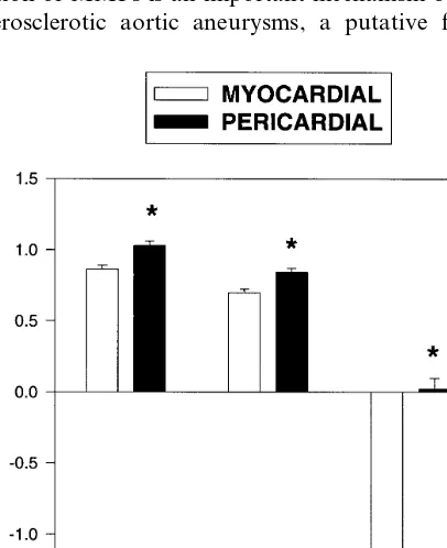
At the reference site mean VA, PA and LA were similar in the Pericardial (P) and myocardial (M) groups. Consistent with their presentation for revascu-larization, LA at the lesion site was similar in both groups. However, VA and PA were significantly larger in the pericardial than the myocardial group (Table 2). NVA was significantly greater in the pericardial group (Fig. 2). Positive, intermediate and negative remodeling was present in nine, nine and seven lesions in the pericardial group and in four, 10 and 26 lesions in the myocardial group respectively (PB0.01 by x2). While normalized plaque area was also greater in the pericar-dial group, comparison of the remodeling index be-tween groups showed there was significantly more outward remodeling for every mm2of plaque growth in
the pericardial group (Fig. 2). By multivariate analysis plaque orientation was a highly significant predictor of NVA (PB0.001) and RI (P=0.007).
4. Discussion
In this study it has been shown that eccentric coro-nary atherosclerotic lesions on the myocardial side of the vessel wall undergo less outward remodeling than Table 2
Intravascular ultrasound (IVUS) measurementsa
Pericardial
Cross-sectional area (mm2) Myocardial
(n=40)
aAll mean9S.E.M.
*PB0.01 vs. pericardial.
1. Normalized vessel area (NVA) — VAlesion/
VAreference. Remodeling was also categorized by this
measure as positive (NVA\1.05) intermediate (0.95BNVAB1.05) or negative (NVAB0.95) as previously described [1].
2. Remodeling index — (VAlesion−VAreference)/
(PAlesion−PAreference).
While the former is a more frequently used measure of remodeling, it may be misleading when two groups of lesions are compared with different proportions of the vessel occupied by plaque, as remodeling has been shown to be proportional to plaque burden. Therefore, plaque areas adjusted for vessel size (normalized plaque area, NPA, PAlesion/VAreference) were compared, and as
plaque burden was significantly different between the two groups, the latter methodology was employed to confirm the results.
2.3. Statistics
those on the free or pericardial side. It suggests that mechanical resistance or splinting from perivascular tissues may influence arterial remodeling.
The mechanisms underlying atherosclerotic remodel-ing have been uncertain. Remodelremodel-ing in normal vessels is predominantly shear-responsive and endothelium de-pendent [9]. Such events may be markedly attenuated by endothelial dysfunction in the presence of atherosclerosis, and it has been proposed that flow-de-pendent arterial remodeling does not occur once signifi-cant plaque has accumulated [10]. This has prompted some authors to propose that localized outward remod-eling may result from the effect of outward radial pressure gradient on a vessel wall weakened by the inflammation from the lipid laden plaque which overlies it. Certainly there has been circumstantial evidence to support such a proposal. Both histopathological and IVUS studies have shown that atherosclerotic remodel-ing with eccentric lesions occurs in a localized manner directly beneath the plaque and is associated with me-dial thinning [2,3]. These morphological changes likely result from enzymatic degradation of vessel wall com-ponents by matrix metalloproteinases (MMPs), which are central components of flow-induced vascular re-modeling [11], and are induced by the inflammatory response to lipids [12]. Furthermore, inflammatory in-duction of MMPs is an important mechanism by which atherosclerotic aortic aneurysms, a putative form of
overexhuberent outward remodeling, form [13], and recent studies have also shown a correlation between markers of inflammation in the vessel wall and outward remodeling [14]. In addition metalloproteinase activity may also be increased by vessel wall stretch [15,16].
If these mechanisms were important one would ex-pect that extravascular splinting would attenuate out-ward remodeling. External splinting of coronary arteries by the myocardium is sufficient to significantly influence both atherosclerosis and response to angio-plasty. It is well known that mechanical external splint-ing reduces atherogenesis: atherosclerotic stenoses are rare in arterial segments surrounded by bone [17], while intima formation in saphenous vein grafts is reduced by externally stenting [18]. External splinting of the coro-nary arteries which are wholly intramyocardial, such as the septal perforators, is sufficient to prevent atherosclerosis even when the epicardial arteries are severely diseased [19]. In the epicardial coronary arter-ies, where only half of the vessel abuts myocardium, extravascular resistance is sufficiently heterogeneous within the circumference of the vessel to influence the orientation of angioplasty-induced coronary dissec-tions. Intimal tears are far more frequent on the free or pericardial side of the vessel than on the myocardial side [20]. It therefore seems quite likely that the differ-ences observed in remodeling are due to heterogeneity in splinting.
4.1. Limitations
However, it is also quite possible that other factors that influence remodeling may be different in pericar-dial and myocarpericar-dial lesions. Myocarpericar-dial lesions were twice as common in this study and the unknown local factors responsible for the preponderance in lesion loca-tion may also influence remodeling. For example, shear stress would be slightly less on the inner curve or myocardial side of the vessel [21] and any flow-respon-sive remodeling might be attenuated. However, these difference in shear stress are small and of doubtful significance when flow dependent remodeling is already attenuated in the presence of advanced atherosclerosis. Secondly, this study was limited to lesions in the LAD, and the effect of myocardial splinting may be less in the right and circumflex coronary arteries, which abut the atria in their proximal segments as well as the peri-cardium and myoperi-cardium. The right coronary also predominantly abuts right ventricular myocardium which is thinner and lower pressure than the left ventri-cle and may not splint the artery so effectively. How-ever, the importance of the observations do not relate to their direct clinical applicability, but in the implica-tions for the mechanisms which influence atheroscle-rotic remodeling.
Fig. 2. Remodeling in eccentric coronary lesions which are predomi-nantly pericardial and myocardial in orientation. Graphs represent mean (9S.E.M.) normalized vessel area (NVA, VAlesion/VAreference),
mean normalized plaque area (NPA, PAlesion/VAreference) and
remod-eling index (RI, VAlesion−VAreference/PAlesion−PAreference). *PB
In summary, it has been demonstrated that extravas-cular structures influence not only atherogenesis, but also the remodeling which accompanies lesion forma-tion. It remains to be determined whether the data may also have relevance to the influence of iatrogenic splint-ing, such as stentsplint-ing, on remodeling.
Acknowledgements
Dr Michael Ward was supported by the National Heart Foundation of Australia. Dr Allen Jeremias was supported by the German Academic Exchange Service, Dr Kiyoshi Hibi was supported by grants from the Getz-Stanford Cardiovascular Research Scholarship Program and the Uehara Memorial Foundation. Dr Niall Herity was supported by the Northern Ireland Council for Postgraduate Medical and Dental Educa-tion and by the Queen’s University of Belfast.
References
[1] Pasterkamp G, Schoneveld AH, van Wolferen W, Hillen B, Clarijs RJ, Haudenschild CC, Borst C. The impact of atheroscle-rotic arterial remodeling on percentage of luminal stenosis varies widely within the arterial system. A postmortem study. Arte-rioscler Thromb Vasc Biol 1997;17:3057 – 63.
[2] Armstrong ML, Heistad DD, Marcus ML, Megan MB, Piegors DJ. Structural and hemodynamic response of peripheral arteries of macaque monkeys to atherogenic diet. Arteriosclerosis 1985;5:336 – 46.
[3] Berglund H, Luo H, Nishioka T, Fishbein MC, Eigler NL, Tabak SW, Siegel RJ. Highly localized arterial remodeling in patients with coronary atherosclerosis: an intravascular ultra-sound study. Circulation 1997;96:1470 – 6.
[4] Oniki T, Iwakami M. Is arterial remodeling truly a compensa-tory biological reaction? A mechanical deformation hypothesis. Atherosclerosis 1997;132:115 – 8.
[5] van der Wal AC, Becker AE, Das PK. Medial thinning and atherosclerosis — evidence for involvement of a local inflamma-tory effect. Atherosclerosis 1993;103:55 – 64.
[6] Mintz GS, Popma JJ, Pichard AD, Kent KM, Satler LF, Chuang YC, DeFalco RA, Leon MB. Limitations of angiogra-phy in the assessment of plaque distribution in coronary artery disease: a systematic study of target lesion eccentricity in 1446 lesions. Circulation 1996;93:924 – 31.
[7] Fitzgerald PJ, Yock C, Yock PG. Orientation of intracoronary ultrasonography: looking beyond the artery. J Am Soc Echocar-diogr 1998;11:13 – 9.
[8] Tsutsui H, Yamagishi M, Uematsu M, Suyama K, Nakatani S, Yasumura Y, Asanuma T, Miyatake K. Intravascular ultra-sound evaluation of plaque distribution at curved coronary segments. Am J Cardiol 1998;81:977 – 81.
[9] Langille BL. Arterial remodeling: relation to hemodynamics. Can J Physiol Pharmacol 1996;74:834 – 41.
[10] Glagov S, Weisenberg E, Zarins CK, Stankunavicius R, Kolettis GJ. Compensatory enlargement of human atherosclerotic coro-nary arteries. N Engl J Med 1987;316:1371 – 5.
[11] Abbruzzese TA, Guzman RJ, Martin RL, Yee C, Zarins CK, Dalman RL. Matrix metalloproteinase inhibition limits arterial enlargements in a rodent arteriovenous fistula model. Surgery 1998;124:328 – 334; discussion 334 – 335.
[12] Aikawa M, Rabkin E, Okada Y, Voglic SJ, Clinton SK, Brinck-erhoff CE, Sukhova GK, Libby P. Lipid lowering by diet reduces matrix metalloproteinase activity and increases collagen content of rabbit atheroma: a potential mechanism of lesion stabilization. Circulation 1998;97:2433 – 44.
[13] Freestone T, Turner RJ, Higman DJ, Lever MJ, Powell JT. Influence of hypercholesterolemia and adventitial inflammation on the development of aortic aneurysm in rabbits. Arterioscler Thromb Vasc Biol 1997;17:10 – 7.
[14] Pasterkamp G, Schoneveld AH, van der Wal AC, Haudenschild CC, Clarijs RJ, Becker AE, Hillen B, Borst C. Relation of arterial geometry to luminal narrowing and histologic markers for plaque vulnerability: the remodeling paradox. J Am Coll Cardiol 1998;32:655 – 62.
[15] Lee RT, Schoen FJ, Loree HM, Lark MW, Libby P. Circumfer-ential stress and matrix metalloproteinase 1 in human coronary atherosclerosis. Implications for plaque rupture. Arterioscler Thromb Vasc Biol 1996;16:1070 – 3.
[16] Fujisawa T, Hattori T, Takahashi K, Kuboki T, Yamashita A, Takigawa M. Cyclic mechanical stress induces extracellular ma-trix degradation in cultured chondrocytes via gene expression of matrix metalloproteinases and interleukin-1. J Biochem (Tokyo) 1999;125:966 – 75.
[17] Thubrikar MJ, Robicsek F. Pressure-induced arterial wall stress and atherosclerosis. Ann Thorac Surg 1995;59:1594 – 603. [18] Izzat MB, Mehta D, Bryan AJ, Reeves B, Newby AC, Angelini
GD. Influence of external stent size on early medial and neointi-mal thickening in a pig model of saphenous vein bypass grafting. Circulation 1996;94:1741 – 5.
[19] Robicsek F, Thubrikar MJ. The freedom from atherosclerosis of intramyocardial coronary arteries: reduction of mural stress — a key factor [see comments]. Eur J Cardiothorac Surg 1994;8:228 – 35.
[20] Wolf A, Fitzgerald PJ. Orientation of coronary dissections rela-tive to extravascular landmarks using intracoronary ultrasound [abstract]. Circulation 1998;98:I – 296.
[21] Krams R, Wentzel JJ, Oomen JA, Vinke R, Schuurbiers JC, de Feyter PJ, Serruys PW, Slager CJ. Evaluation of endothelial shear stress and 3D geometry as factors determining the develop-ment of atherosclerosis and remodeling in human coronary arteries in vivo. Combining 3D reconstruction from angiography and IVUS (ANGUS) with computational fluid dynamics. Arte-rioscler Thromb Vasc Biol 1997;17:2061 – 5.