The Adverse Outcome Pathway approach in nanotoxicology
Kirsten Gerloff, Brigitte Landesmann, Andrew Worth, Sharon Munn, Taina Palosaari, Maurice Whelan
⇑European Commission, Joint Research Centre (JRC), I-21027 Ispra (VA), Italy
a r t i c l e
i n f o
Article history: Received 29 March 2016
Received in revised form 21 July 2016 Accepted 24 July 2016
Available online 10 August 2016
Keywords: AOP Nanoparticle Key Event Liver toxicity Inflammation
a b s t r a c t
An Adverse Outcome Pathway (AOP) is a conceptual construct that describes existing knowledge on the link between a molecular initiating event and an adverse outcome. A sequential chain of causally related events is portrayed at different levels of biological organisation. AOPs are considered to be useful mech-anistic blueprints for the development of novel tools for human and environmental risk assessment. Following OECD guidance, an increasing number of AOPs for chemically-induced adverse effects in humans and environmental species are being proposed. Due to their unique properties, the toxicity of nanomaterials (NMs) and chemicals is often difficult to directly compare since their mechanisms usually differ. While there are still many knowledge gaps in our understanding of NM toxicity, an ever increasing number of mechanistic studies are shedding light on their toxicokinetic and toxicodynamic properties. In this paper, we introduce the concept of AOPs and analyse its possible implementation for nanotoxicology. We illustrate how the AOP framework can be used to rationally combine mechanistic knowledge relating to both NM- and chemically-induced liver toxicity to fill information gaps and guide the development of toxicity testing strategies. The differences between NM and chemically-induced adversity are proposed to be primarily related to differences in toxicokinetics and the nature of the initial Key Events in the AOP. Consequently, much of the mechanistic knowledge captured by AOPs that have been developed from con-sideration of chemically-induced toxicity is also relevant to describe AOPs applicable to NMs, at least in qualitative terms, and thus can be used to inform predictive modelling and risk assessment of NM toxicity. Ó2016 The Author(s). Published by Elsevier B.V. This is an open access article under the CC BY-NC-ND license (http://creativecommons.org/licenses/by-nc-nd/4.0/).
Contents
1. Introduction . . . 4
2. Toxicity mechanisms of metal oxide nanomaterials . . . 5
2.1. NM-induced liver toxicity. . . 6
3. Considerations for the development of AOPs for nanomaterials. . . 6
4. Development of a liver-specific AOP applicable to NMs . . . 7
5. Conclusions. . . 9
Competing interests . . . 9
Transparency Document . . . 9
Acknowledgement . . . 9
References . . . 9
http://dx.doi.org/10.1016/j.comtox.2016.07.001
2468-1113/Ó2016 The Author(s). Published by Elsevier B.V.
This is an open access article under the CC BY-NC-ND license (http://creativecommons.org/licenses/by-nc-nd/4.0/).
Abbreviations:AO, adverse outcome; AOP, Adverse Outcome Pathway; CNTs, carbon nanotubes; H2O2, hydrogen peroxide; IATA, Integrated Approaches to Testing and Assessment; ILSI-RSI, International Life Sciences Risk Sciences Institute; IPCS, International Programme on Chemical Safety; ITS, Integrated Testing Strategies; KE, Key Event; KER, Key Event Relationship; MIE, molecular initiating event; MoA, Mode of Action; MWCNTs, multi-walled carbon nanotubes; NADPH, nicotinamide adenine dinucleotide phosphate; NM, nanomaterial; NLRP-3, NOD-like receptor family, pyrin domain containing 3;1
O2, singlet oxygen; O2, superoxide; OECD, Organisation for Economic
Co-operation and Development; QSAR, quantitative structure–activity relationship; REACH, Registration, Evaluation, Authorisation and Restriction of Chemicals; ROS, reactive oxygen species; SAS, synthetic amorphous SiO2; SCCS, Scientific Committee on Consumer Safety; SiO2, silicon dioxide; TGF-b1, transforming growth factor beta 1; TiO2, titanium dioxide; WHO, World Health Organization.
⇑ Corresponding author.
E-mail addresses:[email protected](K. Gerloff),[email protected](B. Landesmann),[email protected](A. Worth),Sharon.MUNN@ec. europa.eu(S. Munn),[email protected](T. Palosaari),[email protected](M. Whelan).
Contents lists available atScienceDirect
Computational Toxicology
1. Introduction
For the regulatory assessment of chemicals,in vivotesting is
still used extensively to fulfil information requirements, even though animal tests are typically very time-consuming, costly and questionable from an ethical perspective. Moreover, standard guideline tests offer sparse information on the mechanism of tox-icity of a substance and thus provide little help in explaining why a substance might cause an adverse effect of regulatory concern. More than a decade ago, recommendations already emerged to
focus on intelligent testing strategies[1]that move away from a
‘‘generalized, checklist approach” to cover data gaps by acquiring
only essential information[2]. This has led to the development of
Integrated Testing Strategies (ITS) to support the implementation of legislation such as REACH (Registration, Evaluation,
Authorisa-tion and RestricAuthorisa-tion of Chemicals) in the European Union [3,4],
and to more recent efforts within the Organisation for Economic Co-operation and Development (OECD) to develop Integrated Approaches to Testing and Assessment (IATA) which optimally
combine and exploit existing information,in vitroassay data and
computational predictions to satisfy specific information
require-ments[5].
The International Programme on Chemical Safety (IPCS) of the World Health Organization (WHO) and the International Life Sciences Risk Sciences Institute (ILSI-RSI) initiated the Mode of
Action (MoA) human relevance framework[6]for a better
evalua-tion and harmonisaevalua-tion of the assessment of chemical risks. Fol-lowing this, in 2012, a programme for the development of Adverse Outcome Pathways (AOPs) was launched by the OECD which has taken up many of the aspects of the WHO/IPCS work
on MoA[7]. Initially described in the context of ecotoxicological
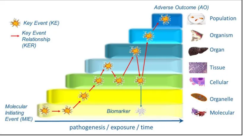
risk assessment, an AOP was defined as ‘‘a conceptual construct that portrays existing knowledge concerning the linkage between a direct molecular initiating event (MIE) and an adverse outcome (AO)”, by capturing the sequential chain of causally-linked Key
Events (KEs) at different levels of biological organisation[8].
Sub-sequently, the AOP concept was extended to support the assess-ment of human health effects. AOPs aim to support regulatory
decision-making by providing the knowledge base to support the development of novel test methods and (OECD) Test Guidelines, QSAR tools and IATA.
In practical terms, the description of an AOP is highly structured and follows well-defined principles and conventions, as described
in OECD guidance, a supplementary ‘User handbook’[9], and in
the scientific literature[10–12]. For example, KEs have to be both
measurable and essential (but not necessarily sufficient) for the AO in question, and the evidence presented to support the causal link-ages between individual KEs, termed Key Event Relationships (KERs), should be based on both biological plausibility and empir-ical data. Evidence can be derived from various sources including in vivoandin vitrostudies, or from computational modelling[8]. An AO can be defined at various levels: for human health effects, an AO seldom relates to whole population level, but rather to indi-vidual organ damage (e.g. liver fibrosis), which has consequences on the individual, whereas in environmental toxicology the AO usually relates to growth inhibition, reduced survival or reproduc-tive impairment of an individual (e.g. a fish) and the consequences on the whole population. The MIE describes the interaction of a material (e.g. chemical) with a biological target, and can be either specific, such as ligand–receptor interaction, or non-specific (e.g. a
toxicant physically residing in a bio-membrane)[9]. By definition,
an AOP consists of a single MIE and a single AO, but can have
mul-tiple causally-linked KEs (Fig. 1). This leads to a simplified and
‘‘lin-ear” representation of an individual AOP, which may be an adequate basis for prediction in certain cases. However, since KEs can be shared by different AOPs, and one MIE can lead to multiple AOs and vice versa, AOP networks generally represent a more
rel-evant basis for toxicity prediction[12]. To facilitate the
develop-ment of AOPs within a network context and to provide a practical collaborative platform for AOP developers to systemati-cally capture, share and integrate their AOP knowledge, the AOP Knowledge Base, including the AOP-Wiki, has been launched in
2014 as publicly accessible tool[13].
Building of networks can be further supported by the emerging concept of Aggregate Exposure Pathways (AEPs), which has been recently introduced to integrate also complex exposure scenarios
and complement AOP development[14]. This would be a useful framework as so far, AOPs primarily focus on direct relationships between a toxicity endpoint resulting from the exposure to the trigger alone. In reality, human exposures are likely to occur as low dose co-exposures with a wide variety of materials, including bacterial agents, chemicals and pharmaceuticals. It is reasonable to suspect that toxicity, especially in the liver, may be driven by sus-ceptibilities associated with such co-exposures.
To date, AOPs have been developed for chemically-induced AOs, although there is increasing awareness of this concept also in the nanotoxicology community. Recently, Vietti and colleagues pub-lished an overview of current knowledge and gaps on KEs involved in lung fibrosis development by carbon nanotubes (CNTs), with the
intention to draft a respective AOP[15]. A large variety of KEs was
described based exclusively on CNT-specific literature, leading to a complex pathway showing the various possibilities of CNT-induced lung fibrosis. Likewise, Labib and colleagues demonstrated the development of an AOP relevant for multi-walled carbon
nanotubes (MWCNTs) based on in vivo-derived transcriptomic
data. This exercise led to a simplified and linear AOP, as foreseen in the AOP guidelines, and demonstrates how transciptomic data
can be used to derive pathway-based points of departure [16].
Again, however, this work is based solely on the literature on MWCNTs. In the present manuscript, we demonstrate how the AOP approach can be applied for nanotoxicology utilising not only NM-specific literature, but also existing knowledge on chemically-induced mechanistic toxicological processes. Following the initiator-agnostic AOP-philosophy, we show how the AOP approach can be used to describe the pathogenesis of NM-induced health effects of regulatory concern where NM-specific information is lacking. However, attention needs to be paid to dis-tinguish NM-specific Key Events from generic ones. The case of liver toxicity induced by chemicals or metal oxide NMs is used as an illustration.
2. Toxicity mechanisms of metal oxide nanomaterials
The possible adverse effects of NMs on the human body are increasingly being discussed and investigated. In 2011, the Euro-pean Commission proposed to define a NM as ‘‘a natural, incidental or manufactured material containing particles, in an unbound state or as an aggregate or as an agglomerate and where, for 50% or more of the particles in the number size distribution, one or more
exter-nal dimensions is in the size range 1 nm–100 nm”[17]. However,
this definition is under continuous discussion[18]. As the use and
possible application areas of NMs for food and food-related prod-ucts are increasing, it is not possible to test their toxicity on a case
by case basis[19–22]. To tackle this, the ITS-NANO project has
pro-posed an Intelligent Testing Strategy for NMs, similar to chemical testing. This strategy will help to identify priority research areas, ultimately limiting individual NM testing and supporting NM risk
assessment[23,24]. The ITS-NANO report already acknowledged
the usefulness of AOPs as a contributor to an ITS for NMs, and first AOP concepts, based on particle-specific literature and describing the species-specific outcomes of sustained particle overload in the lungs, have been described in an ECETOC technical report in
2013[25]and by Morfeld and colleagues in 2015[26].
Some of the major contributors to regular (nano)particle
expo-sure are TiO2or amorphous SiO2, all commonly used in cosmetics,
pharmaceutical products or foods [20,22]. TiO2 in its usual
micron-size (inert) form is used as a whitener (E171), and also
the synthetic amorphous SiO2(SAS) is accepted as a common food
additive (E551), mainly in its micron-sized form, and used for example as an anticaking agent or thickener. A recent approach
to estimate the content of nano-sized SiO2 in food containing
E551 revealed a likely worst case ingestion scenario of
124 mg nano-SiO2per person per day [22], meaning that there
might be continuous exposure and possible bioaccumulation.
Peters and co-workers analysed the amounts of nano-sized SiO2
in several of these E551 containing food products throughout a simulated gastro-intestinal digestion process. They report a disap-pearance of nano-sized particles in gastric pH conditions followed
by their pronounced re-formation in an intestinal medium[27]. A
recent approach to translate knowledge on SAS in food into risk assessment reported low gastrointestinal absorption rates of only 0.03, 0.06 and 0.2%, respectively, depending on the tested material
and the treatment duration or reported study[28]. Interestingly,
the absorption rate seems to decrease with an increase in adminis-tered SAS concentration, which was explained by possible gelation of this material in higher concentrations. This highlights once again that already low doses might result in accumulation and poten-tially in an adverse effect, since the uptake rate cannot necessarily be correlated to the administered dose. It also emphasises the necessity of appropriate kinetic models to accurately predict
uptake rates[28,29].
The ability of NMs to directly induce the formation of reactive
oxygen species (ROS)[19,30,31]is one of the most significant
rea-sons for adverse NM effects. Likewise, oxidative stress is a known
contributor to chemically-induced cell damage and toxicity [32].
A good correlation between cell free and cell-basedin vitroROS
for-mation by NMs with theirin vivoinflammation-generating potency
has been described[33]. Low amounts of ROS can activate various
signalling cascades within the cell, such as the phosphoinositide 30
-kinase/protein kinase B pathway which regulates cellular survival
[34]. Excessive ROS formation however can induce genotoxicity,
leading to DNA strand breakage, oxidative lesions, micronucleus formation or sister chromatid exchanges, and thus can be
poten-tially carcinogenic[35].
In line with this, a classification model was developed to asso-ciate the reactivity of metal oxide NMs with the potential to
gener-ate oxidative stress[36]. The model is based on the ability of the
NMs to exchange electrons with biological redox species in the cell (e.g. antioxidant molecules such as cytochrome C and glutathione). By using this simplified framework, it is possible to predict in a first ranking whether a given metal oxide has the potential to cause oxidative stress by checking whether its band energy levels (conduction and valence bands) overlap with the range of redox potentials of biological reactions occurring inside the cell. The model has been verified by independent experimental studies on
24 metal oxide NMs[37]. It should be kept in mind, however, that
this model was only partially accurate in predicting the capacity of metal oxide NMs to induce oxidative stress, whereas other metal oxides induce similar effects through ion dissolution. This illus-trates the importance of relating QSAR properties to proposed MIEs.
ROS formation is also an important player in the relationship between inflammation and carcinogenesis. The inflammatory phagocyte respiratory burst leads to the indirect formation of ROS, catalysed by nicotinamide adenine dinucleotide phosphate
(NADPH) oxidase [38]. Indeed, activated neutrophils have been
shown to cause oxidative DNA damage in rat lung epithelial cells
[39,40]. Also macrophages are known to play a significant role in NM induced inflammatory processes. They are a key player in
the uptake and elimination of inhaled NMs[41]. Upon the uptake
of a NP by immune cells such as dendritic cells or macrophages, the NLRP-3 (NOD-like receptor family, pyrin domain containing 3) inflammasome can be activated. This process that has been
described for both crystalline and amorphous SiO2, but also for
TiO2 and can ultimately lead to oxidative stress, cell death and
Recent studies underpinned the importance of lysosomal NM uptake for NM-induced toxicity. Once the material is taken up by a cell (such as macrophages or also human astrocytoma cells) and transported to the lysosome by autophagy, the acidic milieu therein can either enhance solubility of a NM, or the material remains in its initial nano-form. Both situations can induce toxic-ity, causing lysosomal swelling, followed by lysosomal disruption
and the release of pro-apoptotic proteins and inflammation[44–
46]. It is known that particles of low solubility and toxicity, such
as TiO2, may cause inflammation in proportion to their specific
sur-face area[47,48]and, as more recently described, their zeta
poten-tial [46]. The zeta potential describes the electric potential
between the surface of a NM (or associated groups thereon) and the suspension medium. It is known that the negatively charged cell membranes can interact more easily with positively charged NMs, making these potentially more toxic than neutral or
nega-tively charged NMs [46]. Acute pulmonary inflammogenicity
in vivois highly correlated with the zeta potential in an acidic envi-ronment (as present in the lysosomes of a cell) for low-solubility NMs. Disruption of the lysosome can trigger an inflammation cas-cade in the target organ. The particle-driven inflammatory response is associated with tissue damage, remodelling and muta-genesis and is referred to as secondary particle toxicity following the exhaustion of antioxidant and DNA damage repair capacities,
as has been described for the lung[30,35,49]. For highly soluble
particles, however, the nature of the ion defines its toxic and/or
inflammogenic potential (e.g. Zn2+as an example of a highly toxic
ion versus Mg2+as an example of a low-toxicity ion)[46]. Thus it is
obvious that solubility is an important aspect in nanotoxicology, and the question arises whether a dissolved material still acts as a ‘‘nanomaterial” in the target tissue. Therefore, here we focus on low-solubility NMs.
Once in a matrix (e.g. a food) or dispersed in biological fluids, such as mucins or the blood plasma, NMs come in contact with a large variety of proteins (e.g. approx. 3700 in the blood plasma), leading to the formation of a protein corona. This is the biomole-cule coating that forms around NMs upon contact with biological molecules and depends on both the size and surface properties of
the NM[50]. The corona consists of a hard and a soft corona: the
hard corona is a tightly bound, near-monolayer of biomolecules around the NM, surrounded by a loose layer of biomolecules, the soft corona. The soft corona can easily be exchanged, whereas the hard corona often retains biomolecules from previous
environ-ments[51]. Therefore, its composition varies in time and depends
on the environmental conditions [52]. The corona is stable for a
longer time than the typical time scale of cellular uptake, thus act-ing as cell ‘‘mediator” in the interaction of the NM with cell
recep-tors[53].
2.1. NM-induced liver toxicity
The liver is one of the main target organs for ingested NMs, but inhaled particles can also reach the liver upon clearance from the lung [54–57]. The mononuclear phagocyte system, also called reticuloendothelial system, is one of the important players in NM uptake and systemic distribution. It consists of the phagocytic monocytes and macrophages which are present in the body, espe-cially in the liver but also in spleen, lymph nodes and bone mar-row. Following recognition and phagocytosis by macrophages of both the mononuclear phagocyte system organs and in the blood, NMs are sequestered to those organs. NMs that are taken up by hepatocytes are potentially excreted into the bile, whereas NMs phagocytosed by Kupffer cells, the resident macrophages of the liver, generally remain in the cells for a long period of time if they
can’t be degraded intracellularly[58–61]. Once in the liver, TiO2
nanoparticles may have the potential to induce DNA damage and
mutagenesis[62,63], but this has been described mainly at high
doses and not following inhalation and has been suggested to be
due to induction of systemic inflammation [62]. In vivo
experi-ments on gavaged or injected (intraperitoneal or intravenously)
TiO2 suggest a wide range of adverse effects on the liver: an
increase in general serum markers for liver damage such as Alanine
Aminotransferase or Aspartate Aminotransferase [64,65], an
increase in inflammatory markers such as pro-inflammatory
cytokines and/or infiltration of inflammatory cells[55,66,67], an
increase of markers for oxidative stress[68,69], apoptosis, necrosis
and also fibrosis[70,71]. It has to be noted, however, that many of
these studies use relatively high particle doses, and the reported effects are usually seen at the highest treatment doses. When low doses of NMs induced an adverse effect, such as an influx of inflammatory cells, recovery to control levels on cessation of NM
exposure was reported[67].
Liver damage and inflammation have also been reported for
other metal oxide particles such as SiO2 [29,72,73] via various
application routes such as intraperitoneal injection or oral admin-istration. Similarly, increased accumulation of the NMs in the liver is often reported. Oral NM administration appeared to induce over-all milder adverse effects than systemic administration, most likely due to the typically limited absorption of NMs in the GI tract.
3. Considerations for the development of AOPs for nanomaterials
As for most risk assessment approaches, the lack of human data is a major difficulty in evaluating the human liver toxicity potential of metal oxide NMs. Risk assessment is based on data from animal
studies, mainly conducted in mice and rats, and from in vitro
experiments with human or rodent cell lines. Moreover, the prop-erties of the nanomaterials tested in various studies vary greatly. The most apparent differences lie within the primary particle size distribution of the materials. NMs of an average primary particle size of 5 nm may lead to a different AO or altered severity of the AO than materials of 100 nm size on average. Furthermore, the tested NMs can vary in their crystalline structure (such as rutile
versus anatase in the case of TiO2) and the ratio of these crystalline
forms, their surface charge, shape or specific surface area. The tox-icokinetics of the NM, including its solubility, is obviously a key factor in describing a material’s toxicity, but are often unknown or only partially described. Another major issue is the lack of suf-ficient physicochemical characterisation especially in early publi-cations. The problem and its consequences have been described
extensively[74]and has led to an increased awareness of
research-ers[75,76]. Scientific journals imposed minimal requirements for adequate NM characterisation and an internationally recognised
guidance has been published by OECD[77]. But even if NMs are
well-described in their pristine form many publications still lack
a thorough characterisation in situ, i.e. the biological fluids the
NM interacts with. Furthermore, tested doses in different studies can differ immensely and are therefore not comparable; some-times the high concentrations used are not relevant for real life exposure conditions. Further, exposure scenarios vary in terms of duration (days versus weeks or months), the route of exposure (for example oral gavage vs injection), and the investigated toxico-logical endpoint.
Understanding the relevant physico-chemical properties of NMs in biological systems is vital when it comes to defining the characteristics of the NM that initiates an event which could potentially result in an AO. Knowledge about the initial fate and
biotransformation of the NMin vivoprior to reaching the biological
to their intestinal uptake, affecting the net surface charge[78]and solubility. The physicochemical properties of the NM and its trans-formation products have a considerable influence on the
absorp-tion, distribution and excretion processes that ultimately
determine the fate of the NM in the body. Moreover, a NM might not only act directly at its target organ, but might also cause toxi-city via second messengers. E.g. inhaled particles are shown to increase the risk of cardiovascular disease indirectly via the induc-tion of a pulmonary acute phase response and enhancing atherosclerotic changes. However, no or only low hepatic acute phase response could be found following inhalation or instillation
[79].
Information on both toxicokinetics and toxicodynamics needs to be combined for hazard and risk assessment purposes. By
defi-nition[9], an AOP is limited to the description of toxicodynamics,
but the kinetics can influence the occurrence of the initiating event.
Another issue in using the AOP framework to describe NM tox-icity is the nature of the MIE. It is plausible, and probably likely, that not all interactions of NMs with cells or cellular components involve a specific molecular interaction or reaction as seen for many chemicals (e.g. pharmaceuticals or pesticides). NMs could in fact induce mechanical/physical damage, e.g. to the cell mem-brane or to the lysosome, which would not be best described as a ‘‘Molecular” Initiating Event. However, AOP principles and guid-ance make provision for describing non-specific interactions with a
biological target[9]and for describing an AOP with unknown MIE.
Therefore, when describing NM-relevant AOPs, it is probably often more appropriate to assign the first KE as initiating event for the respective AOP that could be termed the initial KE.
A large body of knowledge on NM-induced toxicity and under-lying mechanisms exists but for the most part this is fragmented and dispersed across the literature. Different NMs can exhibit tox-icity through different mechanisms, but at least qualitatively com-mon mechanisms are shared by many NMs. In fact, there is also evidence for the comparable toxicological behaviour of nano- and
bulk particles[80]. Developing an AOP is about looking across
dif-ferent material-specific studies and extracting relevant informa-tion, reduced and discretised into a series of causally linked KEs, that is applicable to any NM that has the potential to trigger the (M)IE or initial KE. Restricting this knowledge mining and curation exercise to NM-specific literature makes knowledge gaps evident. Considering that downstream toxicological processes are biology-related rather than substance-specific, the huge chemical-based mechanistic knowledge base can be used to elucidate toxicological processes of NMs. Major differences lie in the initial events that reflect how the chemical or NM perturb the biological system. Using liver toxicity as a case study, we describe how evidence on
chemically-induced toxicity can be used to develop an AOP for NM-induced adverse effects.
4. Development of a liver-specific AOP applicable to NMs
A number of AOPs for adverse liver outcomes are being
elabo-rated within the OECD AOP development programme[13]. These
AOPs are based on MIEs that are induced by chemicals, but never-theless their value for developing NM-specific AOPs should not be
underestimated. As described in Section 3, NM-induced (M)IEs
might differ from chemically-induced ones, as in specific cases they are caused by physical damage rather than molecular interac-tions. However, certain chemicals can also induce similar (M)IEs as NMs. Moreover, the downstream biological effects are essentially the same. To demonstrate this, a well-advanced AOP for (chemically-induced) liver-fibrosis is presented here and used to describe similarities and differences to mechanisms known for NM-derived toxicity.
OECD Project 1.14 ‘‘The Adverse Outcome Pathways from
Pro-tein Alkylation to Liver Fibrosis”, AOP number 38 [81] (Fig. 2)
describes the relevant KEs in the development of liver fibrosis in humans. With chemically-induced protein alkylation being the MIE, hepatocyte injury/apoptosis is described as the subsequent KE. This leads to the activation of Kupffer cells, which account for approximately 15% of the total liver cell population and are involved in the pathogenesis of chemical- or toxin-induced liver injury through the release of inflammatory mediators such as cytokines, chemokines and lysosomal or proteolytic enzymes. Kupffer cells are a main source of the most important profibrogenic
cytokine in this process, TGF-b1, and this is described as KE-3. KE-4
describes the subsequent activation of hepatic stellate cells, which ultimately results in progressive collagen accumulation, the onset of fibrosis. This process is accompanied throughout by oxidative stress, which further promotes the development of fibrosis. More-over, the inflammatory response is a continuous driver in the development of the AO. As a matter of convention, the oxidative stress and inflammatory responses are therefore not described as specific KEs in their own right, but are captured in the KERs. The presented AOP illustrates how a complex biological process can be simplified and described in a linear manner.
To describe adverse effects of NMs on the liver, we have focused
on metal oxide NMs such as TiO2or SiO2. A large number of studies
on general hepatotoxicity endpoints (such as inflammation, liver
damage) have been performed in vivo, whereasin vitro studies
have mainly focused on unveiling the underlying mechanisms. This has led to the development of the AOP ‘‘Lysosomal damage leading to liver inflammation” under the OECD framework (AOP number
144)[82]. As described in Section2, insoluble NMs as well as toxic
Fig. 3.Putative AOP applicable to metal oxide NMs leading to hepatitis. AOP under development in the AOP wiki under the OECD framework titled ‘‘Lysosomal damage leading to liver inflammation”. Metal oxide NMs can induce the AO via lysosomal damage as one relevant (M)IE or initial KE, initiated in hepatocytes. The described AOP leads to hepatitis as the AO, as described in the literature. The formation of ROS can lead to hepatocyte injury/apoptosis induced by mitochondrial damage, ultimately resulting in the infiltration of inflammatory cells, inducing the AO.
ions of soluble NMs can trigger an inflammation cascade by disrup-tion of the lysosomal membrane, which is considered as the initial
KE in the present AOP (Fig. 3). Alternative NM-induced (M)IEs or
initial KEs can also be envisaged; for example direct ROS formation, leading to the induction of oxidative stress and inflammation in the target organ. However, the lysosomal damage in hepatocytes is known to be a relevant starting point for NM-induced toxicity, and thus we describe it here as the initial KE. Once the lysosome is damaged, ROS are formed as a consequence and mitochondrial damage is initiated, both events further amplifying each other, fol-lowed by hepatocyte apoptosis and the inflammatory cascade (Fig. 3).
There are several similarities between KEs of the chemically-induced fibrosis-AOP and NM-chemically-induced toxic effects: liver fibrosis has been described as the AO resulting from repeated treatment
with TiO2or SiO2NMs, following administration via oral treatment
[29,83], i.p.[70,73]or i.v. injection[84]. Exposure of Kupffer cells to
SiO2 NMs leads to their activation via inflammasome activation
[85], causing oxidative stress and inflammation[85,86]. Also, the
appearance of collagen fibres around silicotic nodular like lesions
has been described for NMs[73,84]. However, the downstream
KEs leading to fibrosis development, such as TGF-b1 expression
or hepatic stellate cell activation, are still to be investigated for NMs. Recent findings on renal cells and mouse kidneys describe
an increase of TGF-b expression following nano-TiO2 treatment
[87], further supporting the assumption that excessive exposure
to metal oxide NMs will ultimately lead to liver fibrosis. However, it is currently not possible to describe a full NM-relevant AOP lead-ing to liver fibrosis based on NM toxicology literature alone. Since the development of liver inflammation due to systemic exposure to metal oxides is a well-documented phenomenon, this led us to define hepatitis as the AO, which is in fact a known intermediate event leading to the development of fibrosis (assuming sustained exposure).
In an attempt to learn from the well-developed chemically-induced fibrosis AOP and use this knowledge for the nanotoxicol-ogy domain, the two AOPs were directly compared and merged (Fig. 4). Fig. 4A displays both AOPs in parallel, which highlights similarities and differences. As mentioned above in this section, ‘‘Collagen accumulation” and the AO ‘‘Liver fibrosis” have been described in the literature for NMs, but no direct link to preceding KEs has been established yet.
Based on the KER descriptions and an earlier version of the
chemically-induced fibrosis-AOP graph[88], which includes
oxida-tive stress and inflammation (black arrows,Fig. 4B), it is possible to
directly link the KEs of the AOP relevant to NM toxicity (blue arrows) to the existing fibrosis-AOP. The combined black and blue arrows display KERs common to both AOPs. This merging clearly shows that there are major overlaps of KEs, and that the main dif-ferences between these AOPs lie within the (M)IE or initial KE, and
subsequent (early) KEs. It is very likely that TGF-b1 expression and
hepatic stellate cell activation can be identified as KEs following NM-treatment, leading to liver fibrosis. Based on this assumption, research strategies to confirm or refute this hypothesis can be designed. This also implies that it might be more efficient to base the predictive modelling of NM toxicity on common KEs, rather than on widely varying and poorly characterised (M)IEs.
5. Conclusions
We demonstrate that the mechanistic knowledge captured in AOPs that have been developed to describe chemically-induced toxicity can be utilised to fill knowledge gaps related to the toxicity of poorly soluble NMs. The major differences in the toxicodynam-ics of NMs and chemicals lie in the initial upstream events, and in
particular the (M)IE, although here also overlaps exist. However, chemically- and NM-induced toxicological processes share down-stream events that lead to a particular AO. Of course, NMs and
chemicals differ considerably in theirin vivobiokinetics and in fact
this is the primary factor that sets them apart from a toxicological perspective, at least when considering the current generation of engineered NMs. Considering only the toxicodynamic processes that are described in AOPs, apart from the non-specific nature of NM induced (M)IEs, much of the mechanistic knowledge that is required to describe AOPs relevant to NM toxicity can be found in the chemicals domain. Thus, as AOP networks evolve, the mech-anistic knowledge captured in KEs and KERs can be also used to inform on downstream events related to NM toxicity, and ulti-mately for NM hazard assessment. This provides a tool for a more effective and efficient targeted testing to fill data and knowledge gaps that clearly remain in nanotoxicology.
Competing interests
The authors declare no competing interests.
Transparency Document
TheTransparency documentassociated with this article can be found in the online version.
Acknowledgement
The authors thank Dr. Karin Aschberger for helpful comments on the manuscript.
References
[1]S.P. Bradbury, T.C. Feijtel, C.J. Van Leeuwen, Meeting the scientific needs of ecological risk assessment in a regulatory context, Environ. Sci. Technol. 38 (2004) 463–470.
[2]R. Combes, M. Barratt, M. Balls, An overall strategy for the testing of chemicals for human hazard and risk assessment under the EU REACH system, Altern. Lab. Anim. 31 (2003) 7–19.
[3] European Commission JRC: REACH and the Need for Intelligent Testing Strategies, EUR 21554 EN; http://reach-support.com/download/Intelligent% 20testing.pdf, 2005.
[4]T. Hartung, T. Luechtefeld, A. Maertens, A. Kleensang, Food for thought: integrated testing strategies for safety assessments, ALTEX 30 (2013) 3–18. [5] OECD – Organisation for Economic Co-operation and Development, New
Guidance Document on an Integrated Approach on Testing and Assessment (IATA) for Skin Corrosion and Irritation, ENV/JM/MONO(2014)19, <http:// www.oecd.org/officialdocuments/publicdisplaydocumentpdf/?cote=env/jm/ mono(2014)19&doclanguage=en>, 2014.
[6]M.E.B. Meek, J.R. Bucher, S.M. Cohen, V. Dellarco, R.N. Hill, L.D. Lehman-mckeeman, D.G. Longfellow, T. Pastoor, J. Seed, A framework for human relevance analysis of information on carcinogenic modes of action, Crit. Rev. Toxicol. 33 (2003) 591–653.
[7] OECD, < http://www.oecd.org/chemicalsafety/testing/adverse-outcome-pathways-molecular-screening-and-toxicogenomics.htm> (last accessed 12 July 2016).
[8]G.T. Ankley, R.S. Bennett, R.J. Erickson, D.J. Hoff, M.W. Hornung, R.D. Johnson, D. R. Mount, J.W. Nichols, C.L. Russom, P.K. Schmieder, J.A. Serrrano, J.E. Tietge, D. L. Villeneuve, Adverse outcome pathways: a conceptual framework to support ecotoxicology research and risk assessment, Environ. Toxicol. Chem. 29 (2010) 730–741.
[9] OECD – Organisation for Economic Co-operation and Development, Guidance Document on Developing and Assessing Adverse Outcome Pathways, ENV/ JM/MONO(2013)6, < http://www.oecd.org/officialdocuments/publicdisplay-documentpdf/?cote=env/jm/mono(2013)6&doclanguage=en>, 2013. [10] D.L. Villeneuve, D. Crump, N. Garcia-Reyero, M. Hecker, T.H. Hutchinson, C.A.
LaLone, B. Landesmann, T. Lettieri, S. Munn, M. Nepelska, M.A. Ottinger, L. Vergauwen, M. Whelan, Adverse Outcome Pathway (AOP) development I: strategies and principles, Toxicol. Sci. 142 (2014) 312–320.
[12]S.W. Edwards, Y.-M. Tan, D.L. Villeneuve, M.E. Meek, C.A. McQueen, Adverse outcome pathways – organizing toxicological information to improve decision making, J. Pharmacol. Exp. Ther. 356 (2016) 170–181.
[13] Webpage AOPKB, <www.aopkb.org> (last accessed 12 July 2016).
[14]J.G. Teeguarden, Y.M. Tan, S.W. Edwards, J.A. Leonard, K.A. Anderson, R.A. Corley, M.L. Kile, S.M. Simonich, D. Stone, R.L. Tanguay, K.M. Waters, S.L. Harper, D.E. Williams, Completing the link between exposure science and toxicology for improved environmental health decision making: the aggregate exposure pathway framework, Environ. Sci. Technol. 50 (9) (2016) 4579–4586. [15]G. Vietti, D. Lison, S. van den Brule, Mechanisms of lung fibrosis induced by carbon nanotubes: towards an Adverse Outcome Pathway (AOP), Part. Fibre Toxicol. 13 (2016) 11.
[16]S. Labib, A. Williams, C.L. Yauk, J.K. Nikota, H. Wallin, U. Vogel, S. Halappanavar, Nano-risk science: application of toxicogenomics in an adverse outcome pathway framework for risk assessment of multi-walled carbon nanotubes, Part. Fibre Toxicol. 13 (2016) 15.
[17] Commission E, Commission Recommendation of 18 October 2011 on the Definition of Nanomaterial (2011/696/EU); <https://ec.europa.eu/research/ industrial_technologies/pdf/policy/commission-Recommendation-on-the-Definition-of-Nanomater-18102011_en.pdf>, vol. L275/38, 2011. [18] H. Rauscher, G. Roebben, H. Rauscher, G. Roebben, A.B. Sanfeliu, H. Emons, N.
Gibson, R. Koeber, T. Linsinger, K. Rasmussen, J.R. Sintes, B. Sokull-klüttgen, H. Stamm, European Commission, Joint Research Centre, Towards a Review of the EC Recommendation for a Definition of the Term ‘‘nanomaterial” Part 3, JRC Scientific and Policy Report EUR 27240 EN, 2015.
[19]G. Oberdörster, E. Oberdörster, J. Oberdörster, Nanotoxicology: an emerging discipline evolving from studies of ultrafine particles, Environ. Health Perspect. 113 (2005) 823–839.
[20]Q. Chaudhry, M. Scotter, J. Blackburn, B. Ross, A. Boxall, L. Castle, R. Aitken, R. Watkins, Applications and implications of nanotechnologies for the food sector, Food Addit. Contam. Part A Chem. Anal. Control Expo Risk Assess. 25 (2008) 241–258.
[21]K. Tiede, A.B. Boxall, S.P. Tear, J. Lewis, H. David, M. Hassellov, Detection and characterization of engineered nanoparticles in food and the environment, Food Addit. Contam. Part A Chem. Anal. Control Expo Risk Assess. 25 (2008) 795–821.
[22]S. Dekkers, P. Krystek, R.J.B. Peters, D.P.K. Lankveld, B.G.H. Bokkers, P.H. van Hoeven-Arentzen, H. Bouwmeester, A.G. Oomen, Presence and risks of nanosilica in food products, Nanotoxicology 5 (2011) 393–405.
[23]V. Stone, S. Pozzi-Mucelli, L. Tran, K. Aschberger, S. Sabella, U. Vogel, C. Poland, D. Balharry, T. Fernandes, S. Gottardo, S. Hankin, M.G.J. Hartl, N. Hartmann, D. Hristozov, K. Hund-Rinke, H. Johnston, A. Marcomini, O. Panzer, D. Roncato, A. T. Saber, H. Wallin, J.J. Scott-Fordsmand, ITS-NANO – prioritising nanosafety research to develop a stakeholder driven intelligent testing strategy, Part. Fibre Toxicol. 11 (2014) 9.
[24] V. Stone, S. Pozzi-Mucelli, L. Tran, K. Aschberger, s. Sabella, U.B. Vogel, C.A. Poland, D. Balharry, T.F. Fernandes, S. Gottardo, S. Hankin, M. Hartl, N. Hartmann, D. Hristozov, K. Hund-Rinke, H. Johnston, A. Marcomini, O. Panzer, D. Roncato, A.T. Saber, H. Wallin, J.J. Scott-Fordsmand, Commission E, U.K.V. Stone, D. Balharry, T.F. Fernandes, H. Johnston, L. Munro, M. Hartl, ITS-Nano: Research Prioritisation to Deliver an Intelligent Testing Strategy for the Human and Environmental Safety of Nanomaterials, 2013, p. 128.
[25] European Centre for Ecotoxicology and Toxicology of Chemicals, Poorly Soluble Particles/Lung Overload, Technical Report No. 122, <http://www. ecetoc.org/technical-Reports>, 2013.
[26]P. Morfeld, J. Bruch, L. Levy, Y. Ngiewih, I. Chaudhuri, H.J. Muranko, R. Myerson, R.J. McCunney, Translational toxicology in setting occupational exposure limits for dusts and hazard classification – a critical evaluation of a recent approach to translate dust overload findings from rats to humans, Part. Fibre Toxicol. 12 (2015) 3.
[27]R. Peters, E. Kramer, A.G. Oomen, Z.E.H. Rivera, G. Oegema, P.C. Tromp, R. Fokkink, A. Rietveld, H.J.P. Marvin, S. Weigel, A.C.M. Peijnenburg, H. Bouwmeester, Presence of nano-sized silica during in vitro digestion of foods containing silica as a food additive, ACS Nano 6 (2012) 2441–2451. [28]P.C.E. van Kesteren, F. Cubadda, H. Bouwmeester, J.C.H. van Eijkeren, S.
Dekkers, W.H. de Jong, A.G. Oomen, Novel insights into the risk assessment of the nanomaterial synthetic amorphous silica, additive E551, in food, Nanotoxicology 9 (4) (2015) 442–452.
[29]M. van der Zande, R.J. Vandebriel, M.J. Groot, E. Kramer, Z.E. Herrera Rivera, K. Rasmussen, J.S. Ossenkoppele, P. Tromp, E.R. Gremmer, R.J.B. Peters, P.J. Hendriksen, H.J.P. Marvin, R.L. Hoogenboom, A.A. Peijnenburg, H. Bouwmeester, Sub-chronic toxicity study in rats orally exposed to nanostructured silica, Part. Fibre Toxicol. 11 (2014) 8.
[30]K. Donaldson, L. Tran, L.A. Jimenez, R. Duffin, D.E. Newby, N. Mills, W. MacNee, V. Stone, Combustion-derived nanoparticles: a review of their toxicology following inhalation exposure, Part. Fibre Toxicol. 2 (2005) 10.
[31]A. Nel, T. Xia, L. Mädler, N. Li, Toxic potential of materials at the nanolevel, Science 311 (2006) 622–627.
[32]D.G. Deavall, E.A. Martin, J.M. Horner, R. Roberts, Drug-induced oxidative stress and toxicity, J. Toxicol. 2012 (2012) 64546.
[33]E.K. Rushton, J. Jiang, S.S. Leonard, S. Eberly, V. Castranova, P. Biswas, A. Elder, X. Han, R. Gelein, J. Finkelstein, G. Oberdörster, Concept of assessing nanoparticle hazards considering nanoparticle dosemetric and chemical/ biological response metrics, J. Toxicol. Environ. Health Part A 73 (2010). [34]A. Barthel, L.-O. Klotz, Phosphoinositide 3-kinase signaling in the cellular
response to oxidative stress, Biol. Chem. 386 (2005) 207–216.
[35]R.P.F. Schins, A.M. Knaapen, Genotoxicity of poorly soluble particles, Inhalation Toxicol. 19 (Suppl. 1) (2007) 189–198.
[36]E. Burello, A.P. Worth, A theoretical framework for predicting the oxidative stress potential of oxide nanoparticles, Nanotoxicology 5 (2011) 228–235. [37]H. Zhang, Z. Ji, T. Xia, H. Meng, C. Low-Kam, R. Liu, S. Pokhrel, S. Lin, X. Wang,
Y.-P. Liao, M. Wang, L. Li, R. Rallo, R. Damoiseaux, D. Telesca, L. Mädler, Y. Cohen, J.I. Zink, A.E. Nel, Use of metal oxide nanoparticle band gap to develop a predictive paradigm for oxidative stress and acute pulmonary inflammation, ACS Nano 6 (2012) 4349–4368.
[38]A.M. Knaapen, N. Güngör, R.P.F. Schins, P.J. Borm, F.J. Van Schooten, Neutrophils and respiratory tract DNA damage and mutagenesis: a review, Mutagenesis 21 (2006) 225–236.
[39]A.M. Knaapen, F. Seiler, P.A.E. Schilderman, P. Nehls, J. Bruch, R.P. Schins, P.J. Borm, Neutrophils cause oxidative DNA damage in alveolar epithelial cells, Free Radic. Biol. Med. 27 (1999) 234–240.
[40]A.M. Knaapen, R.P.F. Schins, D. Polat, A. Becker, P.J. Borm, Mechanisms of neutrophil-induced DNA damage in respiratory tract epithelial cells, Mol. Cell. Biochem. 234–235 (2002) 143–151.
[41]K. Donaldson, A. Schinwald, F. Murphy, W.-S. Cho, R. Duffin, L. Tran, C. Poland, The biologically effective dose in inhalation nanotoxicology, Acc. Chem. Res. 46 (2013) 723–732.
[42]M. Winter, H.-D. Beer, V. Hornung, U. Krämer, R.P.F. Schins, I. Förster, Activation of the inflammasome by amorphous silica and TiO2nanoparticles
in murine dendritic cells, Nanotoxicology 5 (2011) 326–340.
[43]V. Hornung, F. Bauernfeind, A. Halle, E.O. Samstad, K.L. Rock, K.A. Fitzgerald, E. Latz, Silica crystals and aluminum salts activate the NALP3 inflammasome through phagosomal destabilization, Nat. Immunol. 9 (2008) 847–856. [44]F. Wang, M.G. Bexiga, S. Anguissola, P. Boya, J.C. Simpson, A. Salvati, K.A.
Dawson, Time resolved study of cell death mechanisms induced by amine-modified polystyrene nanoparticles, Nanoscale 5 (2013) 10868–10876. [45]W.-S. Cho, R. Duffin, S.E.M. Howie, C.J. Scotton, W.A.H. Wallace, W. Macnee, M.
Bradley, I.L. Megson, K. Donaldson, Progressive severe lung injury by zinc oxide nanoparticles; the role of Zn2+
dissolution inside lysosomes, Part. Fibre Toxicol. 8 (2011) 27.
[46]W.-S. Cho, R. Duffin, F. Thielbeer, M. Bradley, I.L. Megson, W. MacNee, C.A. Poland, C.L. Tran, K. Donaldson, Zeta potential and solubility to toxic ions as mechanisms of lung inflammation caused by metal/metal oxide nanoparticles, Toxicol. Sci. 126 (2012) 469–477.
[47]C.L. Tran, D. Buchanan, R.T. Cullen, A. Searl, A.D. Jones, Inhalation of poorly soluble particles. II. Influence of particle surface area on inflammation and clearance, Inhalation Toxicol. 12 (2000) 1113–1126.
[48]C. Monteiller, L. Tran, W. MacNee, S. Faux, A. Jones, B. Miller, K. Donaldson, The pro-inflammatory effects of low-toxicity low-solubility particles, nanoparticles and fine particles, on epithelial cells in vitro: the role of surface area, Occup. Environ. Med. 64 (2007) 609–615.
[49]R. Duffin, L. Tran, D. Brown, V. Stone, K. Donaldson, Proinflammogenic effects of low-toxicity and metal nanoparticles in vivo and in vitro: highlighting the role of particle surface area and surface reactivity, Inhalation Toxicol. 19 (2007) 849–856.
[50]M. Lundqvist, J. Stigler, G. Elia, I. Lynch, T. Cedervall, K.A. Dawson, Nanoparticle size and surface properties determine the protein corona with possible implications for biological impacts, Proc. Natl. Acad. Sci. U.S.A. 105 (2008) 14265–14270.
[51]M.P. Monopoli, C. Aberg, A. Salvati, K.A. Dawson, Biomolecular coronas provide the biological identity of nanosized materials, Nat. Nanotechnol. 7 (2012) 779– 786.
[52]T. Cedervall, I. Lynch, S. Lindman, T. Berggård, E. Thulin, H. Nilsson, K.A. Dawson, Understanding the nanoparticle – protein corona using methods to quantify exchange rates and affinities of proteins for nanoparticles, PNAS 104 (2007) 2050–2055.
[53]F. Wang, L. Yu, M.P. Monopoli, P. Sandin, E. Mahon, A. Salvati, K.A. Dawson, The biomolecular corona is retained during nanoparticle uptake and protects the cells from the damage induced by cationic nanoparticles until degraded in the lysosomes, Nanomedicine 9 (2013) 1159–1168.
[54]H.J. Johnston, G. Hutchison, F.M. Christensen, S. Peters, S. Hankin, V. Stone, A review of the in vivo and in vitro toxicity of silver and gold particulates: particle attributes and biological mechanisms responsible for the observed toxicity, Crit. Rev. Toxicol. 40 (2010) 328–346.
[55]Y. Cui, H. Liu, M. Zhou, Y. Duan, N. Li, X. Gong, R. Hu, M. Hong, F. Hong, Signaling pathway of inflammatory responses in the mouse liver caused by TiO2nanoparticles, J. Biomed. Mater. Res. Part A 96 (2011) 221–229.
[56]L. Geraets, A.G. Oomen, J.D. Schroeter, V.A. Coleman, F.R. Cassee, Tissue distribution of inhaled micro- and nano-sized cerium oxide particles in rats: results from a 28-day exposure study, Toxicol. Sci. 127 (2012) 463–473. [57] SCENIHR (Scientific Committee on Emerging and Newly Identified Health
Risks), Risk Assessment of Products of Nanotechnologies, European Commission, 2009, <http://ec.europa.eu/health/ph_risk/committees/04_ scenihr/docs/scenihr_o_023.pdf>.
[58]J.P.M. Almeida, A.L. Chen, A. Foster, R. Drezek, In vivo biodistribution of nanoparticles, Nanomedicine (Lond.) 6 (2011) 815–835.
[59]M. Longmire, P.L. Choyke, H. Kobayashi, Clearance properties of nano-sized particles and molecules as imaging agents: considerations and caveats, Nanomedicine (Lond.) 3 (2008) 703–717.
[61]E. Sadauskas, G. Danscher, M. Stoltenberg, U. Vogel, A. Larsen, H. Wallin, Protracted elimination of gold nanoparticles from mouse liver, Nanomed. Nanotechnol. Biol. Med. 5 (2009) 162–169.
[62]B. Trouiller, R. Reliene, A. Westbrook, P. Solaimani, R.H. Schiestl, Titanium dioxide nanoparticles induce DNA damage and genetic instability in vivo in mice, Cancer Res. 69 (2009) 8784–8789.
[63]L.P. Sycheva, V.S. Zhurkov, V.V. Iurchenko, N.O. Daugel-Dauge, M.A. Kovalenko, E.K. Krivtsova, A.D. Durnev, Investigation of genotoxic and cytotoxic effects of micro- and nanosized titanium dioxide in six organs of mice in vivo, Mutat. Res. 726 (2011) 8–14.
[64]H. Liu, L. Ma, J. Zhao, J. Liu, J. Yan, J. Ruan, F. Hong, Biochemical toxicity of nano-anatase TiO2particles in mice, Biol. Trace Elem. Res. 129 (2009) 170–
180.
[65]Y. Duan, J. Liu, L. Ma, N. Li, H. Liu, J. Wang, L. Zheng, C. Liu, X. Wang, X. Zhao, J. Yan, S. Wang, H. Wang, X. Zhang, F. Hong, Toxicological characteristics of nanoparticulate anatase titanium dioxide in mice, Biomaterials 31 (2010) 894–899.
[66]L. Ma, J. Zhao, J. Wang, J. Liu, Y. Duan, H. Liu, N. Li, J. Yan, J. Ruan, H. Wang, F. Hong, The acute liver injury in mice caused by nano-anatase TiO2, Nanoscale
Res. Lett. 4 (2009) 1275–1285.
[67]A. Kermanizadeh, Engineered nanomaterial impact in the liver following exposure via an intravenous route—the role of polymorphonuclear leukocytes and gene expression in the organ, J. Nanomed. Nanotechnol. 04 (2012) 1–7.
[68]H. Liu, L. Ma, J. Liu, J. Zhao, J. Yan, F. Hong, Toxicity of nano-anatase TiO2to
mice: liver injury, oxidative stress, Toxicol. Environ. Chem. 92 (2010) 175–186. [69]M.M. Soliman, H.F. Attia, M.M. Hussein, Protective effect of N-acetylcysteine against titanium dioxide nanoparticles modulated immune responses in male albino rats, Am. J. Immunol. 9 (2013) 148–158.
[70]J. Chen, X. Dong, J. Zhao, G. Tang, In vivo acute toxicity of titanium dioxide nanoparticles to mice after intraperitoneal injection, J. Appl. Toxicol. 29 (2009) 330–337.
[71]S. Alarifi, D. Ali, A.A. Al-Doaiss, B.A. Ali, M. Ahmed, A.A. Al-Khedhairy, Histologic and apoptotic changes induced by titanium dioxide nanoparticles in the livers of rats, Int. J. Nanomed. 8 (2013) 3937–3943.
[72]T.R. Downs, M.E. Crosby, T. Hu, S. Kumar, A. Sullivan, K. Sarlo, B. Reeder, M. Lynch, M. Wagner, T. Mills, S. Pfuhler, Silica nanoparticles administered at the maximum tolerated dose induce genotoxic effects through an inflammatory reaction while gold nanoparticles do not, Mutat. Res. 745 (2012) 38–50. [73]T. Liu, L. Li, C. Fu, H. Liu, D. Chen, F. Tang, Pathological mechanisms of liver
injury caused by continuous intraperitoneal injection of silica nanoparticles, Biomaterials 33 (2012) 2399–2407.
[74]H.F. Krug, Nanosafety research—are we on the right track?, Angew Chem. Int. Ed. 53 (2014) 12304–12319.
[75]D.B. Warheit, How meaningful are the results of nanotoxicity studies in the absence of adequate material characterization?, Toxicol Sci. 101 (2008) 183– 185.
[76]C.M. Sayes, D.B. Warheit, Characterization of nanomaterials for toxicity assessment, Wiley Interdiscip. Rev. Nanomed. Nanobiotechnol. 1 (2009) 660–670.
[77] OECD – Organisation for Economic Co-operation and Development, Guidance on Sample Preparation and Dosimetry for the Safety Testing of Manufactured Nanomaterials, ENV/JM/MONO(2012)40, < http://www.oecd.org/officialdocu- ments/publicdisplaydocumentpdf/?cote=ENV/JM/MONO(2012)40&docLangu-age=En>, 2012.
[78]E. Burello, Profiling the biological activity of oxide nanomaterials with mechanistic models, Comput. Sci. Discov. 6 (2013) 014009.
[79]A.T. Saber, N.R. Jacobsen, P. Jackson, S.S. Poulsen, Z.O. Kyjovska, S. Halappanavar, C.L. Yauk, H. Wallin, U. Vogel, Particle-induced pulmonary acute phase response may be the causal link between particle inhalation and cardiovascular disease, Wiley Interdiscip. Rev. Nanomed. Nanobiotechnol. 6 (2014) 517–531.
[80] K. Donaldson, C.A. Poland, Nanotoxicity: challenging the myth of nano-specific toxicity, Curr. Opin. Biotechnol. 24 (2013) 724–734.
[81] B. Landesmann, AOP number 38, <https://aopkb.org/aopwiki/index.php/ Protein_Alkylation_to_Liver_Fibrosis> (last accessed 12 July 2016). [82] K. Gerloff, B. Landesmann, K. Aschberger, AOP number 144, <https://aopwiki.
org/wiki/index.php/Aop:144>, (last accessed 12 July 2016).
[83]H.F. Attia, M.M. Soliman, G.H. Abdel-rahman, M.A. Nassan, S.A. Ismail, M. Farouk, C. Solcan, Hepatoprotective effect of N-acetylcysteine on the toxic hazards of titanium dioxide nanoparticles, Am. J. Pharmacol. Toxicol. 8 (2013) 141–147.
[84]H. Nishimori, M. Kondoh, K. Isoda, S.-I. Tsunoda, Y. Tsutsumi, K. Yagi, Silica nanoparticles as hepatotoxicants, Eur. J. Pharm. Biopharm. 72 (2009) 496–501. [85]S. Kojima, Y. Negishi, M. Tsukimoto, T. Takenouchi, H. Kitani, K. Takeda, Purinergic signaling via P2X7 receptor mediates IL-1bproduction in Kupffer cells exposed to silica nanoparticle, Toxicology 321C (2014) 13–20. [86]Q. Chen, Y. Xue, J. Sun, Kupffer cell-mediated hepatic injury induced by silica
nanoparticles in vitro and in vivo, Int. J. Nanomed. 8 (2013) 1129–1140. [87]K.T. Huang, C.T. Wu, K.H. Huang, W.C. Lin, C.M. Chen, S.S. Guan, C.K. Chiang, S.
H. Liu, Titanium nanoparticle inhalation induces renal fibrosis in mice via an oxidative stress upregulated transforming growth factor-beta pathway, Chem. Res. Toxicol. 28 (2015) 354–364.