PCR-Based Methods for Detecting Single-Locus DNA
Methylation Biomarkers in Cancer Diagnostics,
Prognostics, and Response to Treatment
Lasse Sommer Kristensen1*and Lise Lotte Hansen1
BACKGROUND: DNA methylation is a highly character-ized epigenetic modification of the human genome that is implicated in cancer. The altered DNA methyl-ation patterns found in cancer cells include not only global hypomethylation but also discrete hypermethyl-ation of specific genes. In particular, numerous tumor suppressor genes undergo epigenetic silencing because of hypermethylated promoter regions. Some of these genes are considered promising DNA methylation bio-markers for early cancer diagnostics, and some have been shown to be valuable for predicting prognosis or the response to therapy.
CONTENT: PCR-based methods that use sodium bisulfite–treated DNA as a template are generally ac-cepted as the most analytically sensitive and specific techniques for analyzing DNA methylation at single loci. A number of new methods, such as methylation-specific fluorescent amplicon generation (MS-FLAG), methylation-sensitive high-resolution melting (MS-HRM), and sensitive melting analysis after real-time methylation-specific PCR (SMART-MSP), now com-plement the traditional PCR-based methods and promise to be valuable diagnostic tools. In particular, the HRM technique shows great potential as a diagnos-tic tool because of its closed-tube format and cost-effectiveness.
SUMMARY: Numerous traditional and new PCR-based methods have been developed for detecting DNA methylation at single loci. All have characteristic ad-vantages and disadad-vantages, particularly with regard to use in clinical settings.
© 2009 American Association for Clinical Chemistry
In human cells, DNA methylation occurs almost exclu-sively at the carbon-5 position of cytosine residues of
the CpG dinucleotide, which is particularly abundant in the promoter region of many genes. These regions of high CpG density are referred to as CpG islands and are generally unmethylated in healthy cells. Nevertheless, DNA methylation is important in healthy cells: It is involved in X chromosome inactivation in females(1 ), imprinting(2 ), and inactivation of germ line genes, such as those in theMAGE2(melanoma antigen) gene family(3 ). Furthermore, methylation of CpG dinucle-otides is believed to protect healthy cells from inappro-priate transcription of repetitive elements, such as long interspersed nuclear elements (LINEs)3 and Alu re-peats (4 ), and methylation of CpG dinucleotides is thought to help maintain chromosomal stability(5, 6 ). In fact, 70%– 80% of all CpG dinucleotides of the typ-ical genome are methylated(7 ).
In cancer, many typically unmethylated promoter regions of tumor suppressor genes undergo de novo methylation and transcriptional silencing at an early stage of tumor development, often despite an overall hypomethylation of the cancer genome(8 ). Many im-portant genes undergo silencing in human malignan-cies, and all cellular pathways related to cancer can be affected(9, 10 ). DNA methylation and silencing may affect one or both of the alleles of tumor suppressor genes, and the unmethylated allele may be inactivated by genetic events such as mutation, deletion, or loss of heterozygosity.
Thus, aberrant methylation of the promoters of a number of genes shows great promise as biomarkers for early detection(11, 12 )and predicting prognosis (13–17 ). Furthermore, tumor-derived circulating DNA from apoptotic cancer cells can often be detected in the serum and other body fluids obtained from
can-1Institute of Human Genetics, University of Aarhus, Aarhus, Denmark.
* Address correspondence to this author at: Institute of Human Genetics, Uni-versity of Aarhus, Wilhelm Meyers Alle´, Bygn. 1242, Universitetsparken, Aarhus C, 8000, Denmark. Fax⫹45-86-12-31-73; e-mail [email protected]. Received December 9, 2008; accepted May 27, 2009.
Previously published online at DOI: 10.1373/clinchem.2008.121962
2Human genes:MAGE, melanoma antigen family; MGMT,
O-6-methylguanine-DNA methyltransferase;H19, H19, imprinted maternally expressed transcript (non-protein coding).
3Nonstandard abbreviations: LINE, long interspersed nuclear elements; MIP,
cer patients(18 ). The most studied body fluids are nip-ple aspirate in breast cancer, sputum and bronchoal-veolar lavage in lung cancer, urine in prostate cancer, and plasma and serum in multiple cancers (12 ). In these types of samples, however, tumor-derived DNA is difficult to detect because it is often present at very low concentrations and has been contaminated sub-stantially with DNA from healthy cells. Thus, methods with excellent detection capabilities are often needed to identify aberrantly methylated tumor-derived DNA in body fluids(19 ).
For some applications, tumor-derived material is tested directly and does not require the use of methods with low detection limits. This applies to one of the only DNA methylation biomarkers in clinical use, methylation of the promoter of theMGMTgene (O -6-methylguanine-DNA methyltransferase), which pre-dicts a favorable outcome in glioblastoma patients treated with alkylating agents (20 ). Although many promising DNA methylation biomarkers have been identified, their still limited use in clinical settings is often due to the lack of sufficient diagnostic sensitivity and specificity required for a diagnostic test. The diag-nostic sensitivity of a biomarker is the proportion of individuals with confirmed disease who test positive for the particular biomarker assay, whereas diagnostic specificity is the proportion of healthy control individ-uals who test negative. Otherwise, one uses analytical sensitivity, which we define in this review as the small-est detectable proportion of methylated template in a background of unmethylated template(21 ).
The vast majority of DNA methylation assays are based on a PCR that uses sodium bisulfite–treated DNA as a template. Two different strategies have been used in the design of primers for such reactions. Methylation-independent PCR (MIP) primers are used in most of the available PCR-based methods, which are designed for proportional amplification of methylated and unmethylated DNA; however, meth-ods that provide the highest analytical sensitivity gen-erally use methylation-specific PCR (MSP) primers, which are designed for the amplification of methylated template only.
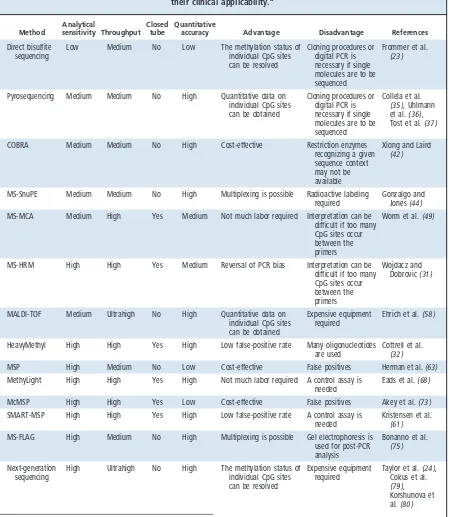
We review a number of the most promising MIP-and MSP-based methods MIP-and discuss the relative ad-vantages and disadad-vantages with regard to their clinical applicability; however, no method is universally supe-rior, because it is impossible to obtain all of the follow-ing objectives with a sfollow-ingle method: quantitative accu-racy, high analytical sensitivity, low false-positive and false-negative rates, high throughput, assessment of single CpG sites, low risk of PCR contamination (closed-tube assay), easily interpretable results, no need for specialized equipment, and cost-effectiveness. Table 1 provides an overview of the methods discussed
and compares them with respect to several parameters that are important for clinical applicability.
Sodium Bisulfite Treatment
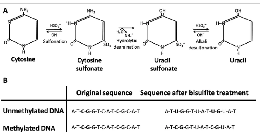
Epigenetic information is lost during the PCR because the DNA polymerase does not distinguish between methylated and unmethylated cytosines; thus, the polymerase incorporates guanine and subsequently unmethylated cytosines in both situations. After the PCR, any originally methylated alleles will be diluted to undetectable concentrations; therefore, the DNA must be modified in a way that allows the methylation infor-mation to be preserved. Treatment with sodium bisul-fite, which deaminates cytosine to uracil (22 )is the method of choice in most laboratories for this purpose. Because the rate of deamination of 5-methylcytosine to thymine is much slower than the conversion of cyto-sine to uracil, it is assumed that the only cytocyto-sines re-maining after sodium bisulfite treatment are derived from 5-methylcytosines. Thus, during subsequent PCRs, uracil residues are replicated as thymine resi-dues, and 5-methylcytosine residues are replicated as cytosines (Fig. 1). The protocol described by Frommer and colleagues(23 )has been widely used for sodium bisulfite treatment, and a variety of commercial kits are now available for this purpose. When the sodium bisul-fite treatment is performed under appropriate condi-tions, the expected conversion rate of unmethylated cytosines is about 99%(24 ). Despite this high conver-sion rate, however, it is possible that a small subset of the DNA copies have a substantially lower conversion rate(25 )and that the distribution of unconverted sites is nonrandom; thus, some promoter regions are more prone to incomplete conversion. The conversion rate may also depend on DNA quality(25 ). This possibility is especially important to keep in mind when looking for low levels of methylation with methods based on MSP primers.
The sense and antisense strands are no longer complementary after sodium bisulfite treatment. Thus, MIP or MSP primers are designed for either strand.
Methods Based on MIP Primers
an-nealing temperature(27 ). In some situations, however, increasing the annealing temperature may be enough to neutralize the bias despite the use of traditional MIP primers without CpG sites(28 ). Amplification of
sin-gle molecules has been shown to overcome the PCR bias phenomenon (29, 30 ). Another limitation of MIP-based methods is the relatively low analytical sen-sitivity, although this sensitivity can be increased by Table 1. Methods discussed in this review compared for several parameters important for
their clinical applicability.a
Method
Analytical
sensitivity Throughput Closed
tube
Quantitative
accuracy Advantage Disadvantage References
Direct bisulfite sequencing
Low Medium No Low The methylation status of individual CpG sites can be resolved
Cloning procedures or digital PCR is necessary if single molecules are to be sequenced
Frommer et al. (23 )
Pyrosequencing Medium Medium No High Quantitative data on individual CpG sites can be obtained
Cloning procedures or digital PCR is necessary if single molecules are to be sequenced
Collela et al. (35 ), Uhlmann et al.(36 ), Tost et al.(37 ) COBRA Medium Medium No High Cost-effective Restriction enzymes
recognizing a given sequence context may not be available
Xiong and Laird (42 )
MS-SnuPE Medium Medium No High Multiplexing is possible Radioactive labeling required
Gonzalgo and Jones(44 ) MS-MCA Medium High Yes Medium Not much labor required Interpretation can be
difficult if too many CpG sites occur between the primers
Worm et al.(49 )
MS-HRM High High Yes Medium Reversal of PCR bias Interpretation can be difficult if too many CpG sites occur between the primers
Wojdacz and Dobrovic(31 )
MALDI-TOF Medium Ultrahigh No High Quantitative data on individual CpG sites can be obtained
Expensive equipment required
Ehrich et al.(58 )
HeavyMethyl High High Yes High Low false-positive rate Many oligonucleotides are used
Cottrell et al. (32 )
MSP High Medium No Low Cost-effective False positives Herman et al.(63 ) MethyLight High High Yes High Not much labor required A control assay is
needed
Eads et al.(68 ) McMSP High High Yes Low Cost-effective False positives Akey et al.(73 ) SMART-MSP High High Yes High Low false-positive rate A control assay is
needed
Kristensen et al. (61 ) MS-FLAG High Medium No High Multiplexing is possible Gel electrophoresis is
used for post-PCR analysis
Bonanno et al. (75 )
Next-generation sequencing
High Ultrahigh No High The methylation status of individual CpG sites can be resolved
Expensive equipment required
Taylor et al.(24 ), Cokus et al. (79 ), Korshunova et al.(80 )
aAnalytical sensitivity and quantitative accuracy are dependent on the specific assay and parameters such as the concentration and quality of input DNA and PCR
introducing CpG sites into the primers(31 )or through the use of oligonucleotide blockers(32 ).
BISULFITE GENOMIC SEQUENCING
The gold standard in DNA methylation analysis has traditionally been the sequencing of bisulfite-modified and PCR-amplified DNA(23 ), because this approach provides information at the level of individual CpG sites. Sequencing is mostly done with MIP primers but can be used to confirm MSP results as well. PCR prod-ucts can be sequenced directly or as single clones. Sequencing of cloned PCR products provides informa-tion on individual molecules, whereas direct sequenc-ing provides an estimate of the average methylation status of each CpG site in all of the molecules. When MSP primers are used, all of the cloned molecules are expected to be methylated, whereas the ratio of meth-ylated to unmethmeth-ylated molecules in a sample can be determined if MIP primers are used and enough clones are sequenced. Unfortunately, sequencing of single clones is too time-consuming and expensive to be used in routine clinical settings(33 ). Bisulfite genomic
se-quencing of single clones can be affected by a cloning bias, and it can be problematic to accurately sequence the longer stretches of thymine that are often encoun-tered in bisulfite-modified DNA(34 ). Recently, a dig-ital bisulfite-sequencing approach that allows sequenc-ing of ssequenc-ingle molecules without clonsequenc-ing procedures has been described(30 ). This approach requires both mul-tiple reactions of the sample and sample dilution to a critical level to minimize the occurrence of more than 2 PCR template molecules per reaction well. At least 96 reactions of the sample should be performed so that a reasonable number of positive wells are available for subsequent sequencing. Nevertheless, this approach saves time and labor compared with subcloning proce-dures for isolating single bisulfite-converted DNA molecules.
PYROSEQUENCING
An attractive alternative to the traditional dideoxy se-quencing approach (Sanger sese-quencing) is pyrose-quencing, which is based on the detection of pyrophos-phate. This method, which has also been adapted to Fig. 1. Sodium bisulfite treatment of genomic DNA.
(A), The procedure is based on the chemical reaction of single-stranded DNA with sodium bisulfite (HSO3⫺) at low pH and high
methylation analysis with bisulfite-modified DNA, yields quantitative information on single CpG sites (35–37 ). Pyrosequencing is based on the detection of emitted light during synthesis of the complementary strand by an exonuclease-deficient DNA polymerase. When nucleotides are incorporated, pyrophosphate is released and converted to ATP by the enzyme ATP sul-furylase. The ATP molecules provide energy for the enzyme luciferase to oxidize luciferin in a reaction that generates light. The 4 different nucleotides are added sequentially to enable base calling.
The instrumentation required for pyrosequencing can be used for many applications(38 ); however, the quantitative accuracy and reliability of the data de-crease with the distance of the CpG from the 3⬘end of the forward primer, a feature that limits the number of bases/CpG sites that can be analyzed in a single se-quencing reaction(39 ). The long stretches of thymine often found in bisulfite-modified DNA are also likely to affect reproducibility. Pyrosequencing is usually car-ried out with MIP products; however, it can also be used to identify false-positive results in MSP assays (40 ).
COMBINED BISULFITE RESTRICTION ANALYSIS
Digestion of PCR products with certain restriction en-zymes can be used to distinguish between methylated and unmethylated DNA(41 ). The differences in se-quence between methylated and unmethylated DNA after bisulfite modification can lead to the creation of new methylation-dependent restriction sites or the maintenance of restriction sites in a methylation-dependent manner. This property was exploited in the development of a quantitative method termed “com-bined bisulfite restriction analysis” (COBRA) (42 ), which relies on the separation of digested PCR prod-ucts by agarose or polyacrylamide gel electrophoresis and subsequent quantitative hybridization. The feature that most limits this technology is that many CpG sites cannot be analyzed because the restriction enzymes that would be appropriate for recognizing the sequence context are not available. Furthermore, apart from the PCR bias phenomenon(26 ), accurate quantification may be compromised by the formation of heterodu-plexes between strands containing the restriction site and strands that do not and by incomplete conversion of unmethylated cytosines during the bisulfite modifi-cation. The method is relatively labor-intensive but is cost-effective.
METHYLATION-SENSITIVE SINGLE-NUCLEOTIDE PRIMER EXTENSION (MS-SnuPE)
The SnuPE assay was originally developed for detection of single-nucleotide mutations(43 )but was later ap-plied to DNA methylation studies(44 ). The product
generated by the MIP is subsequently isolated via gel electrophoresis and annealed to an internal primer that terminates immediately 5⬘of the single nucleotide to be assayed (i.e., the cytosine of a CpG site in methyl-ation studies). The internal primer is then extended with a DNA polymerase that uses32P-labeled dCTP or dTTP. Reaction products are separated on polyacryl-amide gels for visualization, and the relative amounts of the 2 nucleotides present in the MIP product can be quantified with phosphor-imaging analysis. A rela-tively high throughput is possible, especially when multiple internal primers are included in a single primer-extension reaction(45 ). The internal primers should not anneal to sequences that originally con-tained CpG sites to prevent the introduction of a bias at this step, but achieving this goal can be difficult in CpG-dense regions. The method is quite labor-intensive and has the disadvantage of requiring the use of radioactive materials.
Alternatively, the SNaPshot technology from Ap-plied Biosystems can be used as a detection platform, thereby omitting radioactive labeling (46 ). Another variant of MS-SnuPE that uses denaturing HPLC in-stead of radioactivity for separation and quantification of the extended primer products has also been de-scribed(47 ). This approach, SnuPE ion pair reversed-phase HPLC (SIRPH), was recently evaluated for de-tecting MGMT promoter methylation but was not recommended over COBRA and pyrosequencing(33 ). Finally, a microarray-based version was recently intro-duced as a semiquantitative high-throughput method (48 ).
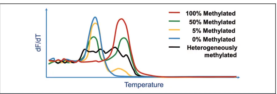
METHYLATION-SENSITIVE MELTING CURVE ANALYSIS
mole-cules is amplified, 2 distinct melting peaks are observed, and interpretation is easy. When heteroge-neously methylated molecules are amplified, however, the melting pattern can be complex and difficult to interpret (Fig. 2).
METHYLATION-SENSITIVE HIGH-RESOLUTION MELTING (MS-HRM)
In general, the development of the HRM technology (50 ) created several methodologic advantages. First, because the HRM approach acquires more data points, the melting peaks are sharper, and subtle differences within the amplicons can be detected. Second, the tem-perature variations produced with HRM instrumenta-tion are generally extremely small, and a relatively high throughput is possible, depending on the instrument used(51 ). Third, most of the software provided with the instruments permits normalization for end-level fluorescence(52 ), temperature shifting, and use of in-ternal oligonucleotide calibrators(53 ). HRM is often performed with a dye that can be used at saturating concentrations without inhibiting the PCR. Nonsat-urating dyes such as SYBR Green I can also be used, and although this dye does not detect heteroduplexes well, this feature may be an advantage in methylation studies because heteroduplexes unnecessarily complicate the melting pattern. Sequence-specific binding of SYBR Green I has been reported(54 ), indicating a potential problem in methylation studies that use melting techniques.
HRM has been used for methylation analysis with traditional MIP primers in the analysis of an imprinted locus (55 ), with MIP primers that include a limited number of CpG sites to correct for PCR bias and to increase the analytical sensitivity, for the analysis of MGMT promoter methylation, and for methylation changes inH19[H19, imprinted maternally expressed transcript (non-protein coding)] (31, 56 ). This ap-proach, MS-HRM, involves including CpG sites in the primer sequences, which pushes the PCR bias toward the methylated allele, and optimization of annealing temperatures has significantly increased the analytical sensitivity(31 )by making the reaction more MSP-like. It has recently been shown that studies of promoters that tend to be heterogeneously methylated can be im-proved with a digital MS-HRM approach(57 ).
MALDI-TOF MASS SPECTROMETRY WITH BASE-SPECIFIC CLEAVAGE AND PRIMER EXTENSION
The use of MALDI-TOF mass spectrometry for DNA methylation analysis has several advantages. The meth-odology is relatively sensitive, and a methylation level of 5% can be detected without including any CpG sites in the MIP primer sequences. Furthermore, very high throughputs are possible, and the methodology is quantitatively accurate(58 ).
The experiments to be performed prior to mass spectrometry analysis can be based on base-specific cleavage or primer extension (59 ). The base-specific cleavage strategy involves amplification with one Fig. 2. Principle of melting analysis for methylation detection.
primer tagged with a T7 promoter sequence to allow in vitro transcription of the PCR product into a single-stranded RNA transcript. Subsequent base-specific cleavage by an endoribonuclease such as RNase A pro-duces different cleavage patterns for methylated and unmethylated CpG sites, depending on the use of non-cleavable nucleotides. The cleavage products are then analyzed by MALDI-TOF mass spectrometry, which permits the relative amounts of methylated and un-methylated DNA to be determined via comparisons of signal intensities.
When a primer-extension strategy is used, a post-PCR primer-extension reaction is performed with a primer designed to anneal immediately adjacent to the CpG site under investigation. The primer is then ex-tended with a mixture of 4 different terminators, such as dideoxy NTPs. Depending on the methylation status of the CpG site, the primer-extension reaction will ter-minate on different nucleotides and generate distinct signals when analyzed by MALDI-TOF mass spec-trometry. Multiplexing of up to 25 different primer-extension reactions is feasible without compromising the quantitative results if primers are designed carefully (59 ). The base-specific cleavage strategy is recom-mended for purposes requiring the analysis of larger regions of unknown methylation content, whereas the primer-extension strategy should be used in routine analyses of a relatively small number of well-characterized informative CpG sites.
The main disadvantages of this methodology are that the required equipment is expensive and the complexity of the methodology can make it chal-lenging to use. For instance, the presence of unex-pected single-nucleotide polymorphisms can lead to misinterpreted results because no actual sequence information is given. Coolen and colleagues recently addressed this problem (60 ) in a publication that also demonstrated that MALDI-TOF mass spec-trometry can determine whether the detected meth-ylation is allele specific.
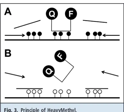
HEAVYMETHYL
In the HeavyMethyl methodology(32 ), oligonucleo-tide blockers are used to discriminate between methyl-ated and unmethylmethyl-ated alleles. The MIP primers are designed to hybridize next to a CpG-rich sequence, for which blockers have been designed to hybridize only to unmethylated DNA. Thus, if the DNA is methylated, the blockers cannot hybridize and leave the primer-binding site accessible for the primers to bind. Ampli-fication will then occur. AmpliAmpli-fication is detected with a probe that contains CpG sites, a fluorophore label, and a quencher. When the exonuclease activity of the polymerase cleaves the probe, the fluorophore is
re-leased from the quencher, and light is emitted (Fig. 3). The emitted light is proportional to the amount of am-plicon in the test tube, allowing accurate quantification of the methylation level.
The use of blocker molecules significantly in-creases the analytical sensitivity, which is comparable to methods that use MSP primers. HeavyMethyl allows a high throughput and is a closed-tube method; how-ever, the main advantage relative to conventional MSP (discussed below) may be that false-positive rates are extremely low. These rates are low because the blockers provide methylation specificity at every cycle of the PCR, whereas in MSP, a false-priming event needs to happen only once to get the amplification going. An-other advantage of HeavyMethyl is that the flexibility of primer and blocker design may allow detection of heterogeneous methylation.
Fig. 3. Principle of HeavyMethyl.
Methods Based on MSP Primers
MSP primers are designed to amplify methylated DNA only, and thus the PCR bias phenomenon associated with MIP-based methods is not an issue. This specific-ity is achieved by including many CpG sites in the primer sequences, preferably at or close to the 3⬘end. In concert with stringent PCR conditions, only ampli-fication of methylated DNA will occur. MSP assays are generally associated with high false-positive rates (40, 61, 62 ), however, especially with a high number of PCR cycles, which can be necessary for obtaining highly analytically sensitive assays. False-priming events (in which amplification happens despite the mismatches of the primer sequences with the template) and incom-pletely bisulfite-converted DNA molecules within the test tube may be responsible for false-positive results. False-priming events can be detected with an appro-priate negative control and prevented by limiting the number of cycles and/or using a higher annealing temperature. Incompletely converted molecules may mimic methylated sequences, because MSP primers contain multiple cytosines derived from CpG sites. Al-though this feature makes such primers highly selective for methylated templates, it also facilitates amplifica-tion of incompletely converted sequences in the bisulfite-treated DNA. Having multiple non-CpG cy-tosines within the MSP primers may limit this problem.
METHYLATION-SPECIFIC PCR
In the traditional MSP methodology(63 ), a second set of primers is often designed (in addition to the MSP primers) for the amplification of unmethylated DNA so that the presence of suitable template can be con-firmed after the sodium bisulfite treatment. Gel elec-trophoresis is used for detecting the PCR products, and in situations in which both unmethylated DNA and methylated DNA are present in the test tube, a compar-ison of band strengths allows a very approximate esti-mate of relative methylation levels. Methylation levels can be estimated more accurately with real-time PCR (see below), but quantitative information can also be obtained with fluorescently labeled amplicons ana-lyzed by a genetic analyzer(64, 65 ). MSP is very cost-effective, but any of the false-positive results men-tioned above cannot be detected. Furthermore, opening of PCR tubes should be avoided, especially in clinical settings, to reduce the risk of PCR contamina-tion. No specialized equipment is needed, however, and the method is simple to use. For these reasons, MSP, which can be performed in almost any labora-tory, is the most widely used method for the analysis of DNA methylation at specific loci.
MSP is known for its high analytical sensitivity. In the original publication(63 ), MSP was reported to de-tect 0.1% methylated template in an excess of unmeth-ylated DNA, but it may detect as little as 0.0002% when a nested approach is used(66 ). The analytical sensitiv-ity of MSP assays is also influenced by primer design, the number of PCR cycles, and annealing temperature. Use of fewer cycles gives fewer false-positive results but also decreases the analytical sensitivity of the assay, whereas the use of appropriately designed primers and a high annealing temperature prevents false-priming events. Thus, many published MSP assays vary tremen-dously in analytical sensitivity(66, 67 ).
QUANTITATIVE MSP: METHYLIGHT
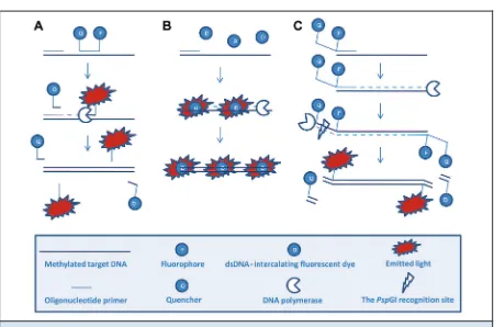
MSP was first made quantitative by the use of fluores-cent hydrolysis probes that enabled real-time detection of the MSP amplification (68 –70 ) (Fig. 4). This ap-proach, which is most often referred to as MethyLight, overcomes most of the problems associated with MSP. First, amplification is observed only when the probe has hybridized between the primers, thus eliminating any signal from nonspecific amplification, such as primer dimer formation. For this reason, the fact that gel electrophoresis is not required makes MethyLight a closed-tube method capable of high throughput. Sec-ond, the additional CpG sites within the probe se-quence make priming events less likely, and false-positives due to incomplete conversion can be limited by having many non-CpG cytosines within the probe sequence. The introduction of a probe complicates as-say design, however, and can cause MSP to miss heter-ogeneously methylated sequences that it would other-wise detect because of the requirement for the probe to hybridize correctly before a signal is observed. The an-alytical sensitivity of MethyLight is similar to that of MSP but has recently been shown to be increased to at least 0.05% when a digital approach for amplifying sin-gle molecules is used (30 ). As with all quantitative MSP-based methods that use sodium bisulfite– modified DNA, a control gene is used to normalize for DNA input.
QUANTITATIVE MSP: SYBR GREEN–BASED
SYBR Green I has also been used for post-PCR melting analysis in the nonquantitative melting curve MSP (McMSP) methodology to avoid the use of gel electrophoresis(73 ); however, the limited resolution of the post-PCR melting analysis did not allow addi-tional information not provided by gel electrophoresis to be obtained. Thus, these melting analyses may be associated with false-positive results, as with conven-tional MSP, but McMSP has the advantage of being a closed-tube method, allowing a high throughput.
QUANTITATIVE MSP: SENSITIVE MELTING ANALYSIS AFTER REAL-TIME MSP (SMART-MSP)
The SMART-MSP methodology(61 )takes advantage of the additional resolution of the HRM technology for
the detection of false-positive results. Thus, SMART-MSP is also based on dsDNA-intercalating fluorescent dyes (Fig. 4). The real-time MSP provides quantitative data that may be analyzed with the relative 2⫺⌬⌬Ct quantification approach if the PCR efficiencies for the gene and the control are approximately equal(74 ). The use of an HRM step after the PCR has improved probe-free quantitative MSP analysis in several ways. This val-idation step provides information that cannot be ob-tained by gel electrophoresis, and false-positive results caused by false-priming events or incomplete conver-sion can often be detected, depending on how the am-plicon is designed(61 ). Notably, HRM analysis may be suitable only for detecting false-positive results, how-ever, and not for situations in which incomplete con-Fig. 4. Three different ways of creating real-time fluorescence signals in quantitative MSP analysis.
version leads to slight overestimation of methylation levels. This is because the signal from the methylated and fully converted molecules will be much stronger in such situations than the signal derived from incom-pletely converted molecules.
SMART-MSP can detect 0.1% methylated tem-plate(61 ). A high throughput is possible, and the risk of PCR contamination is low because of the closed-tube format of the method.
QUANTITATIVE MSP: METHYLATION-SPECIFIC FLUORESCENT AMPLICON GENERATION (MS-FLAG)
Another quantitative MSP approach that circumvents the need for additional probes has recently been intro-duced. In MS-FLAG (75 ), the fluorescence signal is created by cleavage of the MSP primers by the thermo-stable endonucleasePspGI. The primers contain an oli-gonucleotide 5⬘ tail carrying a fluorophore and a quencher separated by the recognition site of the endo-nuclease. The double-stranded recognition site is not created until the primers have annealed and the poly-merase has created a new copy of the target (Fig. 4). The quantitative accuracy of the MS-FLAG methodology can be compromised by primer dimer formation and has to be avoided through optimal design of the prim-ers. Thus, post-PCR analysis is required to confirm that the amplification was specific. Because melting analy-ses are not compatible with this approach, gel electro-phoresis is used for this purpose.
The analytical sensitivity of MS-FLAG is compara-ble to that of other MSP-based methods but is less cost-effective than SMART-MSP because of the use of fluorescently labeled primers and the thermostable en-donuclease. Because additional probes are not re-quired, however, primers can be labeled differently, and so it is possible to design multiplex MS-FLAG as-says to limit costs and labor(75 ).
Discussion
Early detection of cancer often improves the clinical outcome. Methods for relatively early detection exist for breast and prostate cancer. On the other hand, many imaging and cytology-based strategies have failed to achieve early detection of lung and other can-cers (76 ). Thus, there is a need for new molecular methods to detect preneoplastic and small malignant lesions(19 ). A number of specific loci are potential candidates as DNA methylation biomarkers especially directed toward early cancer detection.
There are many reasons for cancer-specific meth-ylated loci being suitable as biomarkers for cancer de-tection(19 ). First, DNA is a stable molecule that can easily be isolated from body fluids and tissues, in con-trast to the RNA needed for reverse-transcription PCR
assays. Second, DNA containing methylation informa-tion can be isolated from formalin-fixed and paraffin-embedded tissues and be used in most PCR-based methods for detecting DNA methylation. Third, the methylation signal to be detected is positive, in contrast to the loss of heterozygosity or changes in gene expres-sion, which can be difficult to detect in the presence of an excess of nonaffected DNA.
Much more work is required to validate the clini-cal use of many DNA methylation biomarkers, how-ever, because existing markers often lack the diagnostic sensitivity and specificity required for a diagnostic test. Lack of diagnostic sensitivity and specificity may be in part a technological problem, because false-positive re-sults will produce a low diagnostic specificity and false-negative results will produce a low diagnostic sensitiv-ity. Nevertheless, numerous new genes that undergo cancer-specific methylation have recently been identi-fied, and the creation of panels of markers with higher diagnostic sensitivity and specificity for particular pur-poses is ongoing(76 ).
The choice of CpG sites to be analyzed is also im-portant, because the methylation status of some sites may prove better than others for distinguishing healthy tissue from malignant tissue (33 ). Furthermore, the degree of interindividual variation is still unknown (77 ), and it is therefore more difficult to define what is typical with respect to DNA methylation, in contrast to genetic events, for which we have a reference sequence. Because tumor-derived material in body fluids of-ten is difficult to detect, highly sensitive methods with low limits of detection are needed for many applica-tions. These methods are generally based on MSP primers; however, they are also associated with false-positive results. The false false-positives can be limited through the use of fluorescent probes in the Methy-Light methodology(68 )or by HRM in the SMART-MSP methodology(61 ). Furthermore, these methods are quantitative. The quantitative data can be used to set a threshold for the methylation level to provide the highest diagnostic sensitivity and specificity for a given biomarker. When the threshold is lowered, the nostic sensitivity typically will increase, and the diag-nostic specificity will decrease.
MS-HRM are quantitative methods that do not require the PCR tubes to be opened. Therefore, the clinical appli-cability of these methods is comparable to that of MethyLight and SMART-MSP (see Table 1).
It is desirable that the results obtained be con-firmed by more than one method; however, because of the innate differences among the various methods, the results obtained with different methods cannot be ex-pected to be identical. Sequencing of a limited number of samples is often used to confirm results in both MIP-and MSP-based methods, but sequencing all samples is too time-consuming and too expensive for most laboratories.
Conclusion and Future Perspectives
A more detailed analysis of individual CpG sites in the studied CpG islands may be necessary to identify the most informative sites (i.e., those providing the highest diagnostic sensitivity and specificity). Currently, there is a tendency in the literature for CpG sites to be chosen for their optimal primer location, depending on the specific assay, because detailed studies of the CpG sites of individual molecules have thus far been expensive and time-consuming. Emerging massively parallel se-quencing methods (78 ), often referred to as “next-generation” sequencing, promise to make the enor-mous amount of information required for this purpose easily obtainable. Currently, 3 platforms are available: the Genome Sequencer FLX system/454 sequencing (named “454” by 454 Life Sciences, now acquired by Roche), the Genome Analyzer system (named “Solexa” by Solexa, now acquired by Illumina), and the SOLiD system (named “SOLiD” by Applied Biosystems). The first of these new methods to be applied to sodium bisulfite–modified DNA was the 454 system based on pyrosequencing. A detailed methylation study of 25 gene-related CpG islands in 40 samples from different nonmalignant and malignant blood cells has been per-formed (24 ). This pilot study confirmed the utility,
robustness, and superiority of the method with bisulfite-modified DNA as the template. In addition to the ultrahigh throughput this approach offers, it is ca-pable of providing accurate sequences, even at the long stretches of thymine often found in bisulfite-modified DNA. This approach eliminates the bias often encoun-tered when PCR products are subcloned in bacteria (34 ), and far more individual “clones” (molecules) can be analyzed relative to the few clones typically analyzed in bisulfite sequencing(23 ). Thus, the 454 approach has the potential to identify the most informative CpG sites within a given CpG island rapidly and with high accuracy and thereby provide guidelines for the devel-opment of analytically sensitive and more cost-effective assays, such as MethyLight, SMART-MSP, HeavyMethyl, and MS-HRM, with sufficient robust-ness for clinical use.
Author Contributions:All authors confirmed they have contributed to the intellectual content of this paper and have met the following 3 re-quirements: (a) significant contributions to the conception and design, acquisition of data, or analysis and interpretation of data; (b) drafting or revising the article for intellectual content; and (c) final approval of the published article.
Authors’ Disclosures of Potential Conflicts of Interest: Upon manuscript submission, all authors completed the Disclosures of Poten-tial Conflict of Interest form. PotenPoten-tial conflicts of interest:
Employment or Leadership:None declared.
Consultant or Advisory Role:None declared.
Stock Ownership:None declared.
Honoraria:None declared.
Research Funding:Region Midtjyllands Sundhedsvidenskabelige forskningsfond.
Expert Testimony:None declared.
Role of Sponsor:The funding organizations played no role in the design of study, choice of enrolled patients, review and interpretation of data, or preparation or approval of manuscript.
Acknowledgments:We thank Eigil Kjeldsen for his input and critical reading of this manuscript.
References
1. Huynh KD, Lee JT. X-chromosome inactivation: a hypothesis linking ontogeny and phylogeny. Nat Rev Genet 2005;6:410 – 8.
2. Lewis A, Reik W. How imprinting centres work. Cytogenet Genome Res 2006;113:81–9. 3. Bodey B. Cancer-testis antigens: promising
tar-gets for antigen directed antineoplastic immuno-therapy. Expert Opin Biol Ther 2002;2:577– 84. 4. Walsh CP, Chaillet JR, Bestor TH. Transcription of
IAP endogenous retroviruses is constrained by cytosine methylation. Nat Genet 1998;20:116 –7. 5. Eden A, Gaudet F, Waghmare A, Jaenisch R. Chromosomal instability and tumors promoted by DNA hypomethylation. Science 2003;300:455. 6. Gaudet F, Hodgson JG, Eden A, Jackson-Grusby L,
Dausman J, Gray JW, et al. Induction of tumors in
mice by genomic hypomethylation. Science 2003; 300:489 –92.
7.Craig JM, Bickmore WA. The distribution of CpG islands in mammalian chromosomes. Nat Genet 1994;7:376 – 82.
8.Baylin SB, Ohm JE. Epigenetic gene silencing in cancer – a mechanism for early oncogenic path-way addiction? Nat Rev Cancer 2006;6:107–16. 9.Esteller M. Aberrant DNA methylation as a cancer-inducing mechanism. Annu Rev Pharma-col ToxiPharma-col 2005;45:629 –56.
10.Esteller M. Epigenetics in cancer. N Engl J Med 2008;358:1148 –59.
11.Laird PW. The power and the promise of DNA methylation markers. Nat Rev Cancer 2003;3: 253– 66.
12.Shi H, Wang MX, Caldwell CW. CpG islands: their potential as biomarkers for cancer. Expert Rev Mol Diagn 2007;7:519 –31.
13.Aggerholm A, Holm MS, Guldberg P, Olesen LH, Hokland P. Promoter hypermethylation of p15INK4B,HIC1, CDH1, and ER is frequent in
myelodysplastic syndrome and predicts poor prognosis in early-stage patients. Eur J Haematol 2006;76:23–32.
14.Fischer JR, Ohnmacht U, Rieger N, Zemaitis M, Stoffregen C, Kostrzewa M, et al. Promoter meth-ylation ofRASSF1A, RARandDAPK predict poor prognosis of patients with malignant mesotheli-oma. Lung Cancer 2006;54:109 –16. 15.Muller HM, Widschwendter A, Fiegl H, Ivarsson L,
serum of breast cancer patients: an independent prognostic marker. Cancer Res 2003;63:7641–5. 16.Wallner M, Herbst A, Behrens A, Crispin A, Stieber P, Goke B, et al. Methylation of serum DNA is an independent prognostic marker in colorectal cancer. Clin Cancer Res 2006;12: 7347–52.
17.Yu J, Cheng YY, Tao Q, Cheung KF, Lam CN, Geng H, et al. Methylation of protocadherin 10, a novel tumor suppressor, is associated with poor prognosis in patients with gastric cancer. Gastro-enterology 2009;136:640 –51.e1.
18.Sidransky D. Emerging molecular markers of can-cer. Nat Rev Cancer 2002;2:210 –9.
19.Cottrell SE, Laird PW. Sensitive detection of DNA methylation. Ann N Y Acad Sci 2003;983: 120 –30.
20.Hegi ME, Diserens AC, Gorlia T, Hamou MF, de Tribolet N, Weller M, et al. MGMT gene silencing and benefit from temozolomide in glioblastoma. N Engl J Med 2005;352:997–1003.
21.Saah AJ, Hoover DR. “Sensitivity” and “specific-ity” reconsidered: the meaning of these terms in analytical and diagnostic settings. Ann Intern Med 1997;126:91– 4.
22.Clark SJ, Harrison J, Paul CL, Frommer M. High sensitivity mapping of methylated cytosines. Nu-cleic Acids Res 1994;22:2990 –7.
23.Frommer M, McDonald LE, Millar DS, Collis CM, Watt F, Grigg GW, et al. A genomic se-quencing protocol that yields a positive display of 5-methylcytosine residues in individual DNA strands. Proc Natl Acad Sci U S A 1992;89: 1827–31.
24.Taylor KH, Kramer RS, Davis JW, Guo J, Duff DJ, Xu D, et al. Ultradeep bisulfite sequencing anal-ysis of DNA methylation patterns in multiple gene promoters by 454 sequencing. Cancer Res 2007; 67:8511– 8.
25.Warnecke PM, Stirzaker C, Song J, Grunau C, Melki JR, Clark SJ. Identification and resolution of artifacts in bisulfite sequencing. Methods 2002; 27:101–7.
26.Warnecke PM, Stirzaker C, Melki JR, Millar DS, Paul CL, Clark SJ. Detection and measurement of PCR bias in quantitative methylation analysis of bisulphite-treated DNA. Nucleic Acids Res 1997; 25:4422– 6.
27.Wojdacz TK, Hansen LL. Reversal of PCR bias for improved sensitivity of the DNA methylation melting curve assay. Biotechniques 2006;41:274, 276, 278.
28.Shen L, Guo Y, Chen X, Ahmed S, Issa JP. Opti-mizing annealing temperature overcomes bias in bisulfite PCR methylation analysis. Biotechniques 2007;42:48, 50, 52 passim.
29.Chhibber A, Schroeder BG. Single-molecule poly-merase chain reaction reduces bias: application to DNA methylation analysis by bisulfite sequenc-ing. Anal Biochem 2008;377:46 –54. 30.Weisenberger DJ, Trinh BN, Campan M, Sharma
S, Long TI, Ananthnarayan S, et al. DNA methyl-ation analysis by digital bisulfite genomic se-quencing and digital MethyLight. Nucleic Acids Res 2008;36:4689 –98.
31.Wojdacz TK, Dobrovic A. Methylation-sensitive high resolution melting (MS-HRM): a new ap-proach for sensitive and high-throughput as-sessment of methylation. Nucleic Acids Res 2007;35:e41.
32.Cottrell SE, Distler J, Goodman NS, Mooney SH, Kluth A, Olek A, et al. A real-time PCR assay for DNA-methylation using methylation-specific blockers. Nucleic Acids Res 2004;32:e10. 33.Mikeska T, Bock C, El-Maarri O, Hubner A,
Ehrentraut D, Schramm J, et al. Optimization of quantitative MGMT promoter methylation analy-sis using pyrosequencing and combined bisulfite restriction analysis. J Mol Diagn 2007;9:368 – 81. 34.Grunau C, Clark SJ, Rosenthal A. Bisulfite genomic sequencing: systematic investigation of critical experimental parameters. Nucleic Acids Res 2001;29:E65.
35.Colella S, Shen L, Baggerly KA, Issa JP, Krahe R. Sensitive and quantitative universal Pyrosequenc-ing methylation analysis of CpG sites. Biotech-niques 2003;35:146 –50.
36.Uhlmann K, Brinckmann A, Toliat MR, Ritter H, Nurnberg P. Evaluation of a potential epigenetic biomarker by quantitative methyl-single nucleo-tide polymorphism analysis. Electrophoresis 2002;23:4072–9.
37.Tost J, Dunker J, Gut IG. Analysis and quantifica-tion of multiple methylaquantifica-tion variable posiquantifica-tions in CpG islands by Pyrosequencing. Biotechniques 2003;35:152– 6.
38.Marsh S. Pyrosequencing applications. Methods Mol Biol 2007;373:15–24.
39.Dupont JM, Tost J, Jammes H, Gut IG. De novo quantitative bisulfite sequencing using the pyro-sequencing technology. Anal Biochem 2004;333: 119 –27.
40.Shaw RJ, Akufo-Tetteh EK, Risk JM, Field JK, Lilo-glou T. Methylation enrichment pyrosequencing: combining the specificity of MSP with validation by pyrosequencing. Nucleic Acids Res 2006;34:e78. 41.Sadri R, Hornsby PJ. Rapid analysis of DNA
meth-ylation using new restriction enzyme sites created by bisulfite modification. Nucleic Acids Res 1996; 24:5058 –9.
42.Xiong Z, Laird PW. COBRA: a sensitive and quan-titative DNA methylation assay. Nucleic Acids Res 1997;25:2532– 4.
43.Kuppuswamy MN, Hoffmann JW, Kasper CK, Spitzer SG, Groce SL, Bajaj SP. Single nucleotide primer extension to detect genetic diseases: ex-perimental application to hemophilia B (factor IX) and cystic fibrosis genes. Proc Natl Acad Sci U S A 1991;88:1143–7.
44.Gonzalgo ML, Jones PA. Rapid quantitation of methylation differences at specific sites using methylation-sensitive single nucleotide primer ex-tension (Ms-SNuPE). Nucleic Acids Res 1997;25: 2529 –31.
45.Gonzalgo ML, Liang G. Methylation-sensitive single-nucleotide primer extension (Ms-SNuPE) for quantitative measurement of DNA methyl-ation. Nat Protoc 2007;2:1931– 6.
46.Kaminsky ZA, Assadzadeh A, Flanagan J, Petronis A. Single nucleotide extension technology for quantitative site-specific evaluation of metC/C in GC-rich regions. Nucleic Acids Res 2005;33:e95. 47.El-Maarri O, Herbiniaux U, Walter J, Oldenburg J. A rapid, quantitative, non-radioactive bisulfite-SNuPE- IP RP HPLC assay for methyl-ation analysis at specific CpG sites. Nucleic Acids Res 2002;30:e25.
48.Wu Z, Luo J, Ge Q, Lu Z. Microarray-based Ms-SNuPE: near-quantitative analysis for a high-throughput DNA methylation. Biosens
Bioelec-tron 2008;23:1333–9.
49.Worm J, Aggerholm A, Guldberg P. In-tube DNA methylation profiling by fluorescence melting curve analysis. Clin Chem 2001;47:1183–9. 50.Wittwer CT, Reed GH, Gundry CN, Vandersteen
JG, Pryor RJ. High-resolution genotyping by am-plicon melting analysis using LCGreen. Clin Chem 2003;49:853– 60.
51.Herrmann MG, Durtschi JD, Wittwer CT, Voelker-ding KV. Expanded instrument comparison of am-plicon DNA melting analysis for mutation scan-ning and genotyping. Clin Chem 2007;53: 1544 – 8.
52.Kristensen LS, Dobrovic A. Direct genotyping of single nucleotide polymorphisms in methyl me-tabolism genes using probe-free high-resolution melting analysis. Cancer Epidemiol Biomarkers Prev 2008;17:1240 –7.
53.Gundry CN, Dobrowolski SF, Martin YR, Robbins TC, Nay LM, Boyd N, et al. Base-pair neutral homozygotes can be discriminated by calibrated high-resolution melting of small amplicons. Nu-cleic Acids Res 2008;36:3401– 8.
54.Giglio S, Monis PT, Saint CP. Demonstration of preferential binding of SYBR Green I to specific DNA fragments in real-time multiplex PCR. Nu-cleic Acids Res 2003;31:e136.
55.White HE, Hall VJ, Cross NC. Methylation-sensitive high-resolution melting-curve analysis of the SNRPN gene as a diagnostic screen for Prader–Willi and Angelman syndromes. Clin Chem 2007;53:1960 –2.
56.Wojdacz TK, Dobrovic A, Algar EM. Rapid detec-tion of methyladetec-tion change at H19 in human imprinting disorders using methylation-sensitive high-resolution melting. Hum Mutat 2008;29: 1255– 60.
57.Candiloro IL, Mikeska T, Hokland P, Dobrovic A. Rapid analysis of heterogeneously methylated DNA using digital methylation-sensitive high res-olution melting: application to the CDKN2B (p15) gene. Epigenetics Chromatin 2008;1:7. 58.Ehrich M, Nelson MR, Stanssens P, Zabeau M,
Liloglou T, Xinarianos G, et al. Quantitative high-throughput analysis of DNA methylation patterns by base-specific cleavage and mass spectrometry. Proc Natl Acad Sci U S A 2005;102:15785–90. 59.van den Boom D, Ehrich M. Mass spectrometric
analysis of cytosine methylation by base-specific cleavage and primer extension methods. Meth-ods Mol Biol 2009;507:207–27.
60.Coolen MW, Statham AL, Gardiner-Garden M, Clark SJ. Genomic profiling of CpG methylation and allelic specificity using quantitative high-throughput mass spectrometry: critical evalua-tion and improvements. Nucleic Acids Res 2007;35:e119.
61.Kristensen LS, Mikeska T, Krypuy M, Dobrovic A. Sensitive melting analysis after real time-methylation specific PCR (SMART-MSP): high-throughput and probe-free quantitative DNA methylation detection. Nucleic Acids Res 2008; 36:e42.
62.Rand K, Qu W, Ho T, Clark SJ, Molloy P. Conversion-specific detection of DNA methylation using real-time polymerase chain reaction (ConLight-MSP) to avoid false positives. Methods 2002;27:114 –20.
assay for methylation status of CpG islands. Proc Natl Acad Sci U S A 1996;93:9821– 6. 64.Mackay DJ, Boonen SE, Clayton-Smith J,
Good-ship J, Hahnemann JM, Kant SG, et al. A maternal hypomethylation syndrome presenting as tran-sient neonatal diabetes mellitus. Hum Genet 2006;120:262–9.
65.Mackay DJ, Temple IK, Shield JP, Robinson DO. Bisulphite sequencing of the transient neonatal diabetes mellitus DMR facilitates a novel diag-nostic test but reveals no methylation anomalies in patients of unknown aetiology. Hum Genet 2005;116:255– 61.
66.Palmisano WA, Divine KK, Saccomanno G, Gilli-land FD, Baylin SB, Herman JG, Belinsky SA. Predicting lung cancer by detecting aberrant pro-moter methylation in sputum. Cancer Res 2000; 60:5954 – 8.
67.Voso MT, Scardocci A, Guidi F, Zini G, Di Mario A, Pagano L, et al. Aberrant methylation of DAP-kinase in therapy-related acute myeloid leukemia and myelodysplastic syndromes. Blood 2004;103: 698 –700.
68.Eads CA, Danenberg KD, Kawakami K, Saltz LB, Blake C, Shibata D, et al. MethyLight: a high-throughput assay to measure DNA methylation. Nucleic Acids Res 2000;28:E32.
69.Eads CA, Danenberg KD, Kawakami K, Saltz LB, Danenberg PV, Laird PW. CpG island hypermeth-ylation in human colorectal tumors is not associ-ated with DNA methyltransferase overexpression. Cancer Res 1999;59:2302– 6.
70.Lo YM, Wong IH, Zhang J, Tein MS, Ng MH, Hjelm NM. Quantitative analysis of aberrant p16 meth-ylation using real-time quantitative methmeth-ylation- methylation-specific polymerase chain reaction. Cancer Res 1999;59:3899 –903.
71.Chu DC, Chuang CK, Fu JB, Huang HS, Tseng CP, Sun CF. The use of real-time quantitative poly-merase chain reaction to detect hypermethylation of the CpG islands in the promoter region flank-ing the GSTP1 gene to diagnose prostate carci-noma. J Urol 2002;167:1854 – 8.
72.Chan MW, Chu ES, To KF, Leung WK. Quantita-tive detection of methylated SOCS-1, a tumor suppressor gene, by a modified protocol of quan-titative real time methylation-specific PCR using SYBR green and its use in early gastric cancer detection. Biotechnol Lett 2004;26:1289 –93. 73.Akey DT, Akey JM, Zhang K, Jin L. Assaying
DNA methylation based on high-throughput melting curve approaches. Genomics 2002;80: 376 – 84.
74.Livak KJ, Schmittgen TD. Analysis of relative gene
expression data using real-time quantitative PCR and the 2⫺⌬⌬CT method. Methods 2001;25:
402– 8.
75.Bonanno C, Shehi E, Adlerstein D, Makrigiorgos GM. MS-FLAG, a novel real-time signal genera-tion method for methylagenera-tion-specific PCR. Clin Chem 2007;53:2119 –27.
76.Anglim PP, Alonzo TA, Laird-Offringa IA. DNA methylation-based biomarkers for early detection of non-small cell lung cancer: an update. Mol Cancer 2008;7:81.
77.Dobrovic A, Kristensen LS. DNA methylation, epimutations and cancer predisposition. Int J Bio-chem Cell Biol 2008;41:34 –9.
78.Park PJ. Epigenetics meets next-generation se-quencing. Epigenetics 2008;3:318 –21. 79.Cokus SJ, Feng S, Zhang X, Chen Z, Merriman
B, Haudenschild CD, et al. Shotgun bisulphite sequencing of theArabidopsis genome reveals DNA methylation patterning. Nature 2008;452: 215–9.