Indonesian Journal of Biotechnology
VOLUME 22(1), 2017, 13–21 | RESEARCH ARTICLEBioac vity and gene c screening of marine ac nobacteria associated
with red algae
Gelidiella acerosa
Maria Ulfah1, Noer Kasanah2,∗, and Niken Satu Nur Handayani1
1Faculty of Biology, Universitas Gadjah Mada, Jl. Teknika Selatan, Sekip Utara, Sinduadi, Mla , Sleman, Daerah Is mewa Yogyakarta 55281,
Indonesia
2Department of Fisheries, Faculty of Agriculture, Universitas Gadjah Mada, Bulaksumur, Caturtunggal, Depok, Sleman, Daerah Is mewa
Yogyakarta 55281 Indonesia
∗Corresponding author:noer.kasanah@ugm.ac.id
ABSTRACTBacterial resistance to exis ng an bio cs has driven a search for new an bio cs from marine ac nobacteria. Bioac vity and gene c screening of ac nobacteria associated with red algaeGelidiella acerosawere conducted to discover new an bacterial compounds againstVibrio alginoly cus. A total of 14 ac nobacteria isolates were obtained fromG. acerosa. The isolates were subjected to gene c screening fornrps(non-ribosomal pep de synthetase) and FADH2-dependent
halo-genase genes. The isolates’ ability to produce secondary metabolites was examined by fermenta on in various media in a six-well mini plate. The bioac vity of the secondary metabolites was screened using a micro ter assay and the agar overlay method. The results showed that all 14 isolates had thenrpsgene, whereas none had the halogenase gene. Meanwhile, eight of the ac nobacteria isolates showed an bacterial ac vity againstV. alginoly cus.
KEYWORDSac nobacteria; an bio c; FADH2-dependent halogenase; NRPS; red algae
1. Introduc on
Antibiotics play a crucial role in treating bacterial infec-tions in both humans and animals. Overuse and inappro-priate prescribing of antibiotics contribute to problems an-tibiotic resistance and emergence of new pathogenic bacte-ria (Luyt et al. 2014). One of the pathogenic bacteria in ma-rine aquaculture and human disease isVibriosp. (Austin 2010). Vibrio spp. are commonly present in seawater, sediments and occupy aquatic environments at tempera-tures ranging from 10 to 30°C and contaminate seafood. Available antibiotic was limited to combat the infection in human (Shaw et al. 2014). New antibiotics is necessary for those cases and due to limited shelf-life of antibiotics (Kasanah and Hamann 2004). In this studyVibrio algi-nolyticuswas used as test strain becauseV. alginolyticus
has high potential as reservoirs of disease-carrying genes in otherVibriosp. and cause gastroenteritis and peritoni-tis due to consumption of seafood contaminated by these bacteria (Gómez-León et al. 2005;Mustapha et al. 2013). The Marine environment is a complex and unique ecosystem characterized by high salinity and pressure, low temperatures and variable oxygen concentrations (Bull et al. 2000). A marine ecosystem consists of huge vari-ety forms of life that allow interactions and associations between themselves. The most frequent associations are
between eukaryotic cells and microorganisms (Egan et al. 2008). Many bioactive compounds such as antibiotics, antitumor, antifouling or antioxidant from the marine mi-croorganism are due to the involvement of specific func-tion in their respective hosts (El-Shatoury et al. 2009).
Red algae, the most potential sources of unique sec-ondary metabolites compared with green and brown al-gae (Schwartsmann et al. 2001). More than 1400 sec-ondary metabolites were successfully isolated from red algae from 1963–2016 (Blunt et al. 2014). Red algae live as host for diverse and abundant symbiotic microor-ganisms. These bacteria protect the host from a preda-tor or others harmful things in pelagic areas (Singh and Reddy 2014) and produce important compounds for hu-man and animal health (Kanagasabhapathy et al. 2006; Pe-nesyan et al. 2010). Various novel compounds with biolog-ical activity have already been identified from seaweed-associated bacteria. Streptomycessp. YM5-799 associ-ated brown algae Analipus japonicus produced Strepto-bactin as an iron chelating activity for treatment human disease (Matsuo et al. 2011). Bacterial strain member of the genusAlteromonaswas associated with the red algae
sec-ondary metabolites of associated microorganism of Geli-diella acerosawas unexplored. Some studies have demon-strated the presence of antibacterial activity from Geli-diella acerosabut not the associated microorganism. A study reported that ethanol extract ofGelidiella acerosa
has antibacterial activity against Staphylococcus aureus
(Elsie et al. 2011).
Marine bacteria as associated microorganisms are pos-sible to participate in the biosynthesis of bioactive com-pound. The use of microorganism is more effective as a source of bioactive compound than its host due to the short life cycle of actinobacteria and the ability to be cul-tured in vitro (Piel et al. 2004). Approximately 80% of antibiotics have been found mainly produced by actinobac-teria especially genusStreptomycesandMicromonospora
(Naikpatil and Rathod 2011). Actinobateria continue to play a significant role in drug discovery and development. Bioactive compounds produced by marine actinobacteria were abysomicin C, diazepinomicin, and salinoporamide A (Lam 2006). Because of the excellent record of acti-nobacteria, a significant effort has been focused on the iso-lation of novel actinobacteria from marine for developing drug discovery especially antibiotics.
A new strategy for selecting potential actinobacteria is through genetic approach by detecting gene target in-volved in the biosynthesis of bioactive compounds. These bioactive compounds can be the result of the activity of non-ribosomal peptide synthetases (NRPS) and FADH2
-dependent halogenase. NRPS is multimodular enzyme consist of three domains, A domain (Adenylation domain) serves as an introduction and activation substrate, domain PCP (Peptidyl Carrier Protein) for transfer to the center catalyst and the C domain (Condensation domain) is con-necting peptide bonds to form the final product (Schwarzer and Marahiel 2001). FADH2-dependent halogenase as
tai-loring enzyme catalyzes genes encoding halogenated com-pounds, this gene is also part of the domain of NRPS gene cluster decisive one functional group of compound and determine the bioactivity (Hur et al. 2012). These gene encode a successful formation of antibiotics compounds such as chloramphenicol, vancomycin, and teicoplanin (Pée and Zehner 2003). Therefore, the objective of this study was to select the potential actinobacteria associated red algaeGelidiella acerosathrough genetic screening of
nrpsandhalgenes, and to examine antibacterial activity againstV. alginolyticus.
2. Materials and methods
2.1. Algae collec on and prepara on
Red algae was collected from Drini Gunungkidul Yo-gyakarta Indonesia on 26th January 2015. Sample was identified asGelidiella acerosa. Sample was rinsed with sterile fresh water, transferred in a sterile zip-lock bag and transported to the laboratory. One g ofGelidiella acerosa
sample was aseptically cut into small pieces and placed
in a sterile mortar with 1 mL of sterile water. Decimal dilutions were made up to 10-3 and plated on different iso-lation media.
2.2. Isola on of ac nobacteria from Gelidiella acerosa
In order to obtain diversity of actinobacteria, five different media were used for the isolation: Actinobacteria Isola-tion Agar (DifcoTM), SC-SW, nutrient agar (Merck),
WAT-SW, and marine agar (Scharlau). The composition of SC-SW media were: 1% soluble starch; 0.003% casein; 0.2% KNO3; 0.2% NaCl; 0.002% CaCO3; 0.005 MgSO4.7H2O;
0.001% FeSO4.7H2O; 1.8% agar in 60:40 sterile
(seawa-ter:distilled water) (Lee and Lee 2008). The composition of WAT-SW media were: 0.05% MgSO4.7H2O; 0.05%
CaCl2.2H2O; 1.5% bacto agar in 60:40 sterile
(seawa-ter:distilled water) (Lee 2006). The plate then incubated at room temperature for 7 days or more. All media were prepared using sterile seawater. After sterilized 25 ug/mL ketoconazole and 25 ug/mL cyprofloxacin were added in order to eliminate fungal contamination and fast growing bacteria.
2.3. 16S rRNA gene amplifica on
Actinobacteria genome was extracted based on DNA extraction kit protocol (Wizard® DNA Purification Kit, Promega). PCR amplification of the 16S rRNA gene of the actinobacteria was performed based on
Stach et al. (2003) using two specific primers: ActF (5’–CGCGGCCTATCAGCTTGTTG–3’) and ActR (5’– CCGTACTCCCCAGGCGGGG–3’). The final volume of reaction mixture of 25 μL contained 12.5 µL GoTaq® Green Master Mix, 1 µL Primer 243F (0.4 mM), 1 µL Primer 1378R (0.4 mM), 1 µL DNA Template, 1 µL ster-ile DMSO, and 8.5 µL Nuclease Free-Water. The PCR program used was pre-denaturation (95°C for 7 min), 30 cycles of denaturation (95°C for 45 s), annealing (59°C for 45 s) and extension (72°C for 60 s), and a final extension (72°C for 5 min). The PCR process stopped at 4°C.
2.4. Screening of Non-Ribosomal Pep de Synthase (NRPS) gene
2.5. Screening of FADH2-dependent halogenase gene
PCR amplification of the FADH2-dependent halogenase
gene was performed based on Hornung et al. (2007) method using set primers Halo-B4-FW (5’- TTCCCSCG-STACCASATCGGS GAG –3’) and Halo-B7-RV (5’-GSGGGATSWMCCAGWACCASC -3’). The final vol-ume of reaction mixture of 25 μL contained 12.5 µL GoTaq® Green Master Mix, 1 µL primer Halo-B4-FW (0.4 mM), 1 µL primer Halo-B7-RV (0.4 mM), 1 µL DNA Template, 1 µL sterile DMSO, and 8,5 µL Nuclease Free-Water. The PCR program used was pre-denaturation (95°C for 3 min), 30 cycles of denaturation (94 °C for 1 min), annealing (58 °C for 90 s) and extension (72 °C for 1 min), and a final extension (72 °C for 5 min).
2.6. UV visualiza on of PCR products
The PCR products were observed on agarose gels, contain-ing 30 mL TAE 1x and 0.75 μL RedSafe was added for electrophoresis staining then visualized on a UV transillu-minator. This process was aimed to ensure that a fragment of the correct size had been amplified.
2.7. Fermenta on and extrac on of metabolites
Production of secondary metabolites of actinobacteria was assessed using 6 well plate. Actinobacteria were inocu-lated in various media and incubated at 28 °C for 6 days. The fermentation product was collected in falcon tube 15 mL and centrifuged to separate the supernatant. This su-pernatant was used for antibacterial assay. The media used for this fermentation were: M-1 consists of glucose 10 g/L, corn dextrin 40 g/L, sucrose 15 g/L, casein hydrolysate 10 g/L, MgSO4.7H2O 1 g/L, and CaCO32 g/L; M-5
contain-ing sucrose 20 g/L, bacto peptone 2 g/L, cane molasses 5 g/L, FeSO4.7H2O 0.1 g/L, MgSO4.7H2O 0.2 g/L,
potas-sium iodide 0.5 g/L, CaCO35 g/L; M-15 consist of sucrose
340 g/L, glucose 10 g/L, bacto peptone 5 g/L, yeast extract 3 g/L, malt extract 3 g/L, and MgCl2.6H2O 1 g/L; M-23
consists of glucose 1 g/L, soluble starch 24 g/L, bacto pep-tone 3 g/L, meat extract 3 g/L, yeast extract 5 g/L, CaCO3
4 g/L; M-43 consists of glucose 20 g/L, lacto peptone 5 g/L, beef extract 5 g/L, NaCl 5 g/L, yeast extract 3 g/L, CaCO3 5 g/L; and M-51 consist of sucrose 10 g/L,
ca-sein hydrolysate 8 g/L, yeast extract 4 g/L, K2HPO43 g/L,
MgSO4.7H2O 0.3g/L.
2.8. An bacterial bioassay of 96 well plate
The antimicrobial activity of actinobacteria was assessed using 96 well plate.V. alginolyticuswas used as test strain for antibacterial activity.V. alginolyticuswas incubated in TSB saline media for 18 h. A final concentration of 5 x 10⁸
cfu/mL was adopted for this assay. A volume of 20 µL of fermentation product of actinobacteria, TSB saline media, andV. alginolyticuswas pipetted into each well. The first row of the plate consist of positive control cyprofloxacin and amphicillin at a dose of 1mg/mL, TSB media andV. al-ginolyticusas negative control. The plate was incubated at
37°C for 16–20 h. After incubation, 10 µL resazurin was diluted as indicator then gently shake the plate. The color change was observed then assessed visually. Any color changes from purple to pink or colourless were recorded as negative bioactivity.
2.9. An bacterial overlay agar
In this study, the antimicrobial activity also conducted us-ing overlayed agar in 6 well plate. V. alginolyticusas test strain with the OD of 0.3 was inoculated in TSA media (0.8% agar) and incubated at 37°C for 16–18 h. Total of 1 mLV. alginolyticuswere overlayed on each plate of 7 days cultured actinobacteria and incubated for 16–18 h. After incubation, 25 mL resazurin was diluted as indicator then gently shake the plate until resazurin cover the entire sur-face. The color change was observed visually.
2.10. Quick detec on of pep de
Peptide as representative compound of product NRPS
gene was detected using on TLC plate and visualization by ninhydrin reagent. Ninhydrin reagent consist of 0.2 g ninhydrin in 100 mL ethanol (Zarzycki 2015). A total of 10 μL product fermentation of actinobacteria was spotted on TLC plate and immersed in ninhydrin reagent. Plate of TLC was heated at 110°C and visually observed the ap-pearance of spots. Peptide was characterized by the pres-ence of pink, purple, until yellow spot on the TLC plate depends on variety aminoacids (Gibbons 2006).
2.11. Sequencing and BLAST sequence analysis
Product of PCR was cleaned using GenElute™ Gel
Ex-traction Kit (Sigma-Aldrich). Product were sequenced by 1st BASE Laboratories Malaysia services.
Sequenc-ing was performed usSequenc-ing BigDye® Terminator v3.1 Cy-cle Sequencing Kit protocol. Technically sequencing performed using the same forward and reverse primer during PCR. DNA sequencing was carried out to de-termine the percent genotype similarity of isolates by 16S rRNA and nrps gene. The sequences were com-pared to some available DNA sequences in GenBank (https://www.ncbi.nlm.nih.gov/genbank/). Sequences of
nrps were translated using the Sequence Manipulation Suite (http://www.bioinformatics.org/sms2/) and com-pared to sequences within the NCBI protein database us-ing BLASTp. Closely related 16S rRNA andnrpsgene sequences were retrived from GenBank database to make phylogenetic trees. The phylogenetic trees were con-tructed using MEGA7 by neighbor-joining algorithm and a bootstrap consisting of 1,000 replications.
3. Results
3.1. Isola on ac nobacteria from Gelidiella acerosa
FIGURE 1Morphology of one of the ac nobacteria isolated from Gelidiella acerosa.
four isolates in SCSW media, three isolates in NB media, five isolates in MA media, and none of isolate grow in WAT-SW media.
Actinobacteria grow as branching hyphae on the agar surface, and mycelium form is prominent. Colonies grown on each media has a different morphology. Actinobacte-ria is able to producing brown, pink, gray, orange, purple, red, or white pigment (Holt 1994). Figure1 showed an example one of isolate actinobacteria morphology white morphology spore-forming.
3.2. Amplifica on of 16S rRNA gene
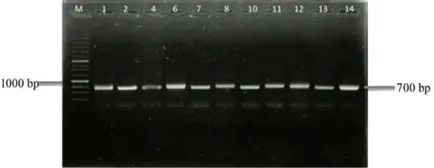
The 16S rRNA gene was amplified using special set primers for actinobacteria ActF/ActR (640bp) for ampli-fying the 16S rRNA gene in actinobacteria (Stach et al. 2003). Based on 16S rRNA actinobacteria gene amplifica-tion, 14 isolates confirmed as actinobacteria as presented in Figure2.
3.3. Screening of FADH2-dependent halogenase gene
Genetic screening of FADH2-dependent halogenase gene
was carried out using primers Halo-B4-FW and Halo-B7-RV (Hornung et al. 2007). Figure3 shows four isolates had 1000 bp as PCR product, while the correct size of FADH2-dependent halogenase gene targeted was 550 bp.
FIGURE 2Visualiza on of 16S rRNA gene representa ve selec ve amplifica on of 640 bp fragments using primers ActF/ActR in 1% (w/v) agarose gel stained with RedSafe.
FIGURE 3Visualiza on of FADH2-dependent halogenase gene
representa ve selec ve amplifica on of 550 bp fragments using primers Halo-B4-FW and Halo-B7-RV in 1% (w/v) agarose gel stained with RedSafe.
Based on BLAST analysis in NCBI database revealed the 1000 bp sequences has no similarity with any halogenase genes. This concludes that none of actinobacteria in this study contain FADH2-dependent halogenase.
3.4. Screening of NRPS gene (non-ribosomal pep de synthase)
Detection of NRPS gene was carried out using set primers A3F/A7R. All of the templates were amplified at 700bp (100%) as represent in Figure4. This results show pres-ence of NRPS gene is dominant. Ayuso-Sacido and Ge-nilloud(2005) screened 210 isolates of actinobacteria and obtained 167 have NRPS gene (79.5%).
3.5. Bioassay an vibrio
Bioassay anti-vibrio was performed using 96 well plate diluted bioassay. This assay was adopted fromGibbons
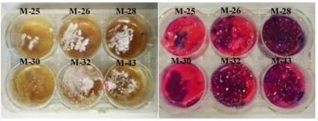
(2006) with slight modifications. Test material used for this assay was fermentation extract of actinobacteria. The changing color from purple to pink or colourless is due to metabolism of bacteria means that the metabolites were inactive. While the absence of color changing means that no bacterial metabolism because the compounds killed the bacteria (Figure5). About eight of isolates actinobaceria showed inhibition againstV. alginolyticus.
Bioassay antivibrio also performed in overlay agar us-ing 6 well plate in 6 different media fermentation. Acti-nobacteria produced secondary metabolites intracellular within cells while extracellular secondary metabolites di-rectly secreted out of the cell and diffused to media. It will determine the purple pattern on antibacterial bioassay. In-tracellular secondary metabolites was showed on purple
FIGURE 5An bacterial qualita ve assay againstVibrio alginoly -cus.
pattern at isolates grown area while the purple pattern of extracellular secondary metabolites was showed around media as represented in Figure6.
3.6. Quick detec on of pep de
Based on detection with the use of ninhydrin as respec-tive visualizing agent showed a total 54 of 84 product fer-mentation of actinobacteria were contained peptide as a bioactive compound. The appearance of pink, purple, or yellow spot on plate showed the compound contained pep-tide with different aminoacid (Figure 7). The pink spot presented the presence of phenylalanine, the purple spot showed the presence of tryptophan, and the yellow spot showed the presence of proline (Gibbons 2006;Zarzycki 2015).
4. Discussion
NRPS are large multimodular enzymes involved in biosyn-thesis of peptide secondary metabolites produced by mi-croorganisms, such as bacteria (Felnagle et al. 2008). Some of antibiotics such as vancomycin and teicoplanin have exquisitely complex structures catalysed by NRPS (Pace and Yang 2006). Indeed, almost all antibiotic pep-tide are catalysed by NRPS. The presence ofnrpsgene in isolates can be proved by molecular screening and the expression of this gene in producing peptide should be checked through chemical screening.
NRPS and FADH2-dependent halogenase are
en-zymes involved in the biosynthesis of important biologi-cal compounds produced by microorganism including
acti-FIGURE 6Results of bioassay an vibrio before (le ) and a er indi-cator (right).
FIGURE 7TLC visualiza on of secondary metabolites of ac nobac-tera with ninhydrin reagent.
nobacteria. A band about 700 bp length was detected in 14 isolates by PCR screening of NRPS gene. The high detection levels of NRPS biosynthetic systems ob-served in our isolates confirmed the wide distribution of these sequences in this bacterial group. While detection of FADH2-dependent halogenase gene was obtained a band
about 1000 bp length.Hornung et al.(2007) mentioned the precise length of this gene was about 550 bp. Analysis of this 1000 bp sequences in NCBI database confirmed this sequences has no similarity with any halogenase genes. In this research none of FADH2-dependent halogenase gene
was detected. Existence of this gene as auxiliary gene of secondary metabolites biosynthesis is rare or even absent. The presence of FADH2-dependent halogenase gene in all
observed isolates was only 4% compared with existence of NRPS and PKS gene as the backbone of biosynthetis secondary metabolites (Hur et al. 2012).
Bioactivity screening was conducted to the fermenta-tion product of actinobacteria that active as antibacterial agent. Fermentation was asessed by growing actinobac-teria in various media to creating diverse environmental conditions. One strain many compound (OSMAC) show one strain of bacteria can produce diverse compounds de-pending on environmental conditions (Bode et al. 2002). A variety compounds are produced during actinobacteria fermentation and due to the nutrient limitation for the de-fense mechanisms.
Resazurin is an oxidation–reduction indicator used for the evaluation of bacterial growth (McNicholl et al. 2007). It is a blue dye that becomes pink when reduced to resofu-rin by oxidoreductases Gram negative bacteria (Leonard et al. 2008). Resofurin is further reduced to hydroresofu-rin (uncolored). The absence of indicator change color in-dicate that the fermentation product of actinobacteria was able to inhibit the growth ofV. alginolyticus. The change color become pink or colorless showed that the fermen-tation product of actinobacteria failure to inhibit the test strain and the cells ofV. alginolyticusstill remain on the well.
Identification of peptide is important to assure that
FIGURE 8Chart of gene c, bioac vity, and pep de screening of ac nobacteria associated red algaeGelidiella acerosa.
amino acids and ninhydrin produces reddish color as rep-resented of most amino acids except proline and hydrox-yproline produce yellow color. The result of chemical screening revealed that almost all isolates produced sec-ondary metabolites containing peptide. The genetic, chem-ical, and bioactivity result of 14 isolates actinobacteria was showed in Figure8. There are 47% isolates havenrps
gene, produce peptide and active againstV. alginolyticus, while the same number of isolates havenrpsgenes, pro-duce peptide but inactive againstV. alginolyticus. Only 6% isolates havenrpsgene, no peptide but active against
V. alginolyticus.
In this study, we identify one potential isolate of acti-nobacteria. Based on sequences analysis of 16S rRNA gene sequences of isolates DR-2S-115-5 has similarity
to Nocardiopsis alba PCM 2702 (97%). Nocardiopsis
species are able to prevail under different environmen-tal conditions mainly because of their versatile genetic make-up, secretion of enzymes, production of compati-ble solutes and surfactants (Li et al. 2013;Bennur et al.
N N H
H N
N H O
O O
O
O N N
S NH
NH
O N
NH
N S
NH S N
NH
NH O
O
O O O O
HO
TP-1161
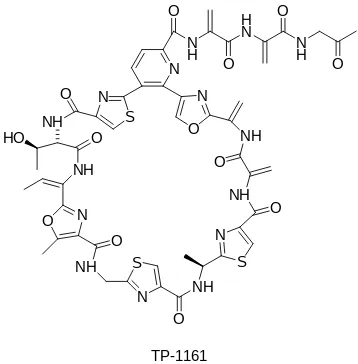
FIGURE 9Chemical structure of macrocyclic thiopep de an bi-o cs TP-1161 prbi-oduced byNocardiopsis.
FIGURE 10Phylogene c tree of isolates DR-2S-115-5 based on 16S rRNA gene sequence by neighbour joining method with 1000x bootsrap replica on.
2014). In addition to these features, they produce an array of bioactive compounds that may help their survival un-der these conditions. Nocardiopsishas been isolated from marine sediments, marine invertebrates association, and hyper saline environments (He et al. 2015).Nocardiopsis
produce bioactive compounds with various activities such as antibiotics, anticancer, and immunomodulator agent (Bennur et al. 2016). Macrocyclic thiopeptide antibiotics TP-1161 produced by marineNocardiopsissp. TFS65-07 isolated from sediments at a depth of 24 m collected in Trondheim Fjord, Norway. TP-1161 showed antibacterial activity against Gram-positive bacteria (Engelhardt et al. 2010). Peptidolipins, peptide antibiotics produced by No-cardiasp. WMMB 215 isolated from ascidian Trididem-num orbiclatum. These compounds showed bacteriostatic activity agaisnt MRSA and MSSA (methicillin sensitive
Staphylococcus aureus) (Wyche et al. 2012).
Analysis of the NRPS sequences with registered pro-tein in the NCBI database confirmed that the NRPS gene of isolate DR-2R-115-35 has similarity to the genus Strep-tomyces. This genus has commercial interest due to their unique capacity to produce novel metabolites. Total of 80% antibiotics have been discovered fromStreptomyces
(Naikpatil and Rathod 2011) included antibiotic on the market such as streptomycin, tetracycline, chlortetracy-cline, neomycin, spiramycin, and daptomycin. The pres-ence of NRPS gene of isolate DR-2R-115-35 have a high
potential to produce antibiotic.
5. Conclusions
Our result revealed that actinobacteria associated red algae
Gelidiella acerosaare potential as producer of antibacte-rial compound. Genetic based screening yielded that 14 actinobacteria containednrpsgene associated with marine peptide. The bioactivity assay showed that eight isolates of actinobacteria were active againstV. alginolyticus. In conclusion, isolate actinobacteria are wealth sources of bioactive compounds.
Acknowledgments
The authors would like to thank Universitas Gadjah Mada for supporting our research under scheme Hibah Kompe-tensi (HIKOM) 2014: LPPM-UGM/1191/UT/2014, New Zealand Community Resilience and Economic Develop-ment (CaRED) Program and Grant Student Publication BPP UGM 2016: 955/BPP/2016 to Noer Kasanah. The authors are thankful to Research Group of Marine Biotech-nology for the assistance.
Authors’ contribu ons
MU carried out the laboratory work and wrote the manuscript. NK as the first adviser designed the study, wrote the manuscript, and reviewed the manuscript. NSNH reviewed the manuscript. All authors read and ap-proved the final version of the manuscript.
Compe ng interests
Authors declare no conflict of interest related to this manuscript.
References
Austin B. 2010. Vibrios as causal agents of zoonoses. Vet Microbiol. 140(3-4):310–317. doi:10.1016/j.vetmic.2009.03.015.
Ayuso-Sacido A, Genilloud O. 2005. New PCR primers for the screening of NRPS and PKS-I systems in acti-nomycetes: detection and distribution of these biosyn-thetic gene sequences in major taxonomic groups. Microb Ecol. 49(1):10–24. doi: 10.1007/s00248-004-0249-6.
Bennur T, Kumar AR, Zinjarde S, Javdekar V. 2014.
Nocardiopsisspecies as potential sources of diverse and novel extracellular enzymes. Appl Microbiol Biotechnol. 98(22):9173–9185. doi: 10.1007/s00253-014-6111-y.
Bennur T, Ravi Kumar A, Zinjarde S, Javdekar V. 2016.
Nocardiopsis species: a potential source of bioac-tive compounds. J Appl Microbiol. 120(1):1–16. doi:10.1111/jam.12950.
Blunt JW, Copp BR, Keyzers RA, Munro MHG, Prinsep MR. 2014. Marine natural products. Nat Prod Rep. 31(2):160. doi:10.1039/c3np70117d.
Bode HB, Bethe B, Höfs R, Zeeck A. 2002. Big effects from small changes: possible ways to explore nature’s chemical diversity. ChemBioChem 3(7):619–627. doi: 10.1002/1439-7633(20020703)3:7<619::AID-CBIC619>3.0.CO;2-9.
Bull AT, Ward AC, Goodfellow M. 2000. Search and discovery strategies for biotechnology: the paradigm shift. Microbiol Mol Biol Rev. 64(3):573–606. Egan S, Thomas T, Kjelleberg S. 2008.
Unlock-ing the diversity and biotechnological potential of marine surface associated microbial commu-nities. Curr Opin Microbiol. 11(3):219–225. doi:10.1016/j.mib.2008.04.001.
El-Shatoury SA, El-Shenawy NS, El-Salam IMA. 2009. Antimicrobial, antitumor and in vivo cytotoxic-ity of actinomycetes inhabiting marine shellfish. World J Microbiol Biotechnol. 25(9):1547–1555. doi:10.1007/s11274-009-0040-4.
Elsie BH, Dhanarajan MS, Sudha PN. 2011. Invitro screening of secondary metabolites and antimicrobial activities of ethanol and acetone extracts from red sea-weedGelidium acerosa. J Chem Res. 2(2):1–3. Engelhardt K, Degnes KF, Zotchev SB. 2010.
Iso-lation and characterization of the gene cluster for biosynthesis of the thiopeptide antibiotic TP-1161. Appl Environ Microbiol. 76(21):7093–7101. doi:10.1128/AEM.01442-10.
Felnagle EA, Jackson EE, Chan YA, Podevels AM, Berti AD, McMahon MD, Thomas MG. 2008. Nonriboso-mal peptide synthetases involved in the production of medically relevant natural products. Mol Pharmaceu-tics 5(2):191–211. doi:10.1021/mp700137g.
Gibbons S. 2006. An introduction to planar chromatogra-phy. In: SD Sarker, Z Latif, AI Gray, editors. Methods in biotechnology-natural products isolation. Totowa, NJ: Humana Press. p. 77–116. doi: 10.1385/1-59259-955-9:77.
Gómez-León J, Villamil L, Lemos ML, Novoa B, Figueras A. 2005. Isolation of Vibrio alginolyticus and Vib-rio splendidus from aquacultured carpet shell clam (Ruditapes decussatus) larvae associated with mass mortalities. Appl Environ Microbiol. 71(1):98–104. doi:10.1128/AEM.71.1.98-104.2005.
He ST, Zhi XY, Jiang H, Yang LL, Wu JY, Zhang YG, Hozzein WN, Li WJ. 2015. Biogeography of Nocar-diopsisstrains from hypersaline environments of Yun-nan and Xinjiang Provinces, western China. Scientific Reports 5:13323. doi:10.1038/srep13323.
Holt JG. 1994. Bergey’s manual of determinative bacteri-ology. Lippincott Williams & Wilkins.
Nichol-son GJ, Bechthold A, Süssmuth RD, Vente A, Pelzer S. 2007. A genomic screening approach to the structure-guided identification of drug candidates from natural sources. ChemBioChem 8(7):757–766. doi:10.1002/cbic.200600375.
Hur GH, Vickery CR, Burkart MD. 2012. Explorations of catalytic domains in non-ribosomal peptide syn-thetase enzymology. Nat Prod Rep. 29(10):1074– 1098. doi:10.1039/c2np20025b.
Kanagasabhapathy M, Sasaki H, Haldar S, Yamasaki S, Nagata S. 2006. Antibacterial activities of marine epibiotic bacteria isolated from brown algae of Japan. Ann Microbiol. 56(2):167–173.
Kasanah N, Hamann MT. 2004. Development of antibi-otics and the future of marine microorganisms to stem the tide of antibiotic resistance. Curr Opin Investig Drugs 5(8):827–837.
Lam KS. 2006. Discovery of novel metabolites from ma-rine actinomycetes. Curr Opin Microbiol. 9(3):245– 251. doi:10.1016/j.mib.2006.03.004.
Lee DW, Lee SD. 2008.Tessaracoccus flavescenssp. nov., isolated from marine sediment. Int J Syst Evol Micro-biol. 58(4):785–789. doi:10.1099/ijs.0.64868-0. Lee SD. 2006. Phycicoccus jejuensis gen. nov., sp.
nov., an actinomycete isolated from seaweed. Int J Syst Evol Microbiol. 56(10):2369–2373. doi:10.1099/ijs.0.64271-0.
Leonard B, Coronel J, Siedner M, Grandjean L, Caviedes L, Navarro P, Gilman RH, Moore DAJ. 2008. Inter-and intra-assay reproducibility of microplate ala-mar blue assay results for isoniazid, rifampicin, ethambutol, streptomycin, ciprofloxacin, and capre-omycin drug susceptibility testing ofMycobacterium tuberculosis. J Clin Microbiol. 46(10):3526–3529. doi:10.1128/JCM.02083-07.
Li HW, Zhi XY, Yao JC, Zhou Y, Tang SK, Klenk HP, Zhao J, Li WJ. 2013. Comparative ge-nomic analysis of the genus Nocardiopsis provides new insights into its genetic mechanisms of envi-ronmental adaptability. PLOS ONE 8(4):e61528. doi:10.1371/journal.pone.0061528.
Luyt CE, Bréchot N, Trouillet JL, Chastre J. 2014. Antibi-otic stewardship in the intensive care unit. Crit Care 18(5):480. doi:10.1186/s13054-014-0480-6.
Matsuo Y, Kanoh K, Jang JH, Adachi K, Matsuda S, Miki O, Kato T, Shizuri Y. 2011. Streptobactin, a tricatechol-type siderophore from marine-derived
Streptomycessp. YM5-799. J Nat Prod. 74(11):2371– 2376. doi:10.1021/np200290j.
McNicholl BP, McGrath JW, Quinn JP. 2007. Development and application of a resazurin-based biomass activity test for activated sludge plant management. Water Res. 41(1):127–133. doi:10.1016/j.watres.2006.10.002.
Mustapha S, Mustapha EM, Nozha C. 2013. Vibrio
al-ginolyticus: an emerging pathogen of foodborne dis-eases. Int J Sci Technol. 2:302–309.
Naikpatil SV, Rathod JL. 2011. Selective isolation and antimicrobial activity of rare actinomycetes from man-grove sediment of Karwar. J Ecobiotechnol. 3(10):48– 53.
Pace JL, Yang G. 2006. Glycopeptides: Update on an old successful antibiotic class. Biochem Pharmacol. 71(7):968–980. doi:10.1016/j.bcp.2005.12.005. Penesyan A, Kjelleberg S, Egan S. 2010.
Develop-ment of novel drugs from marine surface associ-ated microorganisms. Mar Drugs 8(3):438–459. doi:10.3390/md8030438.
Piel J, Hui D, Wen G, Butzke D, Platzer M, Fuse-tani N, Matsunaga S. 2004. Antitumor polyke-tide biosynthesis by an uncultivated bacterial sym-biont of the marine sponge Theonella swinhoei. Proc Natl Acad Sci USA 101(46):16222–16227. doi:10.1073/pnas.0405976101.
Pée KHv, Zehner S. 2003. Enzymology and molecular genetics of biological halogenation. In: Natural pro-duction of organohalogen compounds. The handbook of environmental chemistry. Berlin: Springer. p. 171– 199. doi:10.1007/b10457.
Schwartsmann G, Brondani da Rocha A, Berlinck RG, Jimeno J. 2001. Marine organisms as a source of new anticancer agents. Lancet Oncol. 2(4):221–225. doi:10.1016/s1470-2045(00)00292-8.
Schwarzer D, Marahiel MA. 2001. Multimod-ular biocatalysts for natural product assem-bly. Naturwissenschaften 88(3):93–101. doi:10.1007/s001140100211.
Shaw KS, Goldstein RER, He X, Jacobs JM, Crump BC, Sapkota AR. 2014. Antimicrobial susceptibility of
Vibrio vulnificusandVibrio parahaemolyticus recov-ered from recreational and commercial areas of Chesa-peake Bay and Maryland Coastal Bays. PLOS ONE 9(2):e89616. doi:10.1371/journal.pone.0089616. Singh RP, Reddy CRK. 2014. Seaweed-microbial
interactions: key functions of seaweed-associated bacteria. FEMS Microbiol Ecol. 88(2):213–230. doi:10.1111/1574-6941.12297.
Soria-Mercado IE, Villarreal-Gómez LJ, Rivas GG, Sánchez NEA. 2012. Bioactive compounds from bac-teria associated to marine algae. In: R Sammour, ed-itor. Biotechnology - molecular studies and novel ap-plications for improved quality of human life. InTech. doi:10.5772/27842.
Stach JEM, Maldonado LA, Ward AC, Goodfellow M, Bull AT. 2003. New primers for the class Acti-nobacteria: application to marine and terrestrial environments. Environ Microbiol. 5(10):828–841. doi:10.1046/j.1462-2920.2003.00483.x.
from an ascidian-derivedNocardia sp. J Nat Prod. 75(4):735–740. doi:10.1021/np300016r.