INTRODUCTION
Graves’ disease is a thyroid autoimmune disease with several clinical manifestations such as hyperthyroidism, diffuse goiter, ophtalmopathy and with our without dermopathy.(1)
Incident of Graves’ disease was about 40 cases/100.000 population per year and tend to increase anually. In Wickham study in the United Kingdom, the incidence was reported to be
100 – 200 cases per 100.000 population per year. The incidence in women reported more frequent than men. In Indonesia, incidence of hyperthyroidism caused by Graves’ disease not yet known, but were reported about 44,44% - 48,93% from all of patients with thyroid
disease.(2,3,4) Graves’ diseasewere the most common organ specific autoimmune disease and tend to increase through years. Genetic and environment factors thought to have a role in
increasing of autoimmune disease, such as MHC-II expression, autoreactive B-cell activation
and imbalance of Th1/Th2. Moreover, infection, stress, and excessive consumption of iodium
were also suspected to trigger immune factor that cause Graves’ disease.(5,6)
Hyperthyroidism in Graves’ disease patient cause by increasing of thyroid antibody which cause thyrotoxicosis. This antibody binds to thyroid stimulating hormone (TSH)
receptor and mimics TSH activity in thyroid gland, known as TSH receptor antibody (TRAb).
Antibody activation trigger increasing of thyroid hormone which cause hyperthyroidism
manifestation in Graves’ disease. (7,8,9,10) TSH receptor antibody (TRAb) found 100% on Graves’disease compared to other thyroid antibody such as thyroperoxidase antibody (TPO-Ab) and thyroglobuline, so that increased of TRAb was one of diagnostic point in Graves’ disease. Previous study also shown decreasing of TRAb in patients who already received anti
thyroid medication, so it is often used as remission marker of disease activity.(11,12)
classified thyroid gland enlargement into 3 (three) gradation : grade I (goitre only found from
palpable); grade II (goitre is palpative and can be easily seen); and grade III (goitre is very
big and retrosternal).(6) Ling Chen (2008) shown that increasing of thyroid gland size related
to severity and progresivity of the disease.(13) Aim of this study was to investigated the
association between TSH receptor antibody (TRAb) with goiter size.
MATERIAL AND METHODS Study population
Blood samples were obtained from 30 patients with Graves’ disease. Diagnosis of Graves’ disease was based on the clinical manifestations found such as hyperthyroidism, diffuse goiter, ophtalmopathy and dermopathy with increasing of thyroid hormone (FT4) and
decreasing of TSH level. Exclusion criteria were as follows : using anti-thyroid medications
before the blood sample is checked. Patients were recruited from Metabolic Endocrinology
Outpatient of Internal Medicine Department, M.Djamil Padang Hospital.
Methods
Serum blood of patients were collected about 3 cc to measure thyroid hormone (FT4),
TSH and TRAb. Free thyroxin (FT4), TSH and TRAb were measured by using ELISA
method. Size of goiter were measured using WHO classification which classified to 3 (three)
gradation : I - goiter in normal posture of head it cannot be seen, only found when palpating;
II - goiter is palpative and can be easily seen; III - goiter is very big and is retrosternal.
Furthermore, thyroid ultrasonography were also performed to measure thyroid volume.
Statistical analysis
Data analysis were determined by correlation analysis using SPSS version 20 (SPSS
Inc). Pearson analysis were using when data normally distributed, or Spearman analysis when
non-normally distributed. Chi-square test analysis were use to analyze categorical data.
RESULT
The aim of this study was to investigate the correlation between antibody receptor
TSH (TRAb) and goiter size in Graves’ disease patients. The study is cross sectional, composed of 30 patients of hyperthyroidism caused by Graves’ disease. Table 1 summarized baseline characteristic of this study, which is 66,7% patients were female and mean of age of
this study were 40,7 ± 14,81 years, range from 19 to 74 years old.
The mean serum concentration of free T4 (fT4) was found increase significantly
(74,28 ± 61,1 nmol/L), with reference value was 9-20 nmol/L. Mean serum concentration of
TSH were decrease significantly with 0,1 ± 0,09 IU/ml (normal reference 0,25-5 IU/ml).
Increasing of TRAb and lower serum of TSH values were found to be 100% as shown in
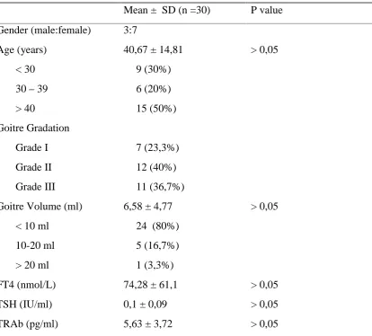
Table 1. Baseline characteristic of Graves’ disease patients.
Mean ± SD (n =30) P value
Gender (male:female) 3:7
Age (years) 40,67 ± 14,81 > 0,05
< 30 9 (30%)
30–39 6 (20%)
> 40 15 (50%)
Goitre Gradation
Grade I 7 (23,3%)
Grade II 12 (40%)
Grade III 11 (36,7%)
Goitre Volume (ml) 6,58 ± 4,77 > 0,05
< 10 ml 24 (80%)
10-20 ml 5 (16,7%)
> 20 ml 1 (3,3%)
FT4 (nmol/L) 74,28 ± 61,1 > 0,05
TSH (IU/ml) 0,1 ± 0,09 > 0,05
TRAb (pg/ml) 5,63 ± 3,72 > 0,05
FT4, free thyroxine; TSH, thyroid stimulating hormone; TRAb, thyroid stimulating hormone
receptor antibody.
Presence of thyroid antibody (TRAb)
Mean serum concentration of TRAb was significantly higher than normal limit (p >
0,05). The reference value of TRAb was > 0,1 pg/ml for positive Graves’ disease.
Goiter gradation and volume
Gradation of goiter in this study were using WHO classification, divided to three (3)
grade, grade I (goiter only found by palpable), grade II (goiter is palpative and easily seen)
most in grade II (40%) than grade I, III (23,3% and 36,7% respectively). Size of goiter were
also measured using ultrasonography by adding left and right goiter lobus volume, and in this
study mean of goiter volume were 6,58 ± 4,77 ml with 80% of goiter volume was less than
10% (table 1). Only 1 case were found > 20 ml (3,3%).
Correlation of thyroid antibody and goiter size
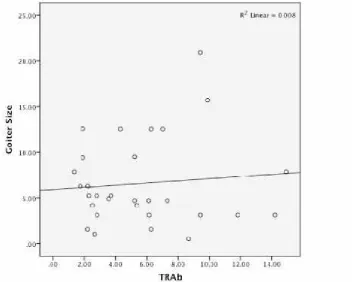
As shown in figure 1 below, we found there is no correlation between thyroid
antibody and goiter volume (r = 0,63; p-value = 0,09). We are also found there was no
significant correlation between TRAb and gradation volume by using Chi-square test analysis
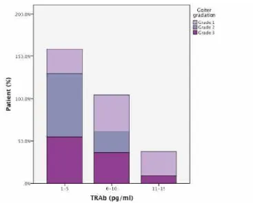
(r = 0,39). Figure 2 described that TRAb level decreasing with advanced of goiter gradation.
Fig 2. Goiter gradation as a function of TRAb (Grade I ; Grade II ; Grade III)
DISCUSSION Gender and Age.
Based on gender, female patients were found more than male with percentage 66,7 :
33,3. Similar result was also found in Marina (2010) with 90% samples were female, and
only 10 % were male patients. Hartono (2010), Mao (2011) and Zhu (2012) were also shown
similar results with our study.(7,10,14) This phenomenon was initially ascribed to hormonal
differences, estrogens exacerbate and androgens inhibit immune responses, which influenced
by gender.(15)
In this study, onset age of Graves’ disease varied from puberty to old age, with range of age from 19 to 74 years old, and mostly found in the age over 40 years (50%) and the
study that 50% of patients were over 40 years old. Mao (2011) shown that mean of age of 77
patient of Graves’ disease were 41,1± 12,7 years old.(7,10)
Correlation of thyroid antibody (TRAb) and goiter size.
Graves’ disease is a Th2 mediated disease with mild lymphocytic infiltration of the thyroid and induce activation of auto-reactive B cells that secrete thyrotropin receptor
(TSH-R) stimulating antibodies (TRAb), which cause hyperthyroidism.(15,16)Enlargement of thyroid
(goiter), ophtalmopathy, and or dermopathy were found as manifestation in Graves’ disease patient which is caused by occurrence of hyperthyroid.(17,18)
The aim of this study was to correlate between thyroid antibody and goiter size, which
were described as goiter gradation and volume. Goiter gradation of this study mostly found in
grade II (40%), followed by grade III (36,7%) and grade I (23,3%). Ling Chen (2008) was
found that from 154 Graves’ disease patient, grade II also found the most, followed by grade III and I. Marina (2010) studied 25 patients, also found grade II the most (60%), followed by
grade I and III. Vos (2009) from 263 patients shown different result with grade I found at
most.(12,13)
Thyroid volume through ultrasonography result shown that mean of thyroid volume
of Graves’ disease patient on this study were 6,58 ± 4,77 ml, with size < 10 ml were found at most. Different from Klatka (2014) that shown mean of thyroid volume were 24,14 ml, with
size range between 10,47–35,5 ml. Remission or relapse mayoccur in Graves’ patients with large thyroid gland.(18,19)
The hyperactive of the thyroid gland is due to the presence of TRAb, and these are
now known to recognize and activate the thyroid stimulating hormone receptor (TSHR).
These TRAb increase the growth and the function of the thyroid follicular cells leading to the
anxiety and weight loss among others.(8) As seen in results, TRAb on this study were found
increase between 1,36 – 14,93 IU/l with mean 5,63 ± 5,21 IU/l. Skowronck (2013) study showed increased TRAb in 30 patients with Graves’ with range between 7 –462 IU/l. Klatka (2014) study showed almost the same result as our research with mean 7,63 ± 4,62 IU/l.
(19,20,21)While Zhu (2012) showed mean of TRAb 24,21 ± 6,06 IU/l.(14)This difference result
may occurred due to difference in number of sample and characteristic of studied subjects
such as age and method of analysis.
On this study, main finding of our study is that volume goiter and gradation is not
related to the TRAb (TSHR antibody) level (p > 0,05) as seen in figure 1. However, as seen
in figure 2, we can see clearly that TRAb level decreased with advanced of goiter gradation,
which described that the more of TRAb level didn’t correlate with enlargement of thyroid. Previous study by Ertek (2010) also shown no correlation between goiter volume and thyroid
antibody in Graves’ disease patients, but TSH levels was significantly correlated with thyroid antibodies (p = 0.0000 for TRAb), which is not described in our study.(20,21)Laurberg (2014)
found different result that there was correlation between goiter size and TRAb level (r = 0.25,
p < 0.001).(22)
Contribution of genetic and environment factors to Graves’ disease such as iodine consumption, drugs, infection, smoking and stress may influenced level of TRAb.(23)
Complex interaction between these factors may alter central and peripheral immune tolerance
that affect to goiter size and level of TRAb.(24,25) Limitation of this study is that we are not
measure iodine consumption in our study; thus we cannot see if there is one of this factor
contribute to our result. Research of T-cell role in Graves’ disease, such as T regulator and cytokines begin to grow. In Graves’ disease, both Th1 mediating cell mediated immunity and Th2 mediating humoral immunity occur. Studies have shown that active inflammatory phase
switches to a Th2 phenotype in long standing Graves' disease. Cytokine expression profiles in
sera and thyroid tissues from Graves’ disease patients indicate a mixed Th1/Th2 status at any time. Further studies, it’s necessary to examine the role of cytokines that provide by T-cell on Graves’ disease.(26,27)
CONCLUSION
There is no correlation between goiter gradation and volume to TRAb serum levels. Further
studies, the interaction between genetic and environmental factors such as iodine
consumption, infection, smoking and stress should be investigated considering influence of
these factors in development of the disease.
REFERENCES
1. Schott M, Scherbaum WA. Autoimmune thyroid disease: review article. Dtsch Arztebl 2006;103(45):A 3023-32.
2. Swain M, Swain T, Mohanty BK. Autoimmune thyroid disorders-an update. Indian Journal of Clinical Biochemistry 2005;20(1):9-17.
3. Bahn RS, Burch HB, Cooper DS. Hyperthyroidism and other causes of thyrotoxicosis: management guidelines of the American Thyroid Association and American of Clinical Endocrinologists. Thyroid 2011;21(6):593-646.
4. Vanderpump M. The epidemiology of thyroid disease. British Med Bulletin 2011;99(1):39-51.
5. Ai J, Leonhardt JM, Heymann WR. Autoimmune thyroid disease: Etiology, pathogenesis and dermatologic manifestations. J Am Acad Dermatol 2003;48(5):641-656.
6. Delange F, Bastani S, Benmiloud M, De Maeyer E, Isayama MG, Koutras D, Muzzo S, Niepomniszcze H, Pandav CS, Riccabona G. Definitions of endemic goiter and cretinism, classification of goiter size and severity of endemias, and survey techniques. In: Towards the Eradication of Endemic Goiter, Cretinism and Iodine Deficiency, (ed.) Dunn JT, Pretell E, Daza CH, Viteri FE, Washing- ton, PAHO Sci Publ 1986; 5:373–376.
7. Mao C, Wang S, Xiao Y, Xu J, Jiang Q, Jin M. Impairment of regulatory capacity of CD4+CD25+ regulatory T cells mediated by dendritic cell polarization and hyperthyroidism in Graves’ disease. Journal of Immunology 2011;1-10.
8. Morshed SA, Latif R, Davies TF. Delineating the autoimmune mechanisms in Graves’ disease. Immunol Res2012;1-13.
9. Morshed SA, Latif R, Davies TF. Immunopathogenesis of Graves’ disease. In George S Eisenbarth (ed) Immunoendocrinology: Scientific and Clinical Aspects, Contemporary Endocrinology. Springer Science Business Media. 2011: 457-481. 10. Hartono A. Tesis: Hubungan kadar interleukin-4 dengan Triiodotironin (T3), Tiroksin
11. Dalan R, Shwleaw K. Immune manipulation for Graves’ disease: re-exploring an unfulfilled promised with modern traditional research. European Journal of Internal Medicine. 2012;23:682-92.
12. Vos XG, Smit N, Endert E, Brosschot JF, Tijssen JG, Wiersinga WM. Low stress level is not causally related to biochemically less severe Graves’ disease in older patients. Eur J Endocrinol 2009;160:193-9.
13. Ling C, Hong-qing W, Yan-yan Y, Jun L, Men W, Jie B et al. Comparison of Methimazole/Hydrocortison ointment with Graves disease: A prospective, randomized, open-label, parallel-group, 18-month study. Current Therapeutic Research 2008;69(4):305-317.
14. Zhu C, Ma J, Liu Y, Tong J, Tian J, Chen J. Increased frequency of follicular helper T cells in patients with autoimmune thyroid disease. J Clin Endocrin Metab 2012;97:943-950.
15. Lichiardopol C, Mota M. The thyroid and autoimmunity: review. Rom. J. Intern. Med. 2009;47(3):207-15.
16. Hongxiang W, Shi Z, Xiaoqiong T et al. Changes of regulatory T cells in Graves’ disease. Journal of Huazhong University of Science and Technology.2006;26(5):545-7.
17. El-Kaissit, Wall JR. Determinants of extraocular muscle volume in patients with Graves’ disease. Journal of Thyroid Research 2012;1-4.
18. Kamath C, Adlan MA, Premawardhana LD. The role of thyrothropin receptor antibody assays in Graves’ disease. Journal of Thyroid Research. 2012;1-8
19. Klatka M, Grywalska E, Partyka M, Charytanowicz, Kiszczak-Bochynska E, Rolinski J. Th17 and Treg cells in Graves’ disease. Impact of treatment with methimazole on these cell subsets. Autoimmunity 2014;47(3):201-211.
20. Ben-Skowronek I. Graves’ disease-the interaction of lymphocytes and thyroid cells. In Chapter 12 Autoimmune Disorders-Pathogenic Aspects. Intech Pres, Polland. 2010;229-240.
21. Ben-Skowronek I, Szewczyk L, Kulik-Rechberger B. The differences in T and B cell subsets in thyroid with Graves’ disease and Hashimoto’s thyroiditis. World J End 2013;9(3):245-250.
22.Ertek S, Cicero A, Caglar O, Erdogan G. Relationship between serum zinc levels, thyroid
hormones and thyroid volume following successful iodine supplementation. Hormones 2010,9(3):263-268.
23. Laurberg P, Wallin G, Tallstedt L, Abraham-Nordling M, Lundell G, Torring O. TSH-receptor autoimmunity in Graves’ disease after therapy with antithyroid drugs, surgery, or radioiodine: a 5 year prospective randomized study. Eur J Endocrinol 2008;158(1):69-75.
24. Dejaco C, Duffner C, Grubeck-Loebenstein B, Schirner M. Imbalance of regulatory T cells in human autoimmune disease. Immunology. 2005;117:289-300.
25. Hu Y, Tian W, Zhang L, Liu H, Yin G, He B. Function of regulatory T-cells improved by dexamethasone in Graves’ disease. European Journal of Endocrinology 2012;166:641-6.
26. Wilson R, Killop JH, Chopa M, Thomson JA. The effect of anti-thyroid drugs on B and T cell activity in vitro. Clinical Endocrinology 2008;28(4):389-97.